The Effect of Curcumin on Endogenous Neuron Regeneration in Rats after TBI
←
→
Page content transcription
If your browser does not render page correctly, please read the page content below
Revista Argentina de Clínica Psicológica
2020, Vol. XXIX, N°4, 864-871                                                                                             864
DOI: 10.24205/03276716.2020.893

           The Effect of Curcumin on Endogenous Neuron
                   Regeneration in Rats after TBI
                              Yimei Shua, Tao Wangb, Xiaoqiang Tangb, Changjie Panb*

               Abstract
               Objective: To investigate the effect of curcumin on endogenous neuron regeneration in
               rats after traumatic brain injury (TBI) in rats.
               Methods: TBI model was prepared with controlled cortical impact model. SD rats were
               divided into 3 groups: control group, TBI+vehicle group and TBI+curcumin group. Serum
               SOD and MDA levels were detected by ELISA. The cognitive function of the rats was
               observed by Morris water maze (MWM). Then the TUNEL method was used to detect the
               apoptosis of the injured area. NeuN/BrdU immunofluorescence double labeling was used
               to detect the newly matured neurons in the injured area. Western blot was used to detect
               DCX protein in the hippocampus.
               Results: Curcumin decresed the serum MDA level and increased the serum SOD level in
               rats with TBI. MWM test showed curcumin decreased the escape latency and increased
               the number of platform crossover of rats with TBI. The apoptotic cells in the injured area
               of the rats in the TBI+curcumin group were significantly less than those in other TBI rats.
               More NeuN+/BrdU+ double-label positive neonatal mature neurons were observed in the
               cortex of the TBI+curcumin group, which was more significant than those in other TBI rats.
               The expression of DCX protein in the hippocampus of the rats in the TBI+curcumin group
               was dramatically more than that in other TBI rats.
               Conclusion: Curcumin can reduce the level of oxidative stress in TBI rats, protect neurons
               from apoptosis, and promote the development of neurons in the injured cortex and
               hippocampus, thus improving the learning and memory functions of TBI rats.
               Keywords: curcumin, traumatic brain injury, oxidative stress, cognitive function

Introduction
    Traumatic brain injury (TBI) is one of the                             neurons from apoptosis and replace the missing
common diseases in clinical brain surgery, and it is                       neurons has been a hot spot and a difficult point of
caused by external force. According to the World                           the research[5, 6].
Health Organization (WHO), more than 5 million                                 There is endogenous neurogenesis in the adult
people worldwide die each year from TBI, which                             central nervous system, and this view has been
accounts for 9% of all deaths and is 1.7 times the                         agreed. It is currently believed that endogenous
death toll from AIDS, tuberculosis and malaria[1, 2].                      neurogenesis in the adult central nervous system is
More than 1.3 million of these deaths are the result                       mainly in the subventricular zone and the dentate
of traffic trauma, and by 2020, it is expected to                          gyrus of the hippocampus[7]. The neurogenesis of
increase to more than 2 million, which will be one                         the subgranular layer of the hippocampal dentate
of the leading causes of death and disability[3, 4].                       gyrus is closely related to the learning and memory
TBI is characterized by the occurrence of neuronal                         functions. As the research progressed, it was found
loss, which leads to cognitive function changes and                        that the cortex in the injured area also had
the occurrence of disability. How to protect the                           neurogenesis. However, in the absence of external
                                                                           intervention factors, the neurogenesis of the
a. The First School of Clinical Medicine, Nanjing Medical University,      injured cortical area is limited. How to improve
Nanjing, Jiangsu, China
b. Department of Radiology, Changzhou Second People's Hospital             endogenous neurogenesis to promote the
affiliated to Nanjing Medical University, Changzhou, Jiangsu, China        treatment of TBI has become a potential new
* Author for Correspondence: Changjie Pan
Department of Radiology, Changzhou Second People's Hospital                treatment strategy[7-12].
affiliated to Nanjing Medical University, No. 68 Gehu Middle Road,             Curcumin is an active ingredient extracted from
Changzhou 213164, Jiangsu, China
Email: panchangjie@njmu.edu.cn
                                                                           the rhizomes of turmeric. Curcumin has a number
Phone/Fax: +86-13815032301                                                 of pharmacological activities such as improvement

                                                                         REVISTA ARGENTINA
                                         2020, Vol. XXIX, N°4, 864-871   DE CLÍNICA PSICOLÓGICA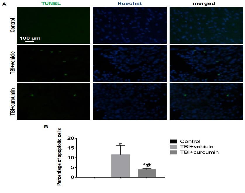
865                                  Yimei Shu, Tao Wang, Xiaoqiang Tang, Changjie Pan

of blood flow, anti-free radicals, and anti-                       Curcumin (Sigma, US) was injected into rats in
inflammatory effects[13-15]. More and more                     TBI+curcumin group at 30 mg / kg immediately after
researches have shown the potential beneficial                 surgery (80 mg curcumin was dissolved in 0.8 ml of
effects of curcumin in the central nervous                     absolute ethanol and afterwards 9.2 ml of normal,
system[13, 16, 17]. It has been reported that                  sterile saline was added in order to produce a
curcumin had the beneficial effects on cerebral                solution of curcumin) once per day, for 14 days at
ischemia[14], TBI[18], and Alzheimer’s disease[19].            the same time. Control and TBI+ group rats were
However, the effect of curcumin on the                         injected by the tail vein with the same quantity of
endogenous neuron regeneration in rats after TBI is            sterile solvent.
not clear. This study aimed to investigate the effect
of curcumin on the endogenous neuron                           Detection of malondialdehyde serum (MDA) and
regeneration in rats after TBI and its possible                SOD levels SOD
mechanism, and provide theoretical and                             1 ml of blood has been collected from the tail
experimental basis for the application of curcumin             vein of rats at 1, 3, 7 and 14 days of surgery. MDA
in the treatment of TBI.                                       and SOD levels in the rat serum were detected in
                                                               MDA and SOD kits (Beyotime Biotechnology,
Methods                                                        China), and strictly in conformity with kit
Pets.                                                          instructions.
    The Nanjing Medical University Animal
Experimental Center has purchased 36,000-220 g                 Morris water labyrinth (MWM) observes the
male and female SD rats. In three groups: control              cognitive function.
group, TBI+vehicle group and the TBI+curcumin                      The cognitive functions of rats were observed
group, the random table method was divided into                with the MWM (Huaibeizhenghua, China) at 15
SD rats. Rats were lodged in groups and sex, free to           days after surgery, as described by Tian[20]. On the
eat and drink, kept in a 12-hour dark / light time             15th day after the operation, the hidden platform
(light on from 8:00am to 8:00pm) at a temperature              testing was initially performed on the rats, and a
of 25±1 ° C with 65±5% humidity. This study has                recording ended after the rats boarded the
been approved by the Nanjing Medical University's              platform for 2 s with a max. recording time of 120 s
Ethics Review Committee. The studies have been                 was recorded for the rats looking for hidden
conducted based on the corresponding guidelines.               platform. Briefly. Rats tested for 4 days three times
                                                               a day. The platform under the water surface was
Preparation of the TBI model                                   removed on the 19th day after the operation and
    The rats had been fixed to a stereotatic                   testing started to detect the number of crossing
instrument after abdominal anesthesiology and the              rats that cross the platform within 120 s.
cranial tops had been cut and prepared, the skin
had been iodine and alcohol disinfested and the                TUNEL Fleece
skin was scalpel-coated and the fascia separated for               After the MWM test, frozen rat brain slice with
the parietal bone exposure. A circlic bone window              a thickness of 15 μm was prepared. The cortical
with a diameter of 5 mm was drilled around the                 apoptotic cells were detected by the TUNEL kit
centre by means of a 3.0 mm left side of the sagittal          (Beyotime Biotechnology, Chinese) and strictly
suture and a 3.5 mm posterior crest, which kept the            followed by the kit instructions. The number of
dura mater intact. Rats were subject to controlled             apoptotic cells under a 400-fold field of view was
cortical impacts using a pneumatic impact device               calculated under a fluorescent microscope (Leica
with 1,5 atm (1 Atm=101,325 kPa) (Model FP302,                 DMR).
AmScien Instruments LLC, USA). The hematoma
appeared immediately in the cortex and the model               Figure of immunofluorescence
was prepared successfully. Layer by layer the                      After MWM test, the rats were prepared with a
incision was sutured. Only the bone window has                 frozen coronate brain slice of 15 μm thickness. At
been opened in the control group and the TBI                   room temperature, the following primary
model has not been prepared. During model                      antibodies were incubated at 6 hours: anti-NeuN
preparation, there was no accidental death in rats.            mouse (mature marker for the neuron) (1.400,
All rats were injected into the bronchial neuron for           Abcam, UK) and anti-BrdU mouse (1:400, Abcam,
5 days intraperitoneically (50 mg / kg, bid).                  UK). The slices were incubated with second-hand
                                                               antibodies for 2 hours after rinsing with the
Curcumin treatment by tail vein injection                      following: Alexa Fluor ® 568-labeled goat anti-

                                                             REVISTA ARGENTINA
                             2020, Vol. XXIX, N°4, 864-871   DE CLÍNICA PSICOLÓGICA
866 Yimei Shu, Tao Wang, Xiaoqiang Tang, Changjie Pan
arbitrary secondary antibody (1:800, Abcam, UK), group. The seral sodium level of TBI+curcumin rats
Alexa Fluor ® 488-labeled goat anti-mouse (1:600, was also reduced at 1 day post-operatively and was
Abcam, UK) with 0,01 mol / L PBS for 3 times. After lower than TBI+ at 3 days, and the sodium level
3 washings of 0.01 mol / L of PBS, Hoechst 33342 rates were reduced at 7 and 14 days to the normal
(1:3000) incubated the slices at room temperature level (Table 2). the memory and learning function of
for 0.5 h. The number of positive cells was rats
measured using a 400-fold field of view with a In comparison to the TBI+ control groups, the
fluorescent microscope (Leica DMR, Germany). rats were considerably proliferated and the exit
latency was also prolonged, but lower than that of
Western blot TBI+ in the TBI+cumin group (Figure 1A). In
The fresh hippocampus tissue was taken from comparison to that of TBI+ vehicles and also in the
the rat after MWM test. The lysate and fully lysed TBI+curcumin group but above that of the TBI+
tissue was added to the hippocampal tissue and vehicle group the platform crossover number of
centribed for 15 minutes at 4 ° C at 12000 g. Each rats was significantly reduced (Figure 1B).
lane has been added 50 μg of the total protein
loading buffer. The protein has been transferred to Rather apoptosis cortical cells
pvdf membrane following SDS / PAGE gel There were almost no apoptotic cells in the rat
electrophoresis. The mouse anti‐β-actin (1:1000, cortex in the control group. The cortex was more
Abcam, UK) were incubated overnight at a apoptotic in rats in the TBI+ car group and the TBI +
temperature of 4 ° C, following blockage, with cumin apoptotical cells was significantly smaller in
rabbit anti-DCX (neuronal precursor cell marker) the cortex of the rats than in the TBI+car (Figure-2).
(1:400, Abcam, UK). HRP tagged goat IgG secondary
antibody (1:1000, Abcam, UK) was then added and Cortex mature neurons neonatal cortex
incubated for one hour at room temperature and There was almost no NeuN+/BrdU+ neonatal
also for a secondary anti-mouse goat HRP tagged mature neurons in the rat cortex within the control
IgG anti-mouse (1:1000, Abcam, UK). Images were group. Many neonatal neurons of NeuN+/BrdU+
taken and the molecular imager ChemiDoc XRS were found in the TBI+ curcumin group (Figure 3),
system was used for densitometric analysis. which was significantly higher than the TBI+ group..
Analysis of statistics Hippocampal DCX level of protein
In order to analyse data in this study, statistical DCX protein expression has decreased
analysis was performed using the Statistical significantly in the TBI+ vehicle group rat
Package for Social Science (SPSS) version 21.0. The hippocampus, composed of the control group, and
data is given as medium ± SD. One-way variance TBI+curcumin expression has decreased, though
analysis (ANOVA) was used to compare the groups. significantly higher than TBI+ vehicle expression
Statistically significant was considered P867 Yimei Shu, Tao Wang, Xiaoqiang Tang, Changjie Pan
was selected in the cortical area above the rat neurons in the injured cortex and hippocampus,
hippocampus, and this area had no effect on the thus improving the learning and memory functions
limb motor function of the rat. On the basis of of TBI rats.
successful modeling, this study first observed the
learning and memory of rats. The results of MWM Conflict of Interest statement
test showed that the learning and memory None.
functions of TBI rats decreased, and the treatment
of curcumin could improve the learning and Acknowledgements
memory functions of TBI rats. What is the possible None.
mechanism of this therapeutic effect of curcumin?
Taking this question into consideration, we have References
observed the antioxidant effect of curcumin. MDA
is a lipid oxidation product, which indicates that the [1] Vander Werff Kathy, R., The Application of the
level of oxidation in the body. SOD is the main International Classification of Functioning,
enzyme for antioxidants, and its level represents Disability and Health to Functional Auditory
the antioxidant capacity in the body. The results in Consequences of Mild Traumatic Brain Injury.
this study showed the rats were in oxidative stress Semin Hear, 2016. 37(3): p. 216-32.
after TBI, and curcumin treatment could decrease [2] Chung, P., S.J. Yun, and F. Khan, A comparison of
the serum MDA level and increase the serum SOD participation outcome measures and the
level, which indicated curcumin had antioxidant International Classification of Functioning,
activity. Next, we used the TUNEL kit to observe the Disability and Health Core Sets for traumatic
apoptosis of the cortex. The result suggested that brain injury. J Rehabil Med, 2014. 46(2): p. 108-
curcumin could protect the injured cortical cells 16.
from apoptosis in TBI rats. [3] Wofford, K.L., D.J. Loane, and D.K. Cullen, Acute
The above results indicated that curcumin had drivers of neuroinflammation in traumatic brain
neuroprotective effect on TBI. What is the effect of injury. Neural Regen Res, 2019. 14(9): p. 1481-
curcumin on endogenous neurogenesis? Taking this 1489.
question into consideration, we used NeuN/BrdU [4] Ntali, G. and S. Tsagarakis, Traumatic brain
immunofluorescence double label to detect the injury induced neuroendocrine changes: acute
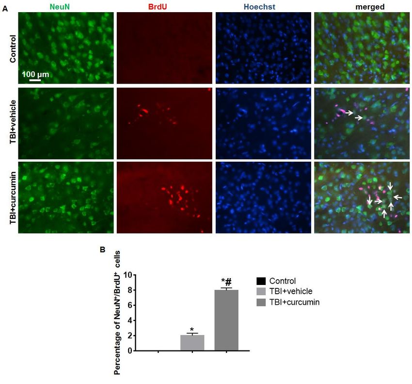
neonatal mature neurons in the injured area. NeuN hormonal changes of anterior pituitary function.
is one of the mature neuron nuclear antigen Pituitary, 2019. 22(3): p. 283-295.
markers[23, 24], and BrdU is one of the markers of [5] Bao, W., et al., Complement cascade on severe
cell proliferation[25]. The result showed that many traumatic brain injury patients at the chronic
NeuN+/BrdU+ neonatal mature neurons were found unconscious stage: implication for pathogenesis.
in TBI+curcumin group, which was significantly Expert Rev Mol Diagn, 2018. 18(8): p. 761-766.
more than that in the TBI+vehicle group, indicating [6] Zhou, Y., et al., Advance of Stem Cell Treatment
that curcumin promoted neuronal regeneration in for Traumatic Brain Injury. Front Cell Neurosci,
the injured cortical area after TBI in rats. In addition, 2019. 13: p. 301.
we used Western blot to detect the expression of [7] Ngwenya, L.B. and S.C. Danzer, Impact of
DCX protein in rat hippocampus. DCX only appears Traumatic Brain Injury on Neurogenesis. Front
in neuronal precursor cells[26]. The result showed Neurosci, 2018. 12: p. 1014.
that the expression of DCX protein in the [8] Liao, R., et al., Histamine H1 Receptors in Neural
hippocampus of the rats in the TBI+vehicle group Stem Cells Are Required for the Promotion of
was significantly decreased compared with control Neurogenesis Conferred by H3 Receptor
group, and that in TBI+curcumin group was also Antagonism following Traumatic Brain Injury.
reduced, but significantly more than that in the Stem Cell Reports, 2019. 12(3): p. 532-544.
TBI+vehicle group, which indicated the number of [9] Li, Q., et al., N-Acetyl Serotonin Protects Neural
newborn neuronal precursor cells in hippocampus Progenitor Cells Against Oxidative Stress-
after TBI was reduced, after treatment with Induced Apoptosis and Improves Neurogenesis
curcumin, the development of neonatal precursor in Adult Mouse Hippocampus Following
cells in hippocampus could be significantly Traumatic Brain Injury. J Mol Neurosci, 2019.
promoted. 67(4): p. 574-588.
In summary, curcumin can reduce the level of [10] Yang, Y., et al., MiR-124 Enriched Exosomes
oxidative stress in TBI rats, protect nerve cells from Promoted the M2 Polarization of Microglia and
apoptosis, and promote the regeneration of Enhanced Hippocampus Neurogenesis After
REVISTA ARGENTINA
2020, Vol. XXIX, N°4, 864-871 DE CLÍNICA PSICOLÓGICA868 Yimei Shu, Tao Wang, Xiaoqiang Tang, Changjie Pan
Traumatic Brain Injury by Inhibiting TLR4 signaling pathways. Recent Pat Food Nutr Agric,
Pathway. Neurochem Res, 2019. 44(4): p. 811- 2019.
828. [19] Thota, R.N., et al., Dietary Supplementation with
[11] Yan, R., et al., Hes1 negatively regulates Curcumin Reduce Circulating Levels of Glycogen
neurogenesis in the adult mouse dentate gyrus Synthase Kinase-3beta and Islet Amyloid
following traumatic brain injury. Exp Ther Med, Polypeptide in Adults with High Risk of Type 2
2018. 16(3): p. 2267-2274. Diabetes and Alzheimer's Disease. Nutrients,
[12] Wu, H., et al., Growth Differentiation Factor 5 2020. 12(4).
Improves Neurogenesis and Functional Recovery [20] Tian, H., et al., Analysis of Learning and Memory
in Adult Mouse Hippocampus Following Ability in an Alzheimer's Disease Mouse Model
Traumatic Brain Injury. Front Neurol, 2018. 9: p. using the Morris Water Maze. J Vis Exp,
592. 2019(152).
[13] Sun, G., et al., Curcumin alleviates [21] Fu, T.S., et al., Health & Economic Burden of
neuroinflammation, enhances hippocampal Traumatic Brain Injury in the Emergency
neurogenesis, and improves spatial memory Department. Can J Neurol Sci, 2016. 43(2): p.
after traumatic brain injury. Brain Res Bull, 2020. 238-47.
162: p. 84-93. [22] Kavosi, Z., et al., The economic burden of
[14] Rocha-Ferreira, E., et al., Curcumin: Novel traumatic brain injury due to fatal traffic
Treatment in Neonatal Hypoxic-Ischemic Brain accidents in shahid rajaei trauma hospital,
Injury. Front Physiol, 2019. 10: p. 1351. shiraz, iran. Arch Trauma Res, 2015. 4(1): p.
[15] Wei, G., et al., Tetrahydrocurcumin Provides e22594.
Neuroprotection in Experimental Traumatic [23] Boekhoorn, K., et al., The microtubule
Brain Injury and the Nrf2 Signaling Pathway as a destabilizing protein stathmin controls the
Potential Mechanism. transition from dividing neuronal precursors to
Neuroimmunomodulation, 2017. 24(6): p. 348- postmitotic neurons during adult hippocampal
355. neurogenesis. Dev Neurobiol, 2014. 74(12): p.
[16] Dai, W., et al., Curcumin provides 1226-42.
neuroprotection in model of traumatic brain [24] Yi, X., et al., Cortical endogenic neural
injury via the Nrf2-ARE signaling pathway. Brain regeneration of adult rat after traumatic brain
Res Bull, 2018. 140: p. 65-71. injury. PLoS One, 2013. 8(7): p. e70306.
[17] Cai, J., et al., Curcumin mitigates cerebral [25] Taupin, P., BrdU immunohistochemistry for
vasospasm and early brain injury following studying adult neurogenesis: paradigms, pitfalls,
subarachnoid hemorrhage via inhibiting limitations, and validation. Brain Res Rev, 2007.
cerebral inflammation. Brain Behav, 2017. 7(9): 53(1): p. 198-214.
p. e00790. [26] Batailler, M., et al., DCX-expressing cells in the
[18] Farkhondeh, T., et al., Impact of curcumin on vicinity of the hypothalamic neurogenic niche: a
traumatic brain injury and involved molecular comparative study between mouse, sheep, and
human tissues. J Comp Neurol, 2014. 522(8): p.
1966-85.
REVISTA ARGENTINA
2020, Vol. XXIX, N°4, 864-871 DE CLÍNICA PSICOLÓGICA869 Yimei Shu, Tao Wang, Xiaoqiang Tang, Changjie Pan
Figure legends
Figure 1. Morris Water Laze (MWM) has detected the learning and memory functions of rats.
(A) The escape latency for TBI+ cars was considerably longer compared to the control group and the escape
latency of the rats was extended but less than that of the TBI+cars in the TBI+curcumin group. (B) The platform
crossover number of rats was substantially decreased compared to the control group in the TBI+ vehicle group
and the TBI+curcumin group also decreased, but more than the TBI+ car group. * VS, P870 Yimei Shu, Tao Wang, Xiaoqiang Tang, Changjie Pan
Figure-3. Immunofluorescence NeuN / BrdU has been detected in mature neurons of the rat cortex.
(A) In the control group of the cortex of rats there were almost no neonatal mature neurons NeuN+/BrdU+. In
a group of TBI+curcumin which is considerably higher than in TBI+ vehicles, many NeuN+/BrdU+ neuronal
mature neurons (white arrow) were found. (B) NeuN+/BrdU+ neonatal neuronal ripeness graph for statistical
analysis. * VS, P871 Yimei Shu, Tao Wang, Xiaoqiang Tang, Changjie Pan
Table 1. The serum MDA level of rats (nmol/mL)
group n 1d 3d 7d 14 d
control 6 33.23±4.85 31.75±4.16 30.97±2.89 31.87±5.41
TBI+vehicle 6 54.89±3.35* 103.37±8.58* 76.52±5.18* 46.31±4.32*
TBI+curcumin 6 43.35±6.31*# 73.36±7.10*# 31.46±2.73# 33.14±2.40#
F value 28.35 164.89 288.89 9.26
P value 0.00 0.00 0.00 0.00
Note: * VS. control, P<0.05; # VS. TBI+ vehicle, P<0.05.
Table 2. The serum SOD level of rats (nmol/mL)
group n 1d 3d 7d 14 d
control 6 151.70±17.69 147.46±9.75 145.87±13.44 155.62±10.36
TBI+vehicle 6 65.52±7.43* 33.60±3.55* 97.14±8.95* 111.61±11.36*
TBI+curcumin 6 93.20±12.34*# 60.36±3.59*# 151.92±17.68# 146.36±14.47#
F value 203.90 103.38 28.33 21.73
P value 0.00 0.00 0.00 0.00
Note: * VS. control, P<0.05; # VS. TBI+ vehicle, P<0.05.
REVISTA ARGENTINA
2020, Vol. XXIX, N°4, 864-871 DE CLÍNICA PSICOLÓGICAYou can also read