The effectiveness of shoe modifications and orthotics in the conservative treatment of Civinini-Morton syndrome: state of art
←
→
Page content transcription
If your browser does not render page correctly, please read the page content below
Acta Biomed 2020; Vol. 91, Supplement 4: 60-68     DOI: 10.23750/abm.v91i4-S.9713                                     © Mattioli 1885

                                                          Review

     The effectiveness of shoe modifications and orthotics
     in the conservative treatment of Civinini-Morton
     syndrome: state of art
     Gabriele Colò1*, Alessandro Rava2*, Elena Manuela Samaila3*, Anna Palazzolo2, Giuseppe
     Talesa4, Marco Schiraldi1, Bruno Magnan3˚, Riccardo Ferracini5˚, Lamberto Felli5˚
     1
      Department of Orthopaedics and Traumatology, Regional Center for Joint Arthroplasty, ASO Alessandria, Italy; 2 Department
     of Orthopaedics and Traumatology, Orthopaedic and Trauma Centre, Città della Salute e della Scienza di Torino, University of
     Turin, Italy; 3 Department of Orthopedics and Trauma Surgery, University of Verona, Surgical Center “P. Confortini”, Verona,
     Italy; 4 Orthopaedic and Traumatologic Clinic, University of Perugia, Santa Maria della Misericordia Hospital, Perugia, Italy; 5
     Orthopaedic Clinic, Department of Surgical Sciences (DISC), University of Genoa, Ospedale Policlinico San Martino, Genoa,
     Italy; *These authors have contributed equally to this work;˚These authors share senior authorship

     Summary. Civinini Morton’s Syndrome (CMS), better known as Morton’s Neuroma, is a benign enlargement
     that typically affects the third common digital branch of the plantar nerve. It is a common cause of metatarsalgia
     leading to debilitating pain. It prefers the female gender, with a female to male ratio of 5:1 and an average age
     of 50 years at time of surgery. Precise aetiology remains under debate, with four etiopathogenetic theories often
     cited in the literature. Clinical symptoms, physical exam and instrumental evidence are important in assessing
     and grading the disease. Biomechanics seem to play an important role, especially regarding the usefulness of
     correct footwear. The first approach in the early stages of this condition usually begins with shoe modifications
     and orthotics, designed to limit the nerve compression. In order to prevent or delay the development of CMS,
     shoes should be sufficiently long, comfortable, broad toe-boxed, should bear a flat heel and a sufficiently thick
     external sole which should not be excessively flexible. Most authors suggested that an insole with medial arch
     support and a retrocapital bar or pad, just proximal to the metatarsal heads, displaces the pressure sites and can
     be beneficial to relieve the pain from the pinched nerve. A threshold period of 4.5 months appears to emerge
     from the results of the analysed studies, indicating that, beyond this period and in neuromas larger than 5-6 mm,
     orthotics and/or shoes modifications do not seem to give convincing results, proving to be more a palliation for
     the clinical condition to allow an acceptable life with pain rather than a real treatment. (www.actabiomedica.it)

     Key words: Morton neuroma; Conservative treatment; Civinini-Morton syndrome; Non-surgical treatment;
     Orthotics

Introduction                                                         prefers the female sex, with a female to male ratio of
                                                                     5:1 (2) and an average age of 50 years at time of sur-
     Civinini Morton’s Syndrome (CMS), better                        gery (3).
known as Morton’s Neuroma, is a benign enlargement                        The pathology is bilateral in 21% of cases, affects
that typically affects the third common digital branch               the third intermetatarsal space (IMS) in 66% of cases,
of the plantar nerve (1).                                            the second in 32%, known as Hauser’s Neuroma, and the
     The magnitude of the problem is still unknown,                  fourth in 2%. Multiple locations are almost rare (1, 4).
although the condition has documented predispos-                          It was first reported by Filippo Civinini in 1835
ing factors. It affects about 30% of the population and              and later by Durlacher in 1845, who described it as a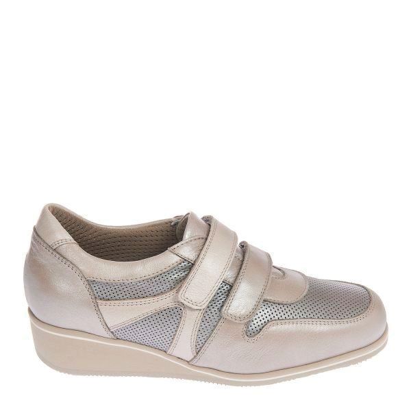
The effectiveness of shoe modifications and orthotics                                                                         61

neuralgic condition. Finally, in 1876 Thomas George                 proximal to the forking of the digital nerves (Figure 1).
Morton wrongly described the pathology as a subluxa-                Entrapment of the interdigital nerve between the
tion of the fourth metatarsophalangeal (MTP) joint                  intermetatarsal ligaments is the principal reason in
(5). Hoadley, in 1883, first excised an interdigital neu-           the occurrence of CMS (14). Macroscopically it has
roma from the third IMS of a foot (6). In 1940, Betts               a typically glossy fusiform shape, from white to yel-
introduced the notion of “neuroma” and compression                  lowish appearance and a relatively soft texture (1). The
of the plantar nerve below the intermetatarsal liga-                common plantar digital nerves are final boughs of the
ment (7).                                                           medial and lateral plantar nerves passing in the IMS,
      Histology shows that it is not really a neuroma,              under the intermetatarsal ligaments. Every common
but a perineural fibrosis with intraneural sclerohyalin-            digital nerve goes through the plantar aponeurosis and
osis (8); therefore, authors as Weinfield and Myerson               splits into 2 branches supplying the plantar skin of the
proposed the more correct term of “interdigital neuri-              toes. Smaller ramifications give innervations to the ad-
tis” (9). Classical symptoms are a tingling and burning             jacent metatarsals, MTP joints and plantar skin, under
sensation in the forefoot with an irregular numbness in             the metatarsal heads (15).
the affected toe (1). It is a common cause of metatar-                    Usually, the third common digital nerve, arising
salgia (10), along with hallux rigidus (11,12) and hal-             from the medial plantar nerve, receives a communicat-
lux valgus (13), in the overload typology.                          ing bough from the lateral plantar nerve, which passes
                                                                    deep to the transverse metatarsal ligament. This is the
                                                                    narrowest space, and for that reason, the nerve there is
Anatomy and pathophysiology                                         less mobile during weight bearing. This might explain
                                                                    why it is a common location for the pathology (16).
     Neuroma consists of a bulge in the interdigital nerve          Female sex is usually the most affected, suggesting that
just distal to the metatarsal transverse ligament and               the high heeled and tight footwear are contributing
                                                                    factors in the aetiology of this disorder (17).
                                                                          The precise cause is still not clear. Until today, four
                                                                    etiopathogenetic theories have been propounded (18):
                                                                    chronic traction damage (16), inflammatory environ-
                                                                    ment due to intermetatarsal bursitis (19), compression
                                                                    by the deep transverse intermetatarsal ligament (20)
                                                                    and ischemia of vasa nervorum (21).
                                                                          Nevertheless, some of these biomechanical con-
                                                                    victions are still debated. The sideboard that CMS
                                                                    should more commonly affected the third IMS in a
                                                                    more pronated foot, because of hyper mobility of the
                                                                    lateral column, is not supported anymore from the
                                                                    last studies. Even the rational hypothesis that subjects
                                                                    with a high body mass index (BMI) should increase
                                                                    pressure in the forefoot during the propulsive phase
                                                                    of walk, which could traumatize plantar IMS nerves,
                                                                    was not confirmed by recent studies on gait analysis.
                                                                    However, a strong association between CMS and re-
                                                                    striction of ankle dorsiflexion was proved (22).
                                                                          It was proposed that the common digital nerve
                                                                    of the third IMS is thicker than the others, as it is
Fig.1 The schematic drawing of the neuroma in the typical
third intermetatarsal space, just distal to the metatarsal trans-
                                                                    the result of an anastomosis between two nerve trunks
verse ligament and before the forking of the digital nerves.        (23). Another possible anatomical consideration is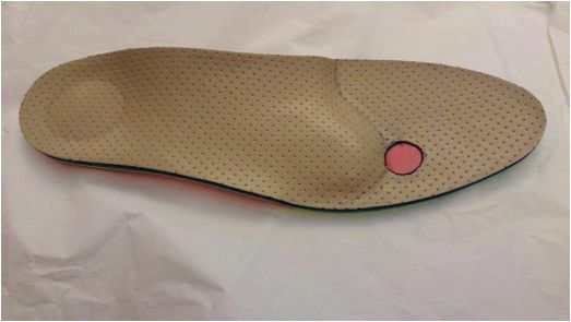
62                                                                                      G. Colò, A. Rava, E. M. Samaila, et al.

the increased mobility of the fourth ray (moving on         Diagnosis
the cuboid), compared to the third (fixed to the cu-
neiform), which could predispose to inflammation. In        Clinical evaluation
addition, some authors affirm that the distal metatar-            CMS evaluation is mostly clinical and requires an
sal transverse ligament may compress the interdigital       accurate physical examination, carried out with the pa-
nerve (24).                                                 tient in the supine position. On inspection, foot is ap-
      In this view, the use of high heels is another pre-   parently normal, with absence of any deformity. Rarely
disposing factor, as it can further increase compression    a diastasis of the digital fornix is not present, however
on it (17). Finally, some studies also described trauma     it is a common sign to all pathologies that create ten-
as a possible cause (25).                                   sion in the IMS, such as bursitis and MTP capsulitis
                                                            (28).
                                                                  The shape of the forefoot, the sub-metatarsal
Signs and symptoms                                          plantar skin and the position of the toes are carefully
                                                            assessed. The metatarsal motility, plantar and dorsal
      When CMS is suspected, medical history and            flexion of the MTP joints are gently tested. Then, the
physical examination play a pivotal role in the diag-       attention moves on to the palpatory exploration of the
nostic process. It is important to listen the patient’s     IMS, where a tenderness and a dorsal bulging with a
symptoms, often described in a very suggestive way.         possible enlargement of the IMS might be appreciated
The foot inspection should search for the exact trig-       (1). It’s very important to identify the pain, usually
ger points, distinguishing between intermetatarsal and      not located on the metatarsal heads, for a differential
metatarsal pain (1).                                        diagnosis (4). In these cases, it will be important to
      The typical symptom is a burning pain between         exclude other factors such as joint instabilities, fore-
the metatarsal heads, often radiating to the two corre-     foot conditions (first ray insufficiency, second metatar-
sponding toes, with cramps and hyperesthesia/dyses-         sal syndrome, bursitis), mid-hindfoot deformities (pes
thesia (26). The pain is intense and so debilitating that   cavus), arthritis in the MTP joint, remote causes that
the patients become afraid and anxious about walking        act mechanically under the forefoot (retraction of gas-
or even putting their foot to the ground. The disorder      trocnemius or Achilles tendon) or even to investigate
is that of a severe, sharp, sometimes piercing pain that    some specific bone condition as Frieberg’s disease (1).
occurs abruptly while walking. At onset, relief of pain           In presence of an atypical presentations such as
can be obtained by massaging the foot or manipulating       a localization in the second space or the presence of
the toes. In the worst cases, pain becomes debilitat-       multiple “neuromas” on the same foot, a mechanical
ing and patients are timorous about walking. In other       “overload” disorder must be ruled out (29).
cases, patients describe milder symptoms of burning or            To help give an accurate clinical evaluation, a full
tingling sensations (27).                                   foot and ankle examination should be performed with
      Description is not always typical, and it is impor-   particular attention to gait, footwear, over-pronation,
tant to thoroughly interrogate the patient; it is to be     sensory disturbances and soft-tissue changes (16).
underlined however, as a discriminating element, the              Subsequently, an accurate examination of digital
disappearance of the pain at rest and the reappearance      sensitivity should be performed, both with a pointed
under weight-bearing. Pathognomonic of the syn-             instrument and with a vibratory tuning fork. The vi-
drome is the so-called “sign of the shop window”, a cu-     bratory sensitivity of the tip of the toe is generally the
rious and picturesque expression used by some special-      most compromised (30). A characteristic sign is the
ists (28) to indicate the urgent need to remove the shoe    “numbness of the toes”, when the opposite surfaces
that covers the affected foot: the patient (frequently a    of the adjacent fingers show a reduced sensation (31);
woman), to be unnoticed in the manoeuvre, stops in          some authors call it “book page hypoesthesia” (28).
front of a shop window pretending to observe it.                  Various clinical tests are described in literature.
                                                            The pressure can be practiced while tightening the
The effectiveness of shoe modifications and orthotics 63
metatarsal heads with the other hand, and this may be by Mulder during the examination (29). The dynamic
associated with a painful and palpable “click” sensation US would be very effective for recognizing masses larger
(Mulder’s sign) (29). This test demonstrated a 94–98% than 3.5 mm using a 10MHz probe (40). The most re-
sensitivity (32). Another useful test is the “Thumb in- cent studies appear to confirm that the high sensitivity
dex finger squeeze” that appears to have the highest of US (0.91) is equal to (p = 0.88) that of the Magnetic
specificity and sensitivity (33). Resonance Imaging (MRI) (0.90) for identification of
Among provocative tests, Cloke and Greiss (34) neuroma and its thickness (41). MRI offers additional
described the “digital nerve stretch test” with 100% of advantages in the diagnosis of CMS, but it must be con-
sensitivity: the toes on either side of the affected IMS sidered a second level investigation with the limit, as in
are passively fully extended, with the ankles in dorsi- the US, to recognize lesions smaller than 4 mm (42).
flexion and the foot on the examiner’s knees. Discom- Certainly, MRI is superior in the differential di-
fort or pain in the IMS of the affected foot indicates a agnosis, for its sensitivity on pathologies such as stress
positive result. fractures, capsulo-synovitis of the MTP, synovial artic-
Other authors (29, 32, 35) describe a similar test, ular forms, or other pathologies of the soft tissues such
the “Lasègue sign of the toes”, in which pain and hy- as lipomas, angiomas, tendon ganglia or connective
poesthesia are evoked by forcing dorsal flexion of the malignancies of the forefoot (43). Main indications for
toes and reduced by proximal interphalangeal joints MRI are unclear clinical assessment and cases when
flexion. more than one IMS is affected (1). Finally, it must be
bear in mind that a negative result does not exclude the
Imaging diagnosis (false negative 17%) (44).
As is often the case in medicine, the diagnosis of
the disease is mainly clinical but rarely physical exami-
nation is considered sufficient; therefore, complemen- Shoe modifications
tary exams are required. The most dependable method
to clarify the diagnosis is a local anaesthetic injection, Shoe modifications and orthotics can play an im-
not always accepted by the patient (1). portant role in the nonsurgical management of fore-
X-rays appears to be essential as a first line imag- foot pathology. Therapeutic footwear may improve pa-
ing approach, to investigate other possible causes of tient gait and increase the level of ambulation; on the
metatarsalgia such as (36): tarsal–metatarsal joint stiff- contrary, inadequate footwear can worsen the symp-
ness, metatarsal hypermetria, Frieberg’s disease, toe toms and be a contributing cause for the development
deformities and MTP instabilities. However, in order of the pathology. (45).
to eliminate possible doubts, sonographic (US) confir- Already in 1897 Bradford (46) noted alterations
mation is usually the instrumental investigation mostly caused by incorrect shoes through an analysis of his-
utilized (14), certainly reliable and easy to prescribe, as torical art. Nowadays, women’s shoes continue to cause
it is fast and inexpensive for the patient. The imaging deformity and predispose to injury, even more so than
must be carried out with plantar and back, transversal in the past. Poorly fitting shoes are a major contribut-
and longitudinal US scans with a 7.5 MHz high fre- ing factor to the difference in incidence of foot disor-
quency probe. The neuroma appears as homogeneously ders between men and women, mostly for those over
hypoechoic mass, well recognizable by the adjacent 61 years of age (47).
hyperechoic fat and by the shadow of the metatarsal Traditionally, men’s shoes tend to be wider and
cortex. Shapiro (37) and Quinn (38) state a diagnostic have lower heels than women’s, and this could con-
reliability around 95% for lesions larger than 5 mm, tribute to explain the different incidence of CMS in
being 2 mm the maximum limit of the normal nerve. men compared to women, as described in some studies
Currently, US should be performed with a dynamic with a female-to-male ratio as high as 18 to 1 (48).
technique (dynamic US), as proposed by Torriani (39) As widely showed in pathophysiology, the prevalence
and then by Perini (40), recreating the “click” described of several musculoskeletal conditions in women are64 G. Colò, A. Rava, E. M. Samaila, et al.
largely the result of biomechanical changes caused by Villeneuve (54) used a small insert to maintain
ill-fitting shoes. In particular, the altered biomechan- postural equilibrium, by stimulating the mechanore-
ics (associated with shoes with a narrowed toe box and ceptors in the plantar surface of the foot. Hayda et al.
high-heeled shoes) has been linked to the genesis of (55), the following year, found that placing a pad just
Freiberg disease, hallux valgus, Haglund’s syndrome, proximal to the metatarsal heads provided significant
hammer toe deformity, metatarsal stress fracture and, reductions in forefoot plantar pressures around the
above all, CMS (17, 48). first and second metatarsal heads. Burgess in 1997 (56)
Thus, the first approach in CMS should consist evaluated a series of 10 non-pathological male patients
in patients’ education to avoid narrow and high-heeled during walking, after one day wearing a pair of oxfords
shoes (4). The objective of shoes modifications is to de- (hard) and running shoes (soft), containing an insert
liver the pressures uniformly over the sole of foot. Shoes of 4 mm in height placed on a 0.8 mm EVA insole. He
should be sufficiently long, comfortable, broad toe- noted that the insert was successful in both shifting
boxed, flat heeled and should bear a sufficiently thick peak pressures from the medial to the lateral forefoot,
external sole, not excessively flexible (Figure 2) (2). whilst reducing peak pressures simultaneously. This
A rocker-bottom sole may be helpful (45). Some au- was only evident in the hard shoe condition however,
thors showed that footwear and padding may be suc- suggesting that the footbed of the running shoe was
cessful in relieving symptoms in 32% of cases after a perhaps too soft to allow the insert to influence sen-
mean of 4.5 months (17, 49) but they seem to achieve sory input sufficiently.
lower satisfaction rates when compared with other More recent studies show that the perceived feel
more invasive methods such as steroid injections (50). is best using wedge angles of 4 degrees and 5 degrees at
In most cases, clinicians will also have to educate a heel height of 25 mm, 10 degrees and 11 degrees at a
the patient on lifestyle changes, such as losing weight heel height of 50 mm and 16 degrees and 18 degrees at
and starting a regular physical activity; both being use- a heel height of 75 mm (57).
ful in most pathologies (51, 52). Footbed shapes appear then to be essential to en-
hanced footwear comfort, regardless of the underly-
ing disease. Thus, when footwear modifications alone
Orthotics are not enough, custom-made insoles could be de-
signed to correct hindfoot malalignments as varus or
Over the years, numerous studies have been car- valgus, support the medial arch, transfer the pressure
ried out to highlight the different effects of plantar just proximal to the metatarsal heads, and reduce the
inserts for the foot comfort. Robbins and Gouw in weights on pressure sites.
1991 (53) proposed that surface irregularities should By decreasing metatarsal head loading and rede-
be added to the insoles of running shoes to improve ploying plantar pressures in a harmonious manner (58,
correct sensory inputs. In 1993, 59), insoles, in selected cases, may alleviate forefoot
problems (60).
In the case of CMS, some authors prefers a cus-
tom orthotics through foam impression methods, in
a neutral subtalar position, with a prolonged longitu-
dinal vault to support the first metatarsal, with a flat
metatarsal support (without olive or bar), in order
to favor the physiological pattern of the metatarsal
weight bearing, from lateral to medial, before the pres-
sure on the big toe (28).
Other authors suggested that a retrocapital bar
Fig. 2 Predisposed and comfortable shoe for orthotics, with a or pad, just proximal to the metatarsal heads displaces
round toe-box and slight heel. the pressure sites and can be beneficial for symptomsThe effectiveness of shoe modifications and orthotics 65
(Figure 3). Metatarsal padding helps to spread and Kilmartin (62) examined 21 patients and states
cushion metatarsal heads to relieve the pain from the that pain associated with CMS was not significantly
pinched nerve. If needed, a cup can be added beneath altered by changing the position of the foot with the
the painful metatarsal head or heads. Custom-made compressed felt orthosis (14% of cases).
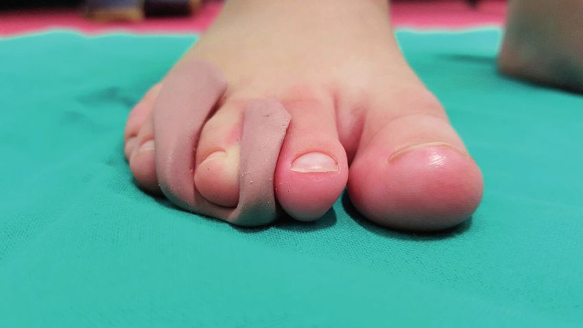
toe inserts modeled in silicone rubber can be added Notwithstanding, this has not been confirmed by
in patients having associated claw-toe deformity latest studies. De Oliveira (63) in his randomized, con-
(Figure 4) (60, 61). trolled, double-blind clinical trial analyzed 72 patients
Bennett (17) reports that 41% of patients treated and proved that customized insole with metatarsal and
conservatively (shoes suitable for extra volume, un- arch support relieved pain during walking (P = 0.048)
loading orthoses, soft metatarsal pads) demonstrate and improved patient-reported measures of function
significant improvements with these non-invasive (in general health domains (P < 0.001) and physical ac-
procedures. However, other authors (28) declare that tivity (P = 0.025)).
these treatments, in case of CMS confirmed by “im- In any cases, when modifications fail or if affected
aging” and larger than 5-6 mm, do not seem to give individuals are no longer willing to make adjustments
convincing results, proving them to be more a clini- to their lifestyle or shoe wear (64, 65), patients may
cal condition to live acceptably with pain rather than always choose to undergo surgery or other non-opera-
a real treatment. tive treatments such as US guided percutaneous radio-
frequency (66), alcohol or corticosteroids injection and
percutaneous electrostimulation-guided alcoholization
with phenol (67-69).
Conclusion
During the years, many therapies have been uti-
lized to treat symptomatic CMS. Among conservative
treatments, orthotics and shoes modifications have
been used to off-load the forefoot and thus reduce
pain from weight-bearing pressure. In order to prevent
Figure 3. Insole with medial arch support, retrocapital bar just or retard the development of CMS, shoes should be
proximal to the metatarsal heads with targeted shock absorber
sufficiently long, comfortable, broad toe-boxed, they
insert in third intermetatarsal space.
should have a flat heel and a sufficiently thick external
sole that is not excessively flexible.
A threshold period of 4.5 months appears to emerge
from the results of the studies, indicating that beyond this
period and in larger neuromas than 5-6 mm, orthotics
and/or shoes modification do not seem to give convinc-
ing results, proving to be more a clinical condition for liv-
ing acceptably with pain than a real treatment.
Despite this, use of orthotics may be considered a
safe and rational treatment before more invasive inter-
ventions, without any complications. Notwithstanding
a good quality of the selected articles, further studies
Figure 4. Custom-made toe insert modeled in silicone rubber with a longer follow-up period and high quality ran-
added in patient affected by Civinini-Morton syndrome and as- domized controlled trials are needed to provide more
sociated with claw deformity of the third toe. solid and accurate proofs.66 G. Colò, A. Rava, E. M. Samaila, et al.
Conflict of interest: Each author declares that he or she has no correction of hallux valgus. Surgical technique. J Bone Joint
commercial associations (e.g. consultancies, stock ownership, equity Surg Am. 2006 Mar;88 Suppl 1 Pt 1:135–48.
interest, patent/licensing arrangement etc.) that might pose a con- 14. Sofka CM, Adler RS, Ciavarra GA, Pavlov H. Ultrasound-
flict of interest in connection with the submitted article guided interdigital neuroma injections: short-term clinical
outcomes after a single percutaneous injection--preliminary
Compliance with Ethical Standards: This article does not contain results. HSS J 2007 Feb; 3(1):44–9. doi: 10.1007/s11420-
any studies with human participants or animals performed by any 006-9029-9.
of the authors. Informed consent: Not applicable.
15. Young G, Lindsey J. Etiology of symptomatic recurrent
interdigital neuromas. J Am Podiatr Med Assoc. 1993
May;83(5):255–8.
References 16. Jain S, Mannan K. The diagnosis and management of Mor-
ton’s neuroma: a literature review. Foot Ankle Spec. 2013
Aug;6(4):307–17. doi: 10.1177/1938640013493464. Epub
1. Di Caprio F, Meringolo R, Shehab Eddine M, Ponziani
2013 Jun 27.
L. Morton’s interdigital neuroma of the foot: A litera-
17. Bennett GL, Graham CE, Mauldin DM. Morton’s inter-
ture review. Foot Ankle Surg. 2018 Apr;24(2):92–98. doi:
digital neuroma: a comprehensive treatment protocol. Foot
10.1016/j.fas.2017.01.007. Epub 2017 Feb 4.
Ankle Int. 1995 Dec;16(12):760–3.
2. Bradley N, Miller WA, Evans JP. Plantar neuroma: analysis
18. Hassouna H, Singh D. Morton’s metatarsalgia: pathogen-
of results following surgical excision in 145 patients. South
esis, aetiology and current management. Acta Orthop Belg.
Med J. 1976Jul;69(7):853–4.
2005 Dec;71(6):646–55.
3. Kasparek M, Schneider W. Surgical treatment of Morton’s
19. Bossley CJ, Cairney PC. The intermetatarsophalangeal bur-
neuroma: clinical results after open excision. Int Orthop.
sa--its significance in Morton’s metatarsalgia. J Bone Joint
2013Sep;37(9):1857–61. doi: 10.1007/s00264-013-2002-6.
Surg Br. 1980 May;62-B(2):184–7.
Epub 2013 Jul 13.
20. Gauthier G. Thomas Morton’s disease: a nerve entrapment
4. Valisena S, Petri GJ, Ferrero A. Treatment of Morton’s
syndrome. A new surgical technique. Clin Orthop Relat
neuroma: A systematic review. Foot Ankle Surg. 2018
Res. 1979 Jul-Aug; (142):90–2.
Aug;24(4):271–281. doi: 10.1016/j.fas.2017.03.010. Epub
21. Nissen KI. Plantar digital neuritis; Morton’s metatarsalgia. J
2017 Apr 5.
Bone Joint Surg Br. 1948 Feb;30B(1):84–94.
5. Pisani G. Is it Morton’s or Civinini’s syndrome? Foot Ankle
22. Naraghi R, Bremner A, Slack-Smith L, Bryant A. The re-
Surg. 2010 Sep;16(3):105–6. doi: 10.1016/j.fas.2009.07.006.
lationship between foot posture index, ankle equinus, body
Epub 2009 Sep 6.
mass index and intermetatarsal neuroma. J Foot Ankle Res.
6. Hoadley A. Six cases of metatarsalgia. Chicago Med Rec.
2016 Dec;9:46. eCollection 2016. doi: 10.1186/s13047-
1893.5:32.
016-0179-9.
7. Delmi M. Métatarsalgies de Morton. In: Valtin B, Leem-
23. Diez EM, Mas SM, Pi JF, Aramburo F. Comparative results
rijsee T, editors. Chirurgie de l’avant-pied. 2005. Cahiers
of two different techniques in the treatment of the Morton’s
d’enseignement de la SOFCOT. Paris: Elsevier.
neuroma. The Foot. 1999 Sep;9(3):134–7.
8. Giannini S, Bacchini P, Ceccarelli F, Vannini F. Interdigi-
24. Alexander IJ, Johnson KA, Parr JW. Morton’s neuroma:
tal neuroma: clinical examination and histopathologic re-
a review of recent concepts. Orthopedics. 1987 Jan;10(1):
sults in 63 cases treated with excision. Foot Ankle Int 2004
103–6.
Feb;25(2):79–84.
25. Litchman MH, Silver CM, Simon SD. Morton’s meta-
9. Weinfeld SB, Myerson MS. Interdigital Neuritis: Di-
tarsalgia and its relationship to trauma. R I Med J. 1964
agnosis and Treatment. J Am Acad Orthop Surg. 1996
Jul;47:328–31.
Nov;4(6):328–335.
26. Ruiz Santiago F, Prados Olleta N, Tomás Muñoz P,
10. Besse JL. Metatarsalgia. Orthop Traumatol Surg Res. 2017
Guzmán Álvarez L, Martínez Martínez A. Short term com-
Feb;103(1S):S29-S39. doi: 10.1016/j.otsr.2016.06.020.
parison between blind and ultrasound guided injection in
Epub 2017 Jan 18.
morton neuroma. Eur Radiol. 2019 Feb;29(2):620–627. doi:
11. Colò G, Alessio-Mazzola M, Dagnino G, Felli L. Long-
10.1007/s00330-018-5670-1. Epub 2018 Jul 31.
Term Results of Surgical Treatment of Valenti Procedures
27. Thomson CE, Gibson JN, Martin D. Interventions for the
for Hallux Rigidus: A Minimum Ten-Year Follow-Up
treatment of Morton’s neuroma. Cochrane Database Syst
Retrospective Study. J Foot Ankle Surg. 2019 Mar; 58(2):
Rev. 2004;(3):CD003118.
291–294. doi: 10.1053/j.jfas.2018.08.055.
28. Volpe A. La sindrome di Morton. LO SCAL. 2011. 25,
12. Colò G, Samaila EM, Magnan B, Felli L. Valenti resection
60–73. doi: 10.1007/s11639-011-0098-2
arthroplasty for hallux rigidus: A systematic review. Foot
29. Mulder JD. The causative mechanism in Morton’s metatar-
Ankle Surg. 2019 Dec 5. pii: S1268-7731(19)30209-7. doi:
salgia. J Bone Joint Surg Br. 1951 Feb;33-B(1):94–5.
10.1016/j.fas.2019.11.009. (Epub ahead of print)
30. Jarde O. Morton’s metatarsalgia. Foot Ankle Surg.
13. Magnan B, Bortolazzi R, Samaila E, Pezzè L, Rossi N,
1998;4:187–191. doi: 10.1046/j.1460-9584.1998.00116.x
Bartolozzi P. Percutaneous distal metatarsal osteotomy forThe effectiveness of shoe modifications and orthotics 67
31. Gougoulias N, Lampridis V, Sakellariou A. Morton’s inter- 46. Bradford EH. The human foot in art. J Bone Joint Surg Am.
digital neuroma: instructional review. EFORT Open Rev. 1897;s1- s10:148–161
2019 Jan;4(1):14–24. doi: 10.1302/2058-5241.4.180025. 47. Frey C. Foot health and shoewear for women. Clin Orthop
eCollection 2019 Jan. Relat Res. 2000 Mar;(372):32–44.
32. Pastides P, El-Sallakh S, Charalambides C. Morton’s neu- 48. Goud A, Khurana B, Chiodo C, Weissman BN. Women’s
roma: A clinical versus radiological diagnosis. Foot Ankle musculoskeletal foot conditions exacerbated by shoe wear:
Surg. 2012 Mar;18(1):22–4. doi: 10.1016/j.fas.2011.01.007. an imaging perspective. Am J Orthop (Belle Mead NJ).
Epub 2011 Feb 16. 2011 Apr;40(4):183–91.
33. Thomas JL, Blitch EL 4th, Chaney DM, et al. Diagnosis 49. Saygi B, Yildirim Y, Saygi EK, Kara H, Esemenli T. Morton
and treatment of forefoot disorders. Section 3. Morton’s in- neuroma: comparative results of two conservative methods.
termetatarsal neuroma. J Foot Ankle Surg. 2009;48(2):251- Foot Ankle Int. 2005 Jul;26(7):556–9.
6. 50. Park HJ, Kim SS, Rho MH, Hong HP, Lee SY. Sono-
34. Cloke DJ, Greiss ME. (2006) The digital nerve stretch graphic appearances of Morton’s neuroma: differences
test: a sensitive indicator of Morton’s neuroma and neu- from other interdigital soft tissue masses. Ultrasound Med
ritis. Foot Ankle Surg. 2006;12:201-203. doi: 10.1016/j. Biol. 2011 Aug;37(8):1204–9. doi: 10.1016/j.ultrasmed-
fas.2006.04.002 bio.2011.05.008. Epub 2011 Jun 16.
35. Owens R, Gougoulias N, Guthrie H, Sakellariou A. (2011) 51. Colò G, Massarini M, Cavagnaro L, Felli L, Ferracini R.
Morton’s neuroma: clinical testing and imaging in 76 Exercise therapy indications in metastatic bone patients.
feet, compared to a control group. Foot Ankle Surg. 2011 Minerva Ortop e Traumatol. 2020;71: 000–000 DOI:
Sep;17(3):197–200. doi: 10.1016/j.fas.2010.07.002. Epub 10.23736/S0394-3410.19.03960-2.
2010 Sep 17. 52. Colò G, Cavagnaro L, Alessio-Mazzola M, Zanirato A,
36. Read JW, Noakes JB, Kerr D, Crichton KJ. Morton’s meta- Felli L, Formica M. Incidence, diagnosis and management
tarsalgia: sonographic findings and correlated histopathol- of sacroiliitis after spinal surgery: a systematic review of the
ogy. Foot Ankle Int. 1999 Mar;20(3):153–61. literature. Musculoskelet Surg. 2019 May 7. doi: 10.1007/
37. Shapiro PP, Shapiro SL. Sonographic evaluation of inter- s12306-019-00607-0. [Epub ahead of print]
digital neuromas. Foot Ankle Int. 1995 Oct;16(10):604–6. 53. Robbins SE, Gouw GJ. Athletic footwear: unsafe due
38. Quinn TJ, Jacobson JA, Craig JG, van Holsbeeck MT. So- to perceptual illusions. Med Sci Sports Exerc. 1991
nography of Morton’s neuromas. AJR Am J Roentgenol. Feb;23(2):217–24.
2000 Jun;174(6):1723–8. 54. Villeneuve, P. Foot and posture. 1993. In 3rd European and
39. Torriani M, Kattapuram SV. Technical innovation. Dynam- First World Congress of Podiatry Programme, p. 14.
ic sonography of the forefoot: The sonographic Mulder sign. 55. Hayda R, Tremaine MD, Tremaine K, Banco S, Teed K. Ef-
AJR Am J Roentgenol. 2003 Apr;180(4):1121–3. fect of metatarsal pads and their positioning: a quantitative
40. Perini L, Del Borrello M, Cipriano R, Cavallo A, Volpe A. assessment. Foot Ankle Int. 1994 Oct;15(10):561–6.
(2006) Dynamic sonography of the forefoot in Morton’s 56. Burgess S, Jordan C, Bartlett R. The influence of a small
syndrome: correlation with magnetic resonance and surgery. insert, in the footbed of a shoe, upon plantar pressure dis-
Radiol Med. 2006 Oct;111(7):897–905. Epub 2006 Oct 11. tribution. Clin Biomech (Bristol, Avon). 1997 Apr;12(3):
41. Bignotti B, Signori A, Sormani MP, Molfetta L, Martinoli S5-S6.
C, Tagliafico A. (2015) Ultrasound versus magnetic reso- 57. Witana CP, Goonetilleke RS, Au EY, Xiong S, Lu X. Footbed
nance imaging for Morton neuroma: systematic review and shapes for enhanced footwear comfort. Ergonomics. 2009
meta-analysis. Eur Radiol. 2015 Aug;25(8):2254–62. doi: May;52(5):617–28. doi: 10.1080/00140130802419503.
10.1007/s00330-015-3633-3. Epub 2015 Mar 26. 58. Chang AH, Abu-Faraj ZU, Harris GF, Nery J, Shereff MJ.
42. Sharp RJ, Wade CM, Hennessy MS, Saxby TS. The role of Multistep measurement of plantar pressure alterations using
MRI and ultrasound imaging in Morton’s neuroma and the metatarsal pads. Foot Ankle Int. 1994 Dec;15(12):654–60.
effect of size of lesion on symptoms. J Bone Joint Surg Br. 59. Holmes Jr GB, Timmerman L. A quantitative assessment
2003 Sep;85(7):999–1005. of the effect of metatarsal pads on plantar pressures. Foot
43. Zanetti M, Strehle JK, Kundert HP, Zollinger H, Hodler J. Ankle. 1990 Dec;11(3):141–5.
Morton neuroma: effect of MR imaging findings on diag- 60. Kang JH, Chen MD, Chen SC, Hsi WL. Correlations be-
nostic thinking and therapeutic decisions. Radiology. 1999 tween subjective treatment responses and plantar pressure
Nov;213(2):583–8. parameters of metatarsal pad treatment in metatarsalgia
44. Torres-Claramunt R, Ginés A, Pidemunt G, Puig L, De patients: a prospective study. BMC Musculoskelet Disord.
Zabala S. MRI and ultrasonography in Morton’s neuroma: 2006 Dec 5;7:95.
Diagnostic accuracy and correlation. Indian J Orthop. 2012 61. Hinz A. Nerve disorders. In: DiGiovanni CW, Greisberg
May;46(3):321–5. doi: 10.4103/0019-5413.96390. J, editors. Foot and ankle: core knowledge in orthopaedics.
45. Janisse DJ, Janisse E. Shoe modification and the use of or- Philadelphia: Elsevier Mosby; 2007. p. 171–6.
thoses in the treatment of foot and ankle pathology. J Am 62. Kilmartin TE, Wallace WA. Effect of pronation and supi-
Acad Orthop Surg. 2008 Mar;16(3):152–8. nation orthosis on Morton’s neuroma and lower extremity68 G. Colò, A. Rava, E. M. Samaila, et al.
function. Foot Ankle Int. 1994 May;15(5):256–62. alternative to surgical excision? Long-term results. Foot
63. de Oliveira HAV, Natour J, Vassalli M, Rosenfeld A, Jen- Ankle Surg. 2019 Apr 17. pii: S1268–7731(19)30057-8.
nings F, Jones A. Effectiveness of customized insoles in doi: 10.1016/j.fas.2019.04.004. [Epub ahead of print]
patients with Morton’s neuroma: a randomized, con- 69. Magnan B, Marangon A, Frigo A, Bartolozzi P. Local phe-
trolled, double-blind clinical trial. Clin Rehabil. 2019 nol injection in the treatment of interdigital neuritis of the
Dec;33(12):1898–1907. doi: 10.1177/0269215519873949. foot (Morton’s neuroma). Chir Organi Mov. 2005 Oct-
Epub 2019 Sep 11. Dec;90(4):371–7.
64. DiPreta JA. Metatarsalgia, lesser toe deformities, and asso-
ciated disorders of the forefoot. Med Clin North Am. 2014
Mar;98(2):233–51. doi: 10.1016/j.mcna.2013.10.003. Epub
2013 Dec 10.
65. Mischitz M, Zeitlinger S, Mischlinger J, Rab M. Nerve de- Received: 10 April 2020
compression according to A.L. Dellon in Morton’s neuroma Accepted: 10 May 2020
- A retrospective analysis. J Plast Reconstr Aesthet Surg. Correspondence:
2020 Jan 22. pii: S1748–6815(20)30027-9. doi: 10.1016/j. Bruno Magnan
bjps.2020.01.008. [Epub ahead of print] Department of Orthopedics and Trauma Surgery,
66. Masala S, Cuzzolino A, Morini M, Raguso M, Fiori R. University of Verona
Ultrasound-Guided Percutaneous Radiofrequency for the Surgical Center “P. Confortini”,
Treatment of Morton’s Neuroma. Cardiovasc Intervent P.le A. Stefani, 1, 37126 Verona (VR), Italy
Radiol. 2018 Jan;41(1):137–144. doi: 10.1007/s00270-017- Tel: + 39 045-8123542/8123510; Fax: + 39 045-8123578
1786-y. Epub 2017 Sep 27. E-mail: bruno.magnan@univr.it
67. Perini L, Perini C, Tagliapietra M, et al. Percutaneous al-
cohol injection under sonographic guidance in Morton’s Gabriele Colò
neuroma: follow-up in 220 treated lesions. Radiol Med. Department of Orthopaedics and Traumatology, Regional
2016 Jul;121(7):597–604. doi: 10.1007/s11547-016-0622- Center for Joint Arthroplasty, ASO Alessandria,
9. Epub 2016 Feb 16. Via Venezia 16, 16121, Alessandria, Italy.
68. Samaila EM, Ambrosini C, Negri S, Maluta T, Valentini Tel: +39 0131 206594
R, Magnan B. Can percutaneous alcoholization of Morton’s Fax: + 39 0131206907
neuroma with phenol by electrostimulation guidance be an E-mail: gabriele.colo@yahoo.itYou can also read


