The Key Muscles of Hatha Yoga - Scientific Keys Volume I Ray Long MD FRCSC
←
→
Page content transcription
If your browser does not render page correctly, please read the page content below
Scientific Keys Volume I

The Key Muscles
of Hatha Yoga
Ray Long MD FRCSC
With Illustrator Chris Macivor
Patanjali, the patron saint of yoga, said that mastery
combines a balance of science and art. Knowledge of
science is like the colors on an artist’s palette – the
greater the knowledge, the more colors available. The
body is the canvas and the asanas are the art we create.
Scientific
         Keys                           Volume I

                                 First Edition: Copyright 2005, Raymond A Long md frcsc
                                Second Edition: Copyright 2006, Raymond A Long md frcsc
                                 Third Edition: Copyright 2006, Raymond A Long md frcsc
                                                 Bandha Yoga Publications
                                                    All rights reserved
         No part of this book may be reproduced, copied or transmitted in any form without the express written
                                                permission of the publisher.
                    Visual and textual content conceived and produced by Raymond A Long, MD FRCSC

             Always consult your healthcare provider before practicing yoga or any other exercise program.
                     Yoga must always be practiced under the supervision of a qualified instructor.
           The author assumes no responsibility for injuries that may occur as a result of the practice of yoga.

                                             Design: Ingrid Patricia Sanchez

                                          www.BandhaYoga.com
About the Author
                         Ray Long
                 Ray Long MD FRCSC is a board
             certified orthopedic surgeon and
              the founder of Bandha Yoga. Ray
                graduated from The University     Chris Macivor
              of Michigan Medical School with
             post-graduate training at Cornell    Chris Macivor is a digital illustrator
                 University, McGill University,   and the visual director of Bandha
                The University of Montreal and    Yoga. Chris is a graduate of Etobicoke
                 Florida Orthopedic Institute.    School of The Arts, Sheridan College
                 He has studied hatha yoga for    and Seneca College. His work has
                    over twenty years, training   spanned many genres from TV and
               extensively with B.K.S. Iyengar    film to videogames and underwater
                       and other of the world’s   videography.
                         leading yoga masters.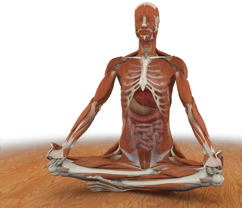
Contents                                     Part Two - The Trunk 113
                                               Chapter 9:
                                               Chapter 10:
                                                             Abdominals 118
                                                             Back Muscles 128
                                               Chapter 11:   Latissimus Dorsi 135
                                               Chapter 12:   Trapezius 139
Introduction 7                                 Chapter 13:   Pectoralis Major & Minor 144

Fundamentals 8                               Part Three - The Shoulder Girdle and Upper Arms 151
  Locations on the Body 8                      Chapter 14:   Rhomboids 157
  Skeleton 10                                  Chapter 15:   Serratus Anterior 162
  Joints 22                                    Chapter 16:   Deltoids 167
  Ligaments 26                                 Chapter 17:   Rotator Cuff 174
  Muscles and Tendons 35                       Chapter 18:   Biceps Brachii 185
  Movement 44                                  Chapter 19:   Triceps Brachii 190
                                               Chapter 20:   Sternocleidomastoid 197

Part One - The Pelvic Girdle and Thighs 49     Chapter 21: Lower Leg and Foot 202
  Chapter 1:     Iliopsoas 57                  Chapter 22: Forearm and Hand 206
  Chapter 2:     Gluteus Maximus 64
  Chapter 3:     Gluteus Medius 69
  Chapter 4:     Tensor Fascia Lata 74         Chapter 23: Myofascial and Organ Planes 210
  Chapter 5a:    Pectineus 79                  Chapter 24: The Breath Connection 212
  Chapter 5b:    Adductor Magnus 84            Chapter 25: Bandhas 220
  Chapter 6:     External Rotators 91          Chapter 26: Chakras 222
  Chapter 7:     Quadriceps 96                 Putting It All Together 224
  Chapter 8:     Hamstrings 103
                                               Appendix of Asanas 230
                                               Index of Asanas 236
                                               Index of Muscles 238
Scientific keys
                                      How to Use This Book
    The images in this book are the keys. We
    present each muscle in the context of
    its function as an agonist, antagonist or
    synergist. Note the interrelated views
    of the muscle in each of its various
    representations.

    Relax and study one muscle at a time.
    Actively apply what you have learned by
    visualizing the muscles as you perform
    the asanas. Consciously contract and
    relax them, as detailed in the images.
    This will consolidate your knowledge.
    Review each studied muscle, first at
    twenty four hours and then again at one
    week. In this way you will master the
    muscles and integrate them into your
    yoga practice.
Introduction
H
         uman anatomy and physiology is a vast subject, as
         is the ar t of hatha yoga. Never theless, combining
         knowledge from both f ields is extremely
         benef icial to the yoga practitioner. Athletes can
improve their per formance and exper ience fewer injur ies
through a basic underst anding of their musculoskelet al
system. Similarly, yoga practitioners can benef it from the
application of Western science to their practice
development.

It is not necessar y to memor ize hundreds of muscles and
bones to exper ience the benef its of applying science to
yoga. What is necessar y is the functional underst anding of
a manageable number of key anatomic structures in their
settings as they relate to hatha yoga. Knowledge of these
structures can be applied immediately to optimize your
practice, break through blockages and avoid injur ies.

This f irst volume presents key muscles in the context of
hatha yoga. For practitioners unf amiliar with the Western
scientif ic terminology of the body, the following section,
“Fundament als,” is recommended.
Joints
     As with the bones, the shape of the joints re-
     flects their function (and their function reflects
     their shape). Joints come in a spectrum of
     shapes, depending on the mobility or stability
                                                           ball and socket       hip
     they require. For example, the hip joint is a ball
     and socket while the knee joint is a hinge. A ball
     and socket type hip joint confers the greatest
     mobility in all planes and is useful for activities
     such changing direction while walking and run-
     ning (or reaching in various directions to grasp
     objects, as with the shoulder). A hinge type
     knee joint provides greater stability and is use-
     ful for propelling the body forward (or drawing
     an object towards the body, as with the elbow).

     Other joints such as the intervertebral between
     the vertebrae allow for limited mobility be-
     tween individual vertebrae but great stability
                                                                                knee
     to protect the spinal cord. Mobility of the spinal
     column comes from combining the limited                   hinge
     movement of individual intervertebral joints as
     a whole.

22
                                                            compressive      lumbar spine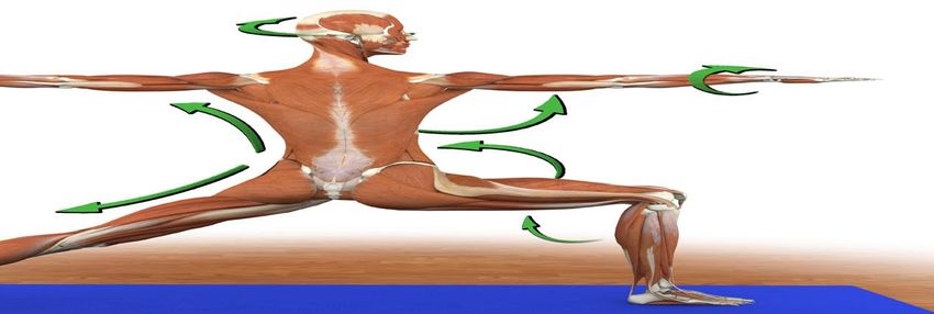
Articular Structure
                            The joint capsule is connective tissue sheath-
                            ing that surrounds and seals synovial joints. It
                            is susceptible to stretch injury when executing
                            extreme movements in yoga postures.

hip articular cartilage     Synovial tissue lines the inside of the joint
                            capsule. This tissue produces synovial fluid,
                            a viscous lubricant for the joint surface that
                            decreases friction during joint movement.
                            Synovial fluid circulates throughout the joint,
                            transporting nutrients to the articular carti-
                            lage and removing debris from the joint space.
                            The various contortions resulting from yoga
                            postures aid flex and expand the joint capsule,
                            stimulating circulation of synovial fluid.

                            Articular cartilage covers the joint surfaces,
                            allowing smooth gliding of one bone over the
                            other. In fact, articular cartilage is one of the
                            smoothest surfaces known to man. Applying
                            excessive pressure to this fragile cartilage can
                            injure it, ultimately resulting in arthritis.

                            The meniscus deepens the articular surface
                            and broadens the contact area of the joint. This
                            aids to stabilize the joint and distributes the
                                                                                knee with
  hip joint capsule with    force of gravity and muscular contraction over a
                            greater surface area. The meniscus is composed
                            of fibrocartilage, giving it a flexible rubbery     menisci
synovium (posterior view)   consistency.
                                                                                        23
Joint reaction forces
Every action has an equal and opposite
reaction. Muscular contraction and
gravity create opposing forces across
the joint surfaces, known as joint reac-
tion forces. It is important to spread
these forces over the greatest possible
joint surface area.
Joint congruency refers to the fit of
a joint’s articular surfaces. A joint is
congruent when its surfaces fit together
perfectly. Movement out of congruency
focuses stress on a small surface area.
A large force focused on a small area of
articular cartilage can injure it, eventu-
ally causing degenerative changes.
Some yoga postures have the capacity
to sublux or take a joint into an incon-
gruent position. Avoid this by using
the joints with a greater range of mo-
tion while protecting those joints with
limited range of motion.
24Joint reaction forces - applied
For example, the ball and socket joint of the hip has greater range of motion
than the hinge joint of the knee. Lotus posture (or padmasana) requires a
large amount of external rotation of the hip joint to bring the foot into posi-
tion on the opposite leg. Obtaining this external rotation from the knee joint
creates incongruency because the knee is a hinge joint with limited capacity to
rotate. This incongruency can result in the abnormal distribution of joint reac-
tion forces, injuring the intra-articular structures of the knee. Therefore it is
essential to first obtain full range of motion of the ball and socket hip joint to
protect the hinge knee joint. (see arrows)
ardha padmasanaStretching Muscles
Static Stretching
Static stretching is the most common technique used in hatha yoga. There are two categories
of static stretching. The first is active static stretching. This involves contracting antagonist
muscles to stretch a target muscle. Contracting the quadriceps, iliopsoas and biceps during
the forward bend paschimottanasana is a form of active static stretching of the hamstrings.
Contracting antagonist muscles in active static stretching results in a phenomenon called
“reciprocal inhibition.” During reciprocal inhibition, the central nervous system signals the
target muscle to relax.
Passive static stretching occurs when we relax into a stretch, using only the force of body
weight (or an externally applied weight) to stretch muscles. The restorative pose setubandha is
an example of passive static stretching of the iliopsoas muscle.
Active Static Stretching Passive Static Stretching
42Facilitated Stretching
Yoga practitioners use facilitated stretching to deepen their postures. This type
of stretching involves contracting the muscle being stretched during an active
static stretching. This action triggers a reflex arc involving the Golgi tendon organ,
resulting in a profound relaxation of the target muscle when the contraction period
ends. This is also known as proprioceptive neuromuscular facilitation (PNF). It is
extremely important to consider the joint reaction forces when using facilitated
stretches, since the force the muscle generates is transmitted to the joints. As a
general rule, gently contract the stretched muscle to avoid excessive joint reaction
forces. These images demonstrate facilitated stretching of the gluteus medius,
maximus and tensor fascia lata.
Dynamic Stretching
Yoga practitioners use dynamic stretching during the vinyasa type
practice. This type of stretching involves repetitive movement of the
body into increasingly deeper stretches. Performing dynamic stretching
in the morning “resets” the resting muscle length for the day.
(Scientific Keys, Volume II covers the physiology of stretching in detail).
43Part
One Pelvic Girdle & Thighs
49External rotators of the hip
1 piriformis
2 gamellus
3 obturator internus
4 obturator externus
1
5 quadratus femoris
2
3
4
5
501
iliopsoas
2
gluteus medius
3
gluteus maximus
4
2
1 2 sartorius
5
tensor fascia lata 3
6
pectineus
6
5 7
8 4 gracilis
8
adductor longus
7
9 7
rectus femoris
9
10
quadriceps
10 11 11
biceps femoris
12 12
semitendonosus 13
13
semimembranosus
14
gastrocnemius
14
51Chapter1 Also known as the psoas muscle, the iliopsoas is
actually a combination of two large muscles:
the psoas major and the iliacus. The psoas
major muscle originates in the lower
Iliopsoas back; the iliacus originates on the inside
of the pelvis. Both muscles combine to
form one tendon that attaches to the
inside of the proximal femur bone.
psoas major The iliopsoas is thus called polyarticular. This
means that it crosses over (and moves) more than
one joint. The iliopsoas also acts like a pulley as
it curves over the front rim of the pelvis on its
way to the femur. Like other pulley systems, this
serves to multiply the force generated when the
iliopsoas contracts. The iliopsoas thus moves
the bones of the lower back, pelvis and hip
in a coupled fashion. This means that when it
contracts, a combination of movements across
several joints is possible.
The iliopsoas first awakens during infancy when
we are learning to sit up and then to walk. Once
iliacus awakened, the iliopsoas becomes constantly
active in activities such as standing and walking.
In spite of this constant use, our awareness of the
iliopsoas quickly becomes unconscious. (Imagine
if we had to think every time we took a step!)
Hatha yoga can be used to reawaken our
consciousness of this large and important muscle.
Once you awaken the iliopsoas, contract or relax it
to transform and deepen your asanas.
iliopsoas
57Origin Innervation & chakra illuminated
1) Psoas major: Tranverse processes, 2) Iliacus: Upper two thirds of the inside
discs and bodies of lumbar vertebrae one surface of the iliac bone up to the inner lip Lumbar nerves 1,2,3,4
through five; body of twelfth thoracic of the iliac crest and anterior sacroiliac Chakra: Second
vertebra. joint.
The second chakra is illuminated by contracting and lengthening
the iliopsoas muscle. This is due to stimulation of the various
sensory nerves at its origin
and insertion, within the
muscle itself, and the skin
surrounding it.
Insertion
Lesser trochanter (the smaller prominence
or knob) of the proximal femur.
58Iliopsoas (il-e-o-SO-us) Synergists
Antagonists
Tensor fascia lata: assists the iliopsoas in
fine-tuning hip flexion.
Gluteus maximus: extends hip and Sartorius: assists the iliopsoas in fine-
trunk resulting in lengthening and tuning hip flexion and external rotation.
stretching of the Iliopsoas, particularly
in backbends.
Rectus femoris: assists the iliopsoas in
fine-tuning hip flexion, also assists the
gluteus maximus in accentuating stretch
of the iliopsoas during back-bending (by
extending the knee).
Hamstrings: extends the hip when
initiating backbends, can be used to
draw the opposite leg iliopsoas into a Pectineus: assists the iliopsoas in fine-
deeper stretch in lunging postures. tuning hip flexion and provides adduction
component to stabilize hip (also balances
abduction action of sartorius).
59Synergy
This illustration uses virabhadrasana II to demonstrate the tensor fascia
lata, sartorius, rectus femoris, and pectineus as synergists of the psoas.
Similarly, the extended back hip demonstrates how the gluteus maximus and
hamstrings act as antagonists to the psoas.
virabhadrasana II
60Synergy
This illustration uses eka pada viparita dandasana to demonstrate the
gluteus maximus and hamstrings stretching the psoas and the synergists
of the psoas in the planted leg. Similarly, the flexed hip of the leg in the
air demonstrates the tensor fascia lata, sartorius, rectus femoris and
pectineus as synergists of the psoas.
eka pada viparita dandasana
61Iliopsoas (il-e-o-SO-us)
Action Awakening
Open chain
(Origin fixed, insertion moving):
Flexes and laterally rotates
the femur at the hip. Ex.
Padangusthasana D
Open chain isometric resistance to Closed chain isometric resistance to
femur flexing. trunk flexing.
Closed chain
(Insertion fixed, origin moving):
Flexes the trunk, anteverts (tilts
forward) the pelvis, straightens
and supports the lumbar spine. Ex.
Virabhadrasana B
Conscious contraction in standing Eccentric contraction in lunging
poses. poses.
62Contracted Stretched
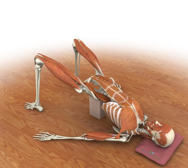
Utthita trikonasana
Ushtrasana stretches the iliopsoas through contraction
optimally contracts the
of the hip and trunk extensors, including the gluteus
psoas major portion of
maximus. Stretch is accentuated by contraction of
the iliopsoas muscle.
the quadriceps (including the rectus femoris, which is
Contraction in this posture
eccentrically contracted).
anteverts the pelvis.
This action draws the
hamstrings’ origin (ischial
tuberosity) away from their
insertion (lower leg), and
accentuates their stretch.
Twisted variations of
utthita trikonasana
preferentially contract
the iliacus portion of the
iliopsoas and complete its
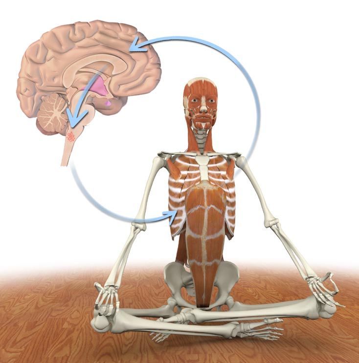
awakening.Chapter24
The Breath Connection
Regions of the brain such as the brain-
stem are highly evolved for survival,
controlling complex functions such as
respiration with speed and precision that
is far beyond the comprehension of the
conscious mind. Great instinctive power is
stored in these regions of the brain. Ha-
tha Yogic breathing techniques “yoke” or
connect the conscious mind to the primal
instinctive regions of the brainstem.
Athletes and martial arts practitioners
access the breath’s primal force by timing
moments of exertion with forced exhala-
tion. Yogis refine this by coordinating the
rhythm of the breath with movements in
the asanas, generally coupling inhalation
with expansion and exhalation with deep-
ening. Pranayama perfects this process.
212Inhalation and Exhalation
The diaphragm is the prime mover for inhalation and exhalation. It is a thin half-dome shaped muscle that separates the thoracic abdominal cavities.
Contracting the diaphragm expands the chest, creating a negative inspiratory pressure in the thorax, and drawing air into the lungs through the
trachea. Contracting the diaphragm also gently massages the abdominal organs.
Unlike most other skeletal muscles, the diaphragm rhythmically contracts and relaxes under the control of the autonomic nervous system, via the
phrenic nerve. We are unaware of the diaphragm, unless we consciously think about its function.
Yogic breathing techniques such as pranayama involve consciously contracting the diaphragm and controlling the breathing, thereby connecting the
conscious and unconscious mind.
These images demonstrate the diaphragm contracting and relaxing. The lungs are elastic and expand when the diaphragm contracts during inhalation.
Like a balloon the lungs passively empty during exhalation as the diaphragm relaxes.
213Ujayi Breath
When we breathe, the air passes through the nasal sinuses and pharynx into the trachea and on to the lungs, oxygenating the blood and
removing carbon dioxide. The pharynx and nasal passages are lined with blood-rich mucosa. The nasal sinuses create turbulence, increas-
ing the amount of air contacting the mucosa. This process warms the air before it passes into the lower parts of the respiratory tract.
The glottis is a muscular aperture below the pharynx and nasal passages. Opening and closing the glottis regulates the flow of air into the
lower respiratory tract. Normally we control the opening and closing of the glottis unconsciously.
Yogic breathing techniques involve consciously regulating airflow through the glottis. For example, we seal the glottis when performing
Nali so that the negative inspiratory pressure generated by contracting the diaphragm draws the abdominal contents upward instead of
drawing breath into the trachea.
Consciously narrowing the opening of the glottis increases the turbulence of the air passing through the nasal and pharyngeal cavities.
This action increases the transfer of heat to the air from the blood-rich mucosal lining, raising the temperature of the air above normal.
Increasing air turbulence also creates an audible vibration similar to that of a flame leaping up from a fire. This process of increasing heat
and creating vibration with the air is known as Ujayi breathing and is fundamental to the practice of Pranayama or “Breath of Fire.”
(See Scientific Keys, Volume II for details on Nali and Pranayama).
1 2 3
214Accessory Muscles of Breath
Accessing the force of the accessory muscles of breath expands
the lung volume and increases the turbulence of air in the respi-
ratory passageways. As with postural muscles, we are generally
not conscious of these accessory breath muscles until awakening
them consciously. Focusing on contracting these muscles brings
them under conscious control with profound effects. The follow-
ing pages illustrate this process in siddhasana, virabhadrasana II,
tadasana and utthanasana.
215Thoracic Bellows
Begin awakening the accessory muscles of
breath by drawing the scapula towards the
midline. Hold this position and then attempt
to roll the shoulders forward by contracting
the pectoralis minor. This closed chain
contraction lifts and opens the lower ribcage
like a bellows and expands the lung volume.
Begin by practicing in siddhasana and then
apply this technique to other postures such
as twists that constrict the volume of the
thoracic cavity.
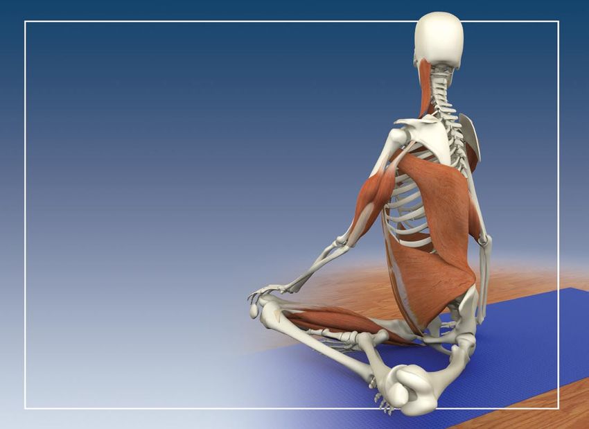
216Accessory 1 2
Muscles of
Breath
1) Straighten the lower back by
contracting the erector spinae and
quadratus lumborum. This draws the
lower posterior ribcage downward.
2) Balance this action by gently
contracting the rectus abdominus.
This draws the lower anterior rib-
cage downward and compresses the
abdominal organs against the dia-
phragm, dynamizing its contraction
and strengthening it.
3) Draw the shoulder blades togeth- 3 4
er by contracting the rhomboids. This
opens the front of the chest.
4) Maintain the contraction of the
rhomboids and simultaneously con-
tract the pectoralis minor and sterno-
cleidomastoid. This lifts and opens the
ribcage like a bellows.
Complete this process by pressing the
hands down on the knees to fully open
the chest (by contracting the latissi-
mus dorsi).
217Exhalation
Access the breath’s primal force when moving into pos-
tures. Gently contract the rectus abdominus, transversus
abdominus and intercostal muscles during exhalation.
Applying this type of contraction rhythmically connects the
conscious and unconscious mind during movement.Synergy
Train the accessory breathing muscles so that they work
synergistically to expand and contract the thorax during
movement.
Increase the lung volume during inhalation by contracting
the accessory breathing muscles in various combinations.
For example, combine the rhomboids with the pectoralis
minor, or the rectus abdominus with the quadratus lumbo-
rum (illustrated here in tadasana).
Expel the residual air in the lungs during exhalation by
contacting the rectus abdominus, transversus abdominus
and intercostal muscles.
Awakening the accessory breathing muscles is an extreme-
ly powerful technique. Begin with very gentle contraction
and progress slowly and with great care. Never force any
yoga technique, especially breathing. Always proceed with
caution under the guidance of an instructor.
219Chapter25
Bandhas
Bandhas are “locks” occuring throughout
the body. The combination of opposing
muscles forms these “locks”, stimulating
nerve conduction and illuminating the
chakras.
Moola bandha
Moola bandha contracts the muscles
of the pelvic floor lifting and toning
the organs of the pelvis including the
bladder and genitalia. The pelvic floor
muscles are recruited and awakened by
contracting associated muscles such as
the iliopsoas. This focuses the mind on the
first chakra.
Simultaneously contracting other muscle groups accentuates moola
bandha. For example, gently squeezing the knees together (by contracting
the adductors) increases contraction of the pelvic floor muscles. Pressing
the hands together has the same effect. This phenomenon is known as
“recruitment.”
220Udyana bandha
Udyana bandha contracts the upper
abdominals in the region approximately two
inches below the solar plexus and focuses
the mind on the third chakra.
rectus abdominus
Jalandhara bandha
Jalandhara bandha contracts the anterior
neck muscles, flexing the neck and drawing
the chin to the sternum. This focuses the
mind on the fifth chakra.
transversus abdominusChapter26
Chakras
The chakras are the subtle energy centers of the body. Like pinwheels, the
chakras spin at the speed of light, emanating the colors of the spectrum, each
bracial plexus
resonating with a particular frequency. These colors combine to form the auras
that surround each of us, connecting us with each other and with the cosmos.
There are seven to eight major and numerous minor chakras in the body. Their
locations correspond to regions of the body where nerves collect and electrical
activity is high, such as the brachial and sacral plexi (major chakras) and the
elbows and knees (minor chakras).
The flow of energy in the chakras can become blocked by life events through
the activity of the autonomic nervous system. For example, when we habitually
assume a defensive posture in response to negative stimuli, we block the flow
of energy in the chakras. Hatha Yoga counteracts this and re-illuminates the
chakras, stimulating them to spin freely.
sacral plexus
Kundalini awakening refers to the “unblocking” of the flow of energy through
and between the chakras. This process can occur instantaneously from contact
with a master (inner or outer) who awakens the student’s awareness of his
or her potential. Classically, this occurs through a touch but can occur with a
glance or even through the mere presence of the master. This is known as Shak-
tipata (the transmission of psycho-spiritual energy). As human consciousness
transitions from the Piscean to the Aquarian Age, more and more people are
spontaneously experiencing varying degrees of Kundalini awakening.
Kundalini awakening is akin to tapping into a high voltage line and requires
careful preparation. Hatha Yoga prepares the practitioner and awakens the
Kundalini at the same time.
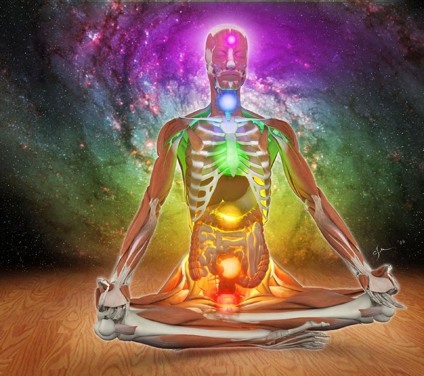
222Asanas connect the body and
mind. Breathing techniques
connect the conscious and
the unconscious. Chakra
meditation connects the
individual to the vibrational
energy of the cosmos. Spend
a few moments gazing at
this image of the chakras
and then meditate as you
visualize them. The chakras
will appear as a subtle but
scintillating light within you.Scientific Keys Volume I The Key
Muscles of
Hatha Yoga
Ray Long MD FRCSC
With Illustrator Chris Macivor
www.BandhaYoga.comYou can also read