The prevalence and physiological impacts of centrilobular and paraseptal emphysema on computed tomography in smokers with preserved ratio impaired ...
←
→
Page content transcription
If your browser does not render page correctly, please read the page content below
ERJ OPEN RESEARCH
                                                                                                                   ORIGINAL RESEARCH ARTICLE
                                                                                                                            Y. SHIRAISHI ET AL.

The prevalence and physiological impacts of centrilobular and
paraseptal emphysema on computed tomography in smokers
with preserved ratio impaired spirometry
Yusuke Shiraishi 1,2,10, Takafumi Shimada 3,10, Naoya Tanabe 1, Kunihiko Terada4, Ryo Sakamoto5,
Tomoki Maetani1, Hiroshi Shima1, Fumi Mochizuki3, Tsuyoshi Oguma1, Kaoruko Shimizu6, Susumu Sato 1,
Shigeo Muro 1,7, Nobuyuki Hizawa8, Motonari Fukui2, Hiroaki Iijima3, Izuru Masuda9 and Toyohiro Hirai1

1
 Dept of Respiratory Medicine, Graduate School of Medicine, Kyoto University, Kyoto, Japan. 2Respiratory Disease Center, Kitano
Hospital, The Tazuke Kofukai Medical Research Institute, Osaka, Japan. 3Dept of Respiratory Medicine, Tsukuba Medical Center,
Tsukuba, Japan. 4Terada Clinic, Respiratory Medicine and General Practice, Himeji, Japan. 5Dept of Diagnostic Imaging and Nuclear
Medicine, Graduate School of Medicine, Kyoto University, Kyoto, Japan. 6Dept of Respiratory Medicine, Faculty of Medicine, Hokkaido
University, Sapporo, Japan. 7Respiratory Medicine, Nara Medical University, Kashihara, Japan. 8Dept of Pulmonary Medicine,
University of Tsukuba, Tsukuba, Japan. 9Medical Examination Center, Takeda Hospital, Kyoto, Japan. 10These authors contributed
equally to this work.

                                                                             Corresponding author: Naoya Tanabe (ntana@kuhp.kyoto-u.ac.jp)

                                        Shareable abstract (@ERSpublications)
                                        Centrilobular and paraseptal emphysema are observed in 43–46% of smokers with preserved ratio
                                        impaired spirometry. Centrilobular emphysema, but not paraseptal emphysema, is closely
                                        associated with air-trapping in these smokers. https://bit.ly/3Ky6LDy

                                        Cite this article as: Shiraishi Y, Shimada T, Tanabe N, et al. The prevalence and physiological impacts
                                        of centrilobular and paraseptal emphysema on computed tomography in smokers with preserved
                                        ratio impaired spirometry. ERJ Open Res 2022; 8: 00063-2022 [DOI: 10.1183/23120541.00063-2022].

                                        Abstract
    Copyright ©The authors 2022         Centrilobular emphysema (CLE) and paraseptal emphysema (PSE) are observed in smokers with preserved
                                        ratio impaired spirometry (PRISm, defined as the ratio of forced expiratory volume in 1 s (FEV1) to forced
    This version is distributed under
    the terms of the Creative
                                        vital capacity (FVC) ⩾0.7 and FEV1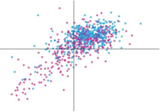
ERJ OPEN RESEARCH                                                                             ORIGINAL RESEARCH ARTICLE | Y. SHIRAISHI ET AL.

                                   persistent respiratory symptoms [2, 5]. However, the management of smokers with PRISm is still
                                   challenging, due to the clinically heterogenous manifestations. Indeed, a subgroup of smokers with PRISm
                                   could develop COPD and show greater mortality than smokers with normal spirometry, but other smokers
                                   with PRISm could remain clinically stable over time and even return to normal on follow-up spirometry [3, 6].
                                   Therefore, further understanding of clinical features is warranted to improve the outcomes of PRISm.

                                   Computed tomography (CT) is widely used to screen for lung cancer in real-world practice, providing
                                   structural information on the airway and parenchyma simultaneously. A prior study showed that
                                   radiological abnormalities of the lungs and chest wall such as paraseptal emphysema (PSE), airway wall
                                   thickening, diaphragm eventration and smaller transverse internal thoracic diameter are more frequent in
                                   smokers with PRISm than those with normal spirometry [7]. Subsequently, the Fleischner Society
                                   published a visual classification system for emphysema [8] to describe the localisation of centrilobular
                                   emphysema (CLE), PSE and panlobular emphysema on CT. The use of this system has enabled showing
                                   that CLE and PSE are common in smokers with and without airflow limitation [9, 10]. Moreover, CLE is
                                   associated with airflow limitation, lung hyperinflation and exacerbation frequency, and even mild signs of
                                   CLE predict poor prognosis, whereas PSE is less associated with physiological impairments than CLE
                                   [10–12]. These results suggest that the differential emphysema subtypes may contribute to the
                                   heterogeneous presentations of smokers with PRISm.

                                   Air-trapping predicts adverse respiratory outcome and progression to COPD in smokers without airflow
                                   limitation [13, 14] and the ratio of forced vital capacity (FVC) to total lung capacity on CT (TLCCT) as a
                                   conceptual surrogate for air-trapping predicts future worsening of symptoms, exacerbation and progression
                                   to COPD in smokers with PRISm [15]. However, little is known about the morphological basis on
                                   air-trapping in these smokers. Therefore, this study tested the hypothesis that CLE and PSE would
                                   differentially affect physiological function in smokers with PRISm. Specifically, the study aimed 1) to
                                   explore whether the prevalence of CLE and PSE in smokers with PRISm differs from that in
                                   never-smokers with PRISm and smokers with normal spirometry and airflow limitation; and 2) to test
                                   whether CLE and PSE are associated with physiological impairments, particularly air-trapping, in smokers
                                   with PRISm.

                                   Material and methods
                                   Study design (subjects)
                                   This was a multicentre retrospective study conducted in three hospitals (Tsukuba Medical Center, Kitano
                                   Hospital and Takeda Hospital) and a general clinic (Terada Clinic) in Japan. In Japan, hospitals and clinics
                                   provide medical checkup programmes in which chest CT scans for lung cancer screening are offered to all
                                   adults regardless of smoking status. In this study, we consecutively enrolled never- and ever-smokers aged
                                   ⩾40 years who underwent inspiratory CT for lung cancer screening and spirometry. Subjects with a history
                                   of lung resection, chest CT abnormalities extending to more than one lobe (such as consolidations,
                                   atelectasis, tumours, pneumothorax and thoracic deformity), missing information on smoking status or
                                   light-smokers (90 days between spirometry and CT scanning or insufficient inspiratory
                                   chest CT (defined as FVC>TLCCT [15]) were excluded (figure 1). The study was conducted in accordance
                                   with the Declaration of Helsinki and approved by the ethics committees of Kyoto University Hospital,
                                   Tsukuba Medical Center, Kitano Hospital and Takeda Hospital (approval R1660-3 and R2751). Written
                                   consent was waived because of the retrospective nature of the study.

                                   CT acquisition, spirometry and TLCCT
                                   All chest CT scans were acquired at full inspiration. Images at 0.5–1.25 mm slice thickness were
                                   reconstructed using sharp kernels. Spirometry was conducted without bronchodilator and evaluated in each
                                   facility by well-trained technicians according to the American Thoracic Society/European Respiratory
                                   Society statement [16]. Predicted FEV1 and FVC values were calculated using the LMS (lambda, mu,
                                   sigma) method reference equations taking age, gender and height into account [17]. Subjects were
                                   classified as having PRISm (FEV1/FVC ⩾0.7 and FEV1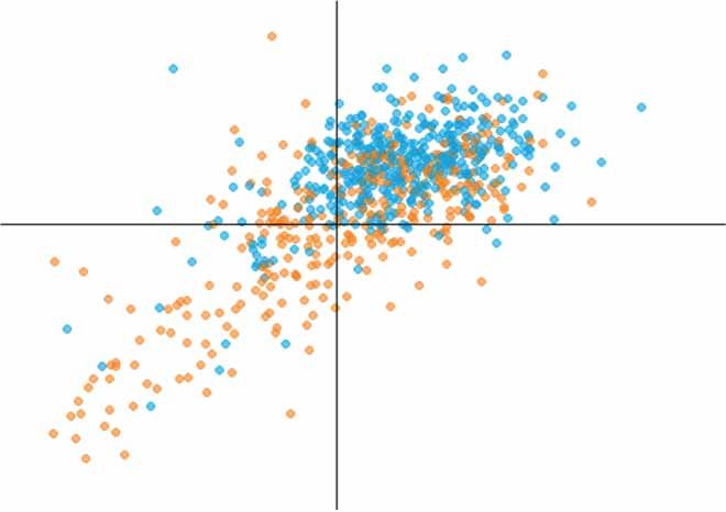
ERJ OPEN RESEARCH                                                                              ORIGINAL RESEARCH ARTICLE | Y. SHIRAISHI ET AL.

                                              2116 subjects with age ≥40 years underwent chest CT for lung cancer screening and spirometry

                                                         298 excluded
                                                          Light-smoker
                                                          Abnormal CT findings
                                                          Lack of demographic data
                                                          Insufficient inspiratory chest CT
                                                          >90 days between CT and spirometry

                                                Never-smoker                                       Smoker with ≥10 pack-years
                                                   n=1046                                                   n=772

                                                           Normal spirometry                                    Normal spirometry
                                                                n=914                                                n=516

                                                                PRISm                                                 PRISm
                                                                 n=86                                                  n=87

                                                           Airflow limitation                                    Airflow limitation
                                                                 n=46                                                  n=169

                                    FIGURE 1 Study population flow chart. CT: computed tomography; PRISm: preserved ratio impaired
                                    spirometry.

                                   expiratory mean lung density and the ratio of expiratory to inspiratory mean lung density ratio were
                                   calculated [19]. In the study, smokers aged ⩾40 years underwent a pair of inspiratory and expiratory chest
                                   CT scans with written informed consent [20].

                                   Visual CT analysis
                                   Six CT-experienced pulmonologists with ⩾5 years’ experience and a chest radiologist with 15 years’
                                   experience performed visual emphysema analysis based on the Fleischner Society classification system [8].
                                   The images were viewed at window width 700 HU and window level −750 HU according to the
                                   Fleischner Society’s recommendation [8]. CLE was classified as trace, mild, moderate, confluent or
                                   advanced destructive, while PSE was classified as mild or substantial [8]. The category of panlobular
                                   emphysema was not used in this study, as it is applied to patients with α1-antitrypsin deficiency [8].
                                   Before assessing visual emphysema score in the study population, the analysts scored training CT datasets
                                   and reviewed substantial discordance to obtain consensus. Each CT scan was assessed by two
                                   CT-experienced pulmonologists without knowledge of clinical information and the discordances were
                                   adjudicated by a chest radiologist. Further details are provided in the supplementary material.

                                   Statistical analysis
                                   The weighted κ-coefficient was calculated for interobserver variability in the visual emphysema
                                   assessments. The severity of CLE (none/trace/mild/moderate/confluent/advanced destructive) was weighted
                                   from 0 to 5, and that of PSE (none/mild/substantial) was weighted from 0 to 2. Data are expressed as
                                   mean±SD unless indicated. Subjects’ characteristics were compared using Fisher’s exact test or Chi-squared
                                   test for categorical data and the t-test for continuous variables. The Bonferroni correction method was used
                                   to adjust for multiple comparisons. Additionally, the question of whether the presence of PSE and CLE
                                   could affect FVC/TLCCT was explored using multivariable linear regression models that included age,
                                   gender, body mass index, smoking pack-years (as a dichotomous variable,
ERJ OPEN RESEARCH                                                                                ORIGINAL RESEARCH ARTICLE | Y. SHIRAISHI ET AL.

                                   smoking status, facilities and the presence of PSE/CLE as independent variables. Statistical analyses were
                                   performed using R statistical software version 4.0.1 and JMP Pro version 16.1.0. A p-value
ERJ OPEN RESEARCH                                                                                               ORIGINAL RESEARCH ARTICLE | Y. SHIRAISHI ET AL.

                    Never-smokers                                                                  Smokers
                    PRISm                                 Normal spirometry                        PRISm                                   Normal spirometry
                    PSE+ 1.2%                                     PSE+ 2.5%                        PSE+ 43.7%                                    PSE+ 36.2%
             0.90                                                                           0.90
  FEV1/FVC

                                                                                 FEV1/FVC
             0.70                                                                           0.70

             0.50                                                                           0.50                                                          PSE–
                                                                                                                                                          PSE+
                                                            Airflow limitation                                                              Airflow limitation
             0.30                                                  PSE+ 4.3%                0.30                                                  PSE+ 71.0%

                    20      40      60     80       100    120      140                            20      40       60      80       100    120     140
                                         FEV1 % pred                                                                      FEV1 % pred

                    Never-smokers                                                                  Smokers
                    PRISm                                 Normal spirometry                        PRISm                                   Normal spirometry
                    CLE+ 4.7%                                     CLE+ 1.9%                        CLE+ 46.0%                                    CLE+ 31.8%
             0.90                                                                           0.90
  FEV1/FVC

                                                                                 FEV1/FVC

             0.70                                                                           0.70

             0.50                                                                           0.50
                                                                                                                                                          CLE–
                                                                                                                                                          CLE+
                                                            Airflow limitation                                                              Airflow limitation
             0.30                                                 CSE+ 10.9%                0.30                                                  CLE+ 79.3%

                    20      40      60     80       100    120      140                            20      40       60      80       100    120     140
                                         FEV1 % pred                                                                      FEV1 % pred

 FIGURE 2 Distribution of emphysema subtypes and lung functions in never-smokers and smokers with ⩾10 pack-years undergoing computed
 tomography lung screening. The horizontal line represents the threshold for airflow limitation (forced expiratory volume in 1 s (FEV1)/forced vital
 capacity (FVC) 0.70), and the vertical line represents the threshold between mild and moderate airflow limitation (FEV1 80% pred). PRISm:
 preserved ratio impaired spirometry; PSE: paraseptal emphysema; CLE: centrilobular emphysema.

                                         and PSE categories in never-smokers and smokers with normal spirometry, PRISm and airflow limitation.
                                         Moreover, as shown in figure 3b, the presence of PSE was significantly associated with the presence of
                                         CLE in smokers with PRISm (Chi-squared p
ERJ OPEN RESEARCH ORIGINAL RESEARCH ARTICLE | Y. SHIRAISHI ET AL.
TABLE 2 The distribution of emphysema subtypes in never- and ever smokers according to lung function categories
Subjects with normal spirometry Subjects with PRISm Subjects with airflow limitation
Never-smoker Smoker p-value Never-smoker Smoker p-value Never-smoker Smoker p-value
Subjects 914 516 86 87 46 169
PSEERJ OPEN RESEARCH ORIGINAL RESEARCH ARTICLE | Y. SHIRAISHI ET AL.
TABLE 3 Characteristics of smokers with preserved ratio impaired spirometry
CLE absent CLE present p-value
Subjects 47 40
Age (years) 63.9±10.8 63.0±11.0 0.71
Male 40 (85.1) 36 (90.0) 0.72
BMI (kg·m−2) 25.5±3.8 23.5±3.3ERJ OPEN RESEARCH ORIGINAL RESEARCH ARTICLE | Y. SHIRAISHI ET AL.
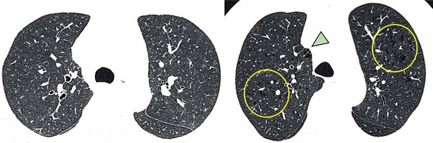
a) b)
FIGURE 4 Representative images of smokers with preserved ratio impaired spirometry emphysema absent or
present. a) 61-year-old male; neither PSE nor CLE is present; b) 66-year-old male presenting both PSE
(arrowhead) and CLE (circles). Both cases had comparable lung function (forced vital capacity (FVC) 74.7%
pred and 79.6% pred, for a) and b), respectively; forced expiratory volume in 1 s (FEV1) 73.5% pred and 71.5%
pred, for a) and b), respectively; and FEV1/FVC 0.80 and 0.75, for a) and b), respectively), but FVC/total lung
capacity measured by computed tomography was lower in case b) than case a) (86.7% and 58.9%,
respectively).
emphysema have been used especially in research [26], the visual assessment complements quantitative
measurements and might be more sensitive in detecting tiny parenchymal changes in smokers without
airflow limitation [27].
The prevalence of PSE and CLE in smokers with PRISm did not differ from those with normal spirometry.
This is not consistent with a previous report showing that PSE was more prevalent in Global Initiative for
Chronic Obstructive Lung Disease unclassified patients (synonymous with PRISm) than smokers with
normal spirometry, while the prevalence of CLE did not differ [7]. Furthermore, the prevalence of PSE and
CLE in normal spirometry and PRISm (PSE 36.2% and 43.7%, respectively; CLE 31.8% and 46.0%,
respectively) in this study was higher than those reported in the previous report (PSE 17% and 33%,
respectively; CLE 22.5% and 27%, respectively). This might be because this study used the Fleischner
Society classification system to detect emphysema subtype more accurately and sensitively, including trace
CLE, which involvedERJ OPEN RESEARCH ORIGINAL RESEARCH ARTICLE | Y. SHIRAISHI ET AL.
In conclusion, the prevalence of both PSE and CLE on CT in smokers with PRISm was higher than that in
never-smokers with PRISm. In smokers with PRISm, the presence of CLE, but not PSE, was associated
with air-trapping, suggesting different physiological roles of PSE and CLE. Visual emphysema subtyping
on CT with the Fleischner Society classification system can help clinicians understand the pathophysiology
of smokers and take a more personalised approach to smokers with PRISm.
Provenance: Submitted article, peer reviewed.
Conflicts of interest: N. Tanabe, S. Sato, T. Oguma and T. Hirai were supported by a grant from FUJIFILM Co., Ltd.
FUJIFILM did not have a role in the design or analysis of the study or in the writing of the manuscript. The other
authors have no conflict of interest to declare.
Support statement: This study was supported by Japan Society for the Promotion of Science grant 19K08624.
Funding information for this article has been deposited with the Crossref Funder Registry.
References
1 Wan ES, Castaldi PJ, Cho MH, et al. Epidemiology, genetics, and subtyping of preserved ratio impaired
spirometry (PRISm) in COPDGene. Respir Res 2014; 15: 89.
2 Wijnant SRA, de Roos E, Kavousi M, et al. Trajectory and mortality of preserved ratio impaired spirometry: the
Rotterdam Study. Eur Respir J 2020; 55: 1901217.
3 Wan ES, Fortis S, Regan EA, et al. Longitudinal phenotypes and mortality in preserved ratio impaired
spirometry in the COPDGene study. Am J Respir Crit Care Med 2018; 198: 1397–1405.
4 Wan ES, Balte P, Schwartz JE, et al. Association between preserved ratio impaired spirometry and clinical
outcomes in US adults. JAMA 2021; 326: 2287–2298.
5 Global Initiative for Chronic Obstructive Lung Disease (GOLD). Global Strategy for the Diagnosis, Management
and Prevention of COPD. 2022. www.goldcopd.org/2022-gold-reports-2/ Date last accessed: 19 January 2022.
6 Wan ES, Hokanson JE, Regan EA, et al. Significant spirometric transitions and preserved ratio impaired
spirometry among ever smokers. Chest 2022; 161: 651–661.
7 Kim SS, Yagihashi K, Stinson D, et al. Visual assessment of CT findings in smokers with nonobstructed
spirometric abnormalities in the COPDGene® study. Chronic Obstr Pulm Dis 2014; 1: 88–96.
8 Lynch DA, Austin JHM, Hogg JC, et al. CT-definable subtypes of chronic obstructive pulmonary disease: a
statement of the Fleischner Society. Radiology 2015; 277: 192–205.
9 Oh AS, Strand M, Pratte K, et al. Visual emphysema at chest CT in GOLD stage 0 cigarette smokers predicts
disease progression: results from the COPDGene study. Radiology 2020; 296: 641–649.
10 El Kaddouri B, Strand MJ, Baraghoshi D, et al. Fleischner Society visual emphysema CT patterns help predict
progression of emphysema in current and former smokers: results from the COPDGene study. Radiology
2020; 298: 441–449.
11 Lynch DA, Moore CM, Wilson C, et al. CT-based visual classification of emphysema: association with mortality
in the COPDGene study. Radiology 2018; 288: 859–866.
12 Smith BM, Austin JHM, Newell JD, et al. Pulmonary emphysema subtypes on computed tomography: the
MESA COPD study. Am J Med 2014; 127: 94.e7–94.e23.
13 Zeng S, Tham A, Bos B, et al. Lung volume indices predict morbidity in smokers with preserved spirometry.
Thorax 2019; 74: 114–124.
14 Fortis S, Comellas AP, Bhatt SP, et al. Ratio of FEV1/slow vital capacity ofERJ OPEN RESEARCH ORIGINAL RESEARCH ARTICLE | Y. SHIRAISHI ET AL.
21 Hogg JC, Chu F, Utokaparch S, et al. The nature of small-airway obstruction in chronic obstructive pulmonary
disease. N Engl J Med 2004; 350: 2645–2653.
22 Kim WD, Eidelman DH, Izquierdo JL, et al. Centrilobular and panlobular emphysema in smokers: two distinct
morphologic and functional entities. Am Rev Respir Dis 1991; 144: 1385–1390.
23 Koo HK, Vasilescu DM, Booth S, et al. Small airways disease in mild and moderate chronic obstructive
pulmonary disease: a cross-sectional study. Lancet Respir Med 2018; 6: 591–602.
24 Tanabe N, Vasilescu DM, Hague CJ, et al. Pathological comparisons of paraseptal and centrilobular
emphysema in chronic obstructive pulmonary disease. Am J Respir Crit Care Med 2020; 202: 803–811.
25 Park J, Hobbs BD, Crapo JD, et al. Subtyping COPD by using visual and quantitative CT imaging features.
Chest 2020; 157: 47–60.
26 Bhatt SP, Washko GR, Hoffman EA, et al. Imaging advances in chronic obstructive pulmonary disease.
Insights from the genetic epidemiology of chronic obstructive pulmonary disease (COPDGene) study. Am J
Respir Crit Care Med 2019; 199: 286–301.
27 Regan EA, Lynch DA, Curran-Everett D, et al. Clinical and radiologic disease in smokers with normal
spirometry. JAMA Intern Med 2015; 175: 1539–1549.
28 Tan WC, Sin DD, Bourbeau J, et al. Characteristics of COPD in never-smokers and ever-smokers in the general
population: results from the CanCOLD study. Thorax 2015; 70: 822–829.
29 Lee H, Hong Y, Lim MN, et al. Inflammatory biomarkers and radiologic measurements in never-smokers with
COPD: a cross-sectional study from the CODA cohort. Chron Respir Dis 2018; 15: 138–145.
30 O’Donnell CR, Bankier AA, Stiebellehner L, et al. Comparison of plethysmographic and helium dilution lung
volumes: which is best for COPD? Chest 2010; 137: 1108–1115.
31 Iwano S, Okada T, Satake H, et al. 3D-CT volumetry of the lung using multidetector row CT. Comparison with
pulmonary function tests. Acad Radiol 2009; 16: 250–256.
https://doi.org/10.1183/23120541.00063-2022 10You can also read


