The Rat Grimace Scale: A partially automated method for quantifying pain in the laboratory rat via facial expressions
←
→
Page content transcription
If your browser does not render page correctly, please read the page content below
Sotocinal et al. Molecular Pain 2011, 7:55
http://www.molecularpain.com/content/7/1/55
                                                                                                                                  MOLECULAR PAIN

 METHODOLOGY                                                                                                                                 Open Access

The Rat Grimace Scale: A partially automated
method for quantifying pain in the laboratory
rat via facial expressions
Susana G Sotocinal1, Robert E Sorge1, Austin Zaloum1, Alexander H Tuttle1, Loren J Martin1, Jeffrey S Wieskopf1,
Josiane CS Mapplebeck1, Peng Wei2, Shu Zhan3, Shuren Zhang3, Jason J McDougall3, Oliver D King2 and
Jeffrey S Mogil1*

  Abstract
  We recently demonstrated the utility of quantifying spontaneous pain in mice via the blinded coding of facial
  expressions. As the majority of preclinical pain research is in fact performed in the laboratory rat, we attempted to
  modify the scale for use in this species. We present herein the Rat Grimace Scale, and show its reliability, accuracy,
  and ability to quantify the time course of spontaneous pain in the intraplantar complete Freund’s adjuvant,
  intraarticular kaolin-carrageenan, and laparotomy (post-operative pain) assays. The scale’s ability to demonstrate the
  dose-dependent analgesic efficacy of morphine is also shown. In addition, we have developed software, Rodent
  Face Finder®, which successfully automates the most labor-intensive step in the process. Given the known
  mechanistic dissociations between spontaneous and evoked pain, and the primacy of the former as a clinical
  problem, we believe that widespread adoption of spontaneous pain measures such as the Rat Grimace Scale
  might lead to more successful translation of basic science findings into clinical application.

Introduction                                                                        spontaneous pain, in many cases it has been difficult
Despite great advances in basic understanding of mole-                              to demonstrate that these behaviors display specificity
cular pain mechanisms and considerable investment by                                and sensitivity as measures of pain [5].
industry, translational achievements in analgesic drug                                Because of the known utility of facial coding scales
development have been extremely limited. Many believe                               (based on the facial action coding system; FACS) [6] for
that the high attrition is due, at least in part, to the                            the quantification of pain in non-verbal human popula-
poor predictivity of current animal models of pain [1].                             tions [see [7]], and the prediction by Darwin that nonhu-
As in vivo animal research remains the mainstay of                                  man animals exhibit similar facial expressions to
analgesic drug development [2,3], much recent effort                                emotional states as do humans [8], we recently developed
has been devoted to reexamining pain testing para-                                  and characterized the Mouse Grimace Scale (MGS) [9]. It
digms in laboratory animals. Of the criticisms directed                             consists of five facial “action units” (orbital tightening,
at the status quo in rodent algesiometry, one of the                                nose bulge, cheek bulge, ear position, and whisker
most common is that the vast majority of preclinical                                change) scored on a 0-2 scale for their prominence in
studies measure withdrawal responses to evoking ther-                               still photographs taken from digital video of mice in
mal and mechanical stimuli instead of the more clini-                               either a baseline or pain condition. We demonstrated
cally important spontaneous pain [4]. Although a                                    that the MGS displays high accuracy and reliability, is
number of rodent behaviors are correlated in time                                   useful for quantifying pain of moderate duration (from
with injuries that presumably also produce                                          several minutes to approximately 1 day), is sensitive to
                                                                                    detecting weak analgesic effects, and may represent a
                                                                                    measure of the animal’s affective response to pain [9].
* Correspondence: jeffrey.mogil@mcgill.ca
1
 Dept. of Psychology and Alan Edwards Centre for Research on Pain, McGill             The purpose of the present work was two-fold. First,
University, Montreal, QC H3A 1B1, Canada                                            despite increasing use of the mouse over the past few
Full list of author information is available at the end of the article

                                      © 2011 Sotocinal et al; licensee BioMed Central Ltd. This is an Open Access article distributed under the terms of the Creative
                                      Commons Attribution License (http://creativecommons.org/licenses/by/2.0), which permits unrestricted use, distribution, and
                                      reproduction in any medium, provided the original work is properly cited.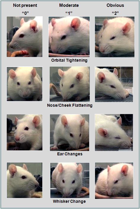
Sotocinal et al. Molecular Pain 2011, 7:55                                                                   Page 2 of 10
http://www.molecularpain.com/content/7/1/55

decades, the rat remains by far the most common sub-         Laparotomy
ject of preclinical pain research [1]. The evolutionary      A laparotomy, designed to mimic a sham ventral ovar-
stability of facial expression [7,8] would clearly predict   iectomy [13], was performed under isoflurane/oxygen
that the MGS could be translated to the rat. Second, the     anesthesia. Following shaving and disinfection, a 1-cm
main practical disadvantage of the MGS is the labor-         midline incision was made using a scalpel. Muscle layers
intensive nature of one step in the process: grabbing        were closed with polydioxanone suture 5-0 (Vicryl ® ;
individual face-containing frames from digital video,        Ethicon, Somerville, NJ) and skin edges apposed using
which is hampered by uncooperative subjects (not look-       tissue glue (Vetbond®; 3M, St. Paul, MN). Rats (n = 6)
ing directly at the camera) or otherwise poor optics due     were tested before, and 1 h, 4 h, 6 h and 12 h post-
to motion blurring. The utility of this method would         surgery.
thus be greatly improved by automated frame grabbing.
We report here the development of the Rat Grimace            Morphine
Scale (RGS), its ability to quantify pain in three common    Morphine sulfate was obtained from Sandoz Canada.
algesiometric assays (intraplantar complete Freund’s         Mice were injected with physiological saline (10 ml/kg)
adjuvant, intraarticular kaolin/carrageenan, and laparot-    or 1, 2, or 5 mg/kg morphine (n = 4-8/dose), adminis-
omy), and the development of Rodent Face Finder® soft-       tered 5.5 h after CFA (see above) and 15 min before the
ware for automated generation of scoring-ready still         start of 30-min digital video recording (see below).
photographs of both mouse and rat faces.
                                                             Digital video
Materials and Methods                                        Rats (two at a time) were placed on a table top in cubicles
In all experiments, male and female rats were used in        (21 × 10.5 × 9 cm high) with walls of transparent Plexi-
equal numbers [10]. No sex differences were observed         glas® and a separating wall of removable stainless steel.
and so data were combined for reported analyses.             One digital video camera was placed on either side of the
                                                             apparatus in order to maximize the opportunity for clear
Animals                                                      head shots. Rats were digitally videotaped using high-reso-
All subjects were Wistar rats, aged 6-8 weeks (200-250       lution (1920 × 1080) digital video cameras (Sony High
g), obtained from Charles River Laboratories (Boucher-       Definition Handycam® Camcorder; model HDR-CX100)
ville, QC). Rats were housed in groups of 2-4, under a       for 30 min immediately prior to injection or surgery (base-
12:12-hour light cycle (lights on at 07:00 h) in a tem-      line or no pain photos), and for 30 min at various time
perature-controlled environment (20 ± 2°C) with ad lib       points after injection or surgery (pain photos).
access to food (Prolab RHM 2500) and tap water. Each
nociceptive assay utilized a separate cohort of rats, such   Automated frame capture using Rodent Face Finder®
that no subject participated in more than one assay. All     Previous to the development of Rodent Face Finder ®
studies were approved by a local animal care and use         (RFF), we extracted images manually from digital video.
committee, and were consistent with national guidelines.     Using Windows Media Player, individual frames of the
                                                             resultant AVCHD video files were “grabbed” and
Inflammatory assays                                          cropped (so that body position was no longer visible)
Inflammatory assays were used in this study since the        using the Windows 7 Snipping Tool whenever a clear,
limited duration of facial grimacing is not appropriate      unobstructed head shot was observed. This process is
for neuropathic assays. Complete Freund’s adjuvant           considerably labor-intensive, and a C++ program (using
(CFA), kaolin and carrageenan were all obtained from         the Open CV2.0 library; http://opencv.willowgarage.
Sigma (St. Louis, MO). In the intraplantar CFA model         com), RFF was developed to automate it.
[11], rats were injected with 50% CFA, in a 150 μl injec-      RFF detects rodent eyes and ears using boosted cascades
tion volume, into the plantar surface of one hind paw.       of Haar classifiers [14], which use differences between
Rats (n = 10) were tested before, and 1 h, 4 h, 6 h, 24 h    pixel intensities in small rectangular regions (Haar-like
and (in a separate cohort; n = 8) 48 h and 7 days post-      features) to capture textural and orientation information,
injection. In the rat intraarticular kaolin/carrageenan      and combine the response from many such regions to
model [12], 2% kaolin and 2% carrageenan were succes-        make predictions on whether a specific sub-region in an
sively injected (separated by 10 min), under isoflurane/     image contains an eye or ear. The precise regions and cut-
oxygen anesthesia, into one knee joint, each in a volume     offs used by the cascades were obtained by Haar training,
of 200 μl. Rats (n = 6) were tested before, and 3 h, 6 h,    using approximately 500 cropped images of ears and eyes
and 12 h post-injection. Group sizes were based on our       from both baseline and pain-experiencing rodents as posi-
experience using similar assays in mice [9].                 tive examples, and a comparable number of non-face-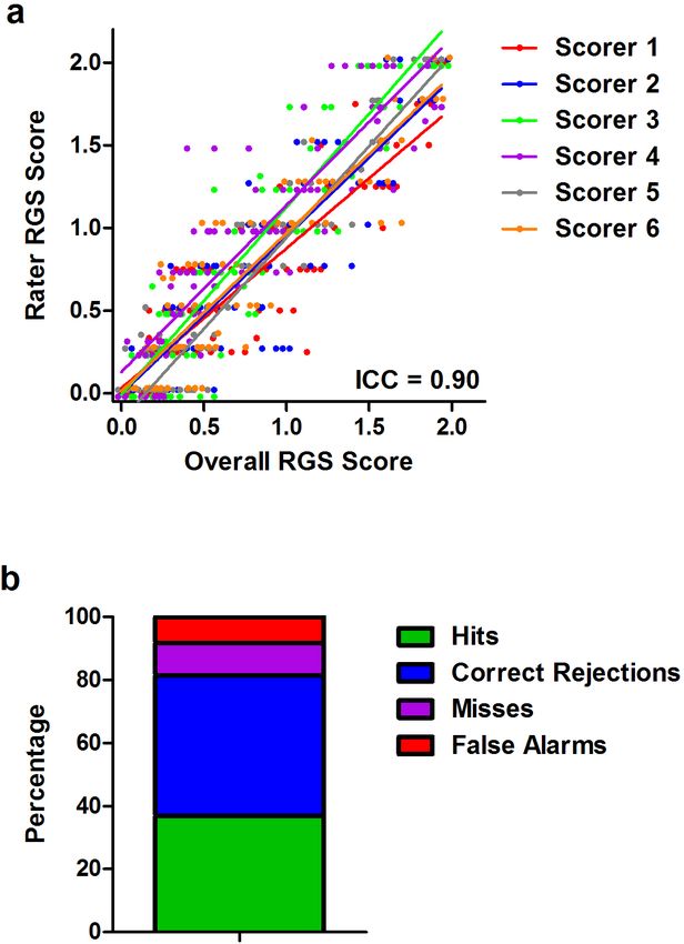
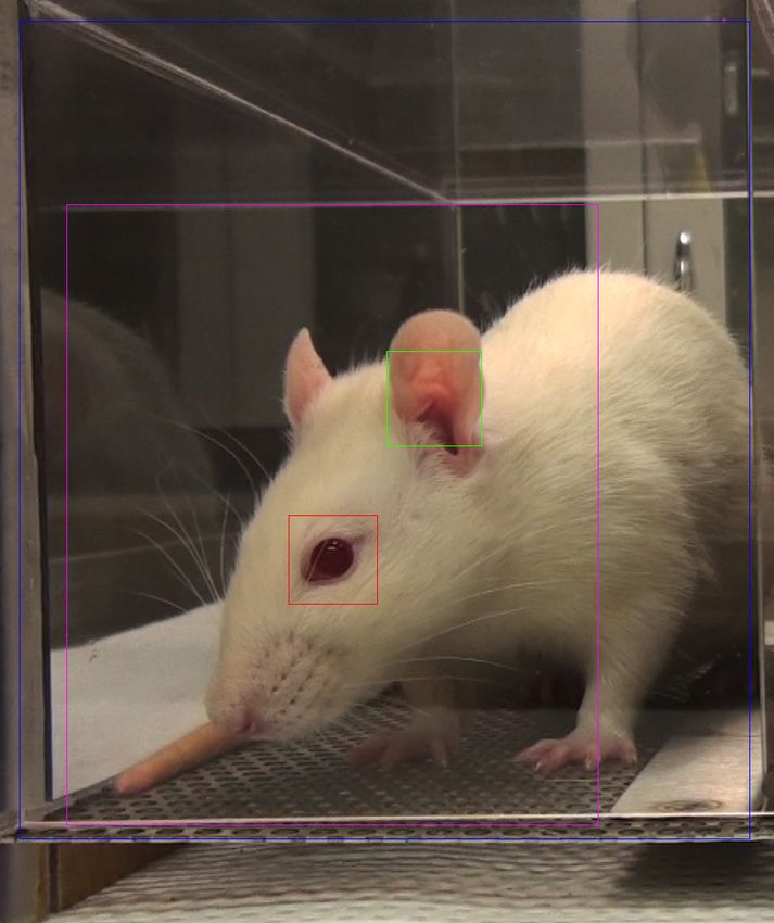
Sotocinal et al. Molecular Pain 2011, 7:55                                                                                   Page 3 of 10
http://www.molecularpain.com/content/7/1/55

containing frames and unrelated images as negative train-             were restricted to zones of the video corresponding to
ing examples. The resulting detectors were used to scan               single cages, with the left and right cages analyzed
each video frame for eyes and ears, at a variety of scales.           separately.
Frames with at least one eye and at least one ear detected,             From each 3-min time interval, the single image most
and satisfying bounds on the distance between them to                 suitable for manual RGS scoring was manually selected
reduce false positives, were flagged as candidates for scor-          from among the candidate images. For some intervals,
ing. Figure 1 illustrates a video frame flagged by RFF for            no candidate images were extracted by RFF, which can
scoring.                                                              occur if the rodent does not face either camera during
  To reduce the number of images for manual scoring,                  this interval, or due to false negatives in the program. In
and to minimize blurring due to rapid motions such as                 some intervals with no candidate images, images were
grooming, among all the candidate video frames in each                extracted manually.
3-min time interval, only the three with the smallest aver-
age absolute pixel difference relative to the previous                RGS coding
video frame (1/30 th of a second earlier) were saved as               Image files were then copied into PowerPoint, one image
images. Both the feature detection and motion estimation              per slide. A PowerPoint macro (http://www.tushar-mehta.

 Figure 1 Uncropped image identified by RFF for RGS scoring. Boxes: blue, total region analyzed; red, detected eye; green, detected ear;
 purple, estimated face region.
Sotocinal et al. Molecular Pain 2011, 7:55                                                                  Page 4 of 10
http://www.molecularpain.com/content/7/1/55

com/powerpoint/randomslideshow/index.htm) was then           causing the whisker pads to flatten. The flattening of
used to randomize the slide order. Identifications were      normal bulging in the nose and cheek appear to always
removed in order to ensure that subsequent coding was        occur together, such that a single action unit, which
performed blind.                                             we call Nose/Cheek Flattening, appears to show the
  Randomized and unlabeled photos were presented on          highest correlation with the presence of pain in the
a large, high-resolution computer monitor, one at a          rat. This major change renders the RGS much more
time. For each photo, the scorer assigned a value of 0, 1    sensitive and accurate in detecting pain in rats than
or 2 for each of the four RGS action units (see section      the MGS.
3.1 and Figure 1). In every case, a score of “0” indicated     Thus, the four action units of the RGS (illustrated in
high confidence of the scorer that the action unit was       Figure 2) are as follows:
absent. A score of “1” indicated either high confidence      1. Orbital Tightening
of a moderate appearance of the action unit, or equivo-      Rats in pain display a narrowing of the orbital area,
cation over its presence or absence. A score of “2” indi-    manifesting either as (partial or complete) eye closure or
cated the detection of an obvious appearance of the          eye “squeezing.”
action unit, with high confidence.                           2. Nose/Cheek Flattening
                                                             Rats in pain display successively less bulging of the nose
Accuracy and reliability determination                       and cheek (see above), with eventual absence of the
A detailed handout was prepared and distributed (by S.       crease between the cheek and whisker pads.
G.S.) to members of the J.S.M. lab, explaining each fea-     3. Ear Changes
ture and providing prototypic photos for each intensity      The ears of rats in pain tend to fold, curl and angle for-
score (0-2) of each action unit. Five postdoctoral, gradu-   wards or outwards, resulting in a pointed shape. The
ate or undergraduate student coders were then given          space between the ears may appear wider.
104 randomized, unlabeled photos (half no pain; half         4. Whisker Change
CFA pain) in order to assess inter-rater reliability and     The whiskers of rats in pain move forward (away from
accuracy of the RGS. Reliability was quantified by com-      the face) from the baseline position, and tend to bunch,
paring average action unit scores across coders, using       giving the appearance of whiskers standing on end.
the intraclass correlation coefficient (ICC) [15]. Accu-       More detailed descriptions may be found in the RGS
racy was determined by global pain vs. no pain dichoto-      training manual, provided as Additional File 1.
mous judgments also made by the scorers.
                                                             Reliability and accuracy of the RGS
Statistical analyses                                         Reliability and accuracy of the RGS in quantifying CFA
All statistical analyses were performed using Systat v.11    pain is shown in Figure 3. The overall ICC was 0.90
(SPSS Inc.), with a criterion a = 0.05, except for the       (Figure 2a), exactly the same as that of the MGS on the
ICC, which was calculated using SPSS v. 17. Time-            abdominal constriction test [9]. Reliability was statisti-
course data were analyzed using repeated measure             cally identical for front-view (two eyes present) versus
ANOVA; group/dose differences by one- or two-way             side-view (one-eye present) photos. All four action units
ANOVA followed where appropriate by Dunnett’s case-          displayed high inter-rater reliability, with ICCs ranging
comparison posthoc test. Half-maximal analgesic doses        from 0.86 (Nose/Cheek Flattening) to 0.96 (Orbital Tigh-
(AD50s) were calculated using the method of Tallarida        tening). On average, the scorers achieved an accuracy
and Murray [16] as implemented by FlashCalc 40.1 ®           rate of 81.6% (Figure 2b); of inaccurate pain/no pain
software (M. Ossipov, University of Arizona).                determinations, false alarms (8.2%) were slightly more
                                                             common than misses (10.3%). Individual scorers’ accu-
Results                                                      racy ranged from 76.0-87.5%. Front-view and side-view
The RGS compared to the MGS                                  photos were scored with equal accuracy.
Preliminary attempts to use the existing MGS to score
pain in rats were broadly successful (data not shown),       Quantification of pain in three nociceptive
but with increasing experience we noticed one striking       assays using the RGS
difference between the “pain face” of the two rodent         The extent and time course of pain in three nociceptive
species. In the mouse, the nose and cheek at baseline        assays was quantified using the RGS (Figure 4). Baseline
have a smooth appearance, whereas in the presence of         RGS scores were the same in each experiment (F2,19 =
pain distinct bulges are noted in both the nose and the      1.7, p = 0.21), and in every case RGS scores increased
cheek regions [9]. By contrast, at baseline the nose and     from baseline levels by 2-4-fold, and then returned to
cheek regions of the rat display distinct bulging, and       baseline levels. Repeated measures ANOVA was per-
with pain the bridge of the nose flattens and elongates,     formed on RGS scores (including baseline), followed by
Sotocinal et al. Molecular Pain 2011, 7:55 Page 5 of 10 http://www.molecularpain.com/content/7/1/55 Figure 2 The four action units of the Rat Grimace Scale (RGS). See text for details. posthoc testing for repeated measures with Bonferroni 7 days post-injection. Although the 48-h time point was correction for multiple comparisons. significantly increased from its own baseline (F2,14 = 9.1, In the CFA assay (analyzed up to 24 h), ANOVA p< 0.005; posthoc test for repeated measures, p< 0.05), revealed a highly significant effect of repeated measures increased variability was observed such that grimacing (F4,36 = 9.2, p< 0.001). Significant increases from base- was observed in some rats but not others. In any case, line were observed at 4 h, 6 h and 24 h post-injection. by day 7 all rats had returned to baseline levels. Because the 24-h time point still showed significant gri- In the kaolin/carrageenan assay, ANOVA revealed a macing, a separate cohort of rats was tested at 48 h and highly significant effect of repeated measures (F 3,15 =
Sotocinal et al. Molecular Pain 2011, 7:55                                                                                             Page 6 of 10
http://www.molecularpain.com/content/7/1/55

 Figure 3 Interrater reliability (a) and accuracy (b) of the RGS in the quantification of pain. In both cases 100 photographs were scored,
 half pain (CFA) and half no pain (baseline). Scorer 1 developed the RGS, and trained the others; the signal detection data represent the average
 of all six scorers. Hits: pain photograph scored as pain; Correct Rejections: no pain photograph scored as no pain; Misses: pain photograph scored
 as no pain; False Alarms: no pain photograph scored as pain. ICC: intraclass correlation coefficient (see text).

11.9, p< 0.001). A significant increase from baseline was                   Individual action units
observed at 3 h post-injection, with a strong trend to                      To compare the utility of the four action units comprising the
increased scores (p = 0.08) observed at 6 h. RGS scores                     RGS, we analyzed difference in scores (pain - no pain) of data
were at baseline levels by 12 h post-injection.                             from all three assays combined. To facilitate a valid compari-
  Finally, in the laparotomy assay, ANOVA revealed a                        son, the single time point showing maximal RGS scores in
highly significant repeated measures effect (F4,20 = 8.0,                   each assay (6 h for CFA, 3 h for kaolin/carrageenan, and 4 h
p< 0.001). A significant increase from baseline was                         for laparotomy) was used to supply pain photos.
observed at 1 h, 4 h and 6 h post-injection, but not at                       No differences in the RGS difference scores were
12 h post-injection.                                                        noted among the four action units (one-way ANOVA:
Sotocinal et al. Molecular Pain 2011, 7:55 Page 7 of 10
http://www.molecularpain.com/content/7/1/55
F3,63 = 1.8, p = 0.16), attesting to their individual utility
(Figure 5a). Correlations between each action unit and
the average (i.e., overall) difference scores ranged from r
= 0.72-0.86 (all p≤ 0.001). Averaging all four action
units appears to improve the signal-to-noise ratio, as a
smaller S.E.M. was observed for the average score (0.06)
than for any of the individual action units (0.07-0.10). A
comparison of action unit difference scores by nocicep-
tive assay (Figure 5b) revealed only one instance of a
significant difference in action unit “strength” between
assays: the Ear Changes action unit was significantly
more prominent after laparotomy than in the other
assays (p< 0.01; corrected). However, this appears to be
just an exaggerated example of a general trend whereby
laparotomy produced higher peak RGS scores (see Fig-
ure 5b “AVERAGE”).
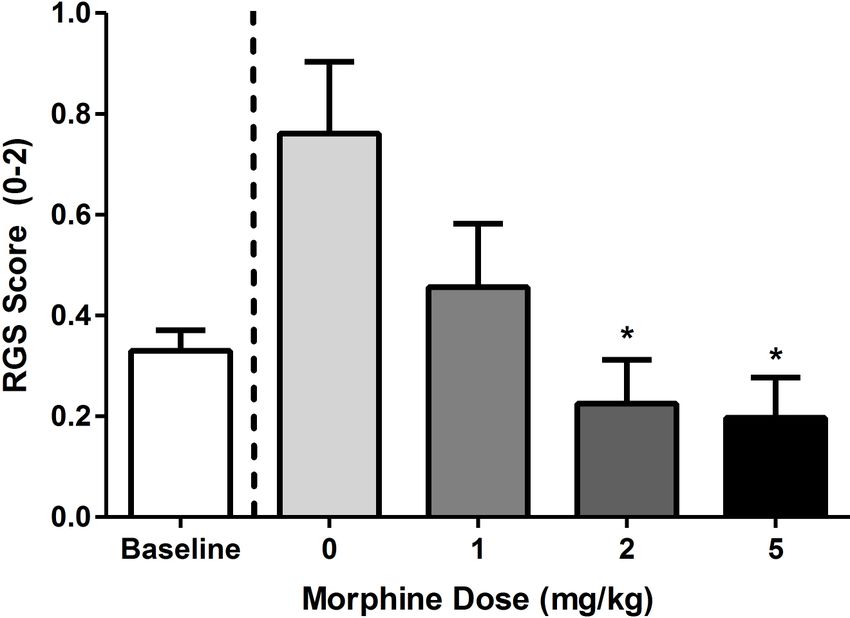
Morphine Analgesia
If the RGS is truly quantifying pain levels, then it must
be able to detect the pain-inhibiting effect of known
analgesics such as morphine. Figure 6 shows dose-
dependent inhibition (F3,16 = 4.4, p< 0.05) of facial gri-
macing caused by CFA (at the 6 h time point) by mor-
phine. The AD50 of morphine was calculated as 0.8 mg/
kg (95% confidence interval: 0.4-1.2 mg/kg).
Discussion
We report here the development, reliability, accuracy,
analgesic sensitivity, and utility of the RGS, a method to
quantify spontaneous pain in the laboratory rat. In addi-
tion, we have developed an automated system–the RFF
software package–that can successfully extract scorable
image files from digital video, previously the most labor-
intensive step in the application of the RGS (or MGS).
The RFF can be obtained directly from one of the
authors (ODK at king@bbri.org) upon request by inter-
ested individuals, at no charge for academic users and
via licensing agreement for corporate users.
RGS vs. MGS
As predicted by the evolutionary conservation of facial
expressions of emotions [8], including pain [7], the
“pain face” of the rat was found to be broadly similar to
that of the mouse, with three of the RGS action units
essentially unchanged from the MGS. A major exception
is the nose and cheek, whereby pain in the mouse
results in bulging, but in the rat bulging occurs naturally
Figure 4 Quantification of spontaneous pain in three
nociceptive assays: intraplantar CFA (a), intraarticular kaolin/
and this characteristic actually diminishes when the rat
carrageenan (b), and postoperative (laparotomy) pain (c). Bars is in pain.
represent mean ± SEM RGS score (n = 6-10 rats/assay). *p< 0.05; Observed accuracy rates for the RGS are lower than
**p< 0.01 compared to baseline (Bonferroni-corrected). those observed in the MGS using similar high-definition
video (97% by S.G.S.) [9], but still far above chanceSotocinal et al. Molecular Pain 2011, 7:55 Page 8 of 10 http://www.molecularpain.com/content/7/1/55 Figure 5 Prominence of individual action units at the peak of apparent spontaneous pain in each assay (see Figure 4). Bars represent mean ± SEM difference scores (pain - no pain; n = 23 rats). Overall, all action units were equally prominent statistically (a), and this was also true in each assay considered separately (b). **p< 0.01 (Bonferroni-corrected) compared to other assays. K/C = kaolin/carrageenan. levels. We note that when testing the accuracy of the Time course of inflammatory pain MGS, another well-validated pain-related behavior Peak RGS scores were observed at 6 h post-CFA, 3 h (abdominal constrictions) was used to verify the exis- post-kaolin/carrageenan, and 4 h post-laparotomy. It is tence of spontaneous pain in each subject, whereas here, tempting to conclude that this represents the peak of using CFA, we were forced to simply assume its exis- spontaneous pain in these assays, as opposed to allody- tence. This fact likely accounts for the lower accuracy nia. There are, of course, very few extant studies where values obtained, since rats in some of the pain photo- spontaneously emitted behaviors have been recorded in graphs may not have been, at that precise moment, these assays, and even then it’s not clear that what is actually in pain, which would artificially inflate the miss being measured is spontaneous pain (as opposed to rate. mechanical allodynia), or even pain at all [1,4,5]. In an Inter-rater reliability of the RGS was very high, as high early study using intraplantar 100% CFA in the rat [17], as on the MGS. We note, however, that this was only a number of behavioral characteristics including food tested in five individuals in one laboratory. We encou- intake, open field behavior, and core body temperature rage others to use the method so that true reliability were altered by CFA, some for over 5 weeks. In con- and accuracy rates can be ascertained. trast, Djouhri and colleagues [18], using spontaneous
Sotocinal et al. Molecular Pain 2011, 7:55                                                                                      Page 9 of 10
http://www.molecularpain.com/content/7/1/55

                                                                     that the disappearance of the facial grimacing may not
                                                                     necessarily represent the disappearance of spontaneous
                                                                     pain, as there are adaptive advantages to inhibiting a
                                                                     “pain face” as soon as possible.

                                                                     New approaches to algesiometry
                                                                     The problematic symptoms of chronic pain in humans
                                                                     include spontaneous pain, numbness, dysesthesias, and
                                                                     evoked (mechanical, heat and cold) hypersensitivity. But
                                                                     these are not equally common, or of equal concern. For
                                                                     both neuropathic and non-neuropathic pain, sponta-
                                                                     neous or ongoing pain (especially deep pain) is far more
                                                                     prevalent than evoked pain, especially touch- and
                                                                     warmth-evoked pain [24,25]. Spontaneous pain is also
 Figure 6 Quantification of morphine analgesia by the RGS.           rated as more bothersome, and more highly correlated
 Morphine was administered 5.5 h after CFA and 15 min before the     with global ratings of pain severity [24]. Despite this
 start of 30-min digital video recording. Bars represent ± SEM RGS   clinical reality, preclinical studies of pain are strongly
 score (n = 4-10 rats/dose). *p< 0.05 compared to saline (0) by      weighted towards the study of mechanical and thermal
 Dunnett’s case-comparison posthoc test (one-way).
                                                                     hypersensitivity states, largely for reasons of practicality
                                                                     and inertia [4].
foot lifting as a measure of spontaneous pain in Wistar                However, new approaches to measuring pain (and/or
rats, noted that all rats displayed foot lifting 1 day after         the impact of pain) appear to be gaining popularity;
injection, but less than 20% did by day 2 and none did at            these include thermal preference/escape models [e.g.,
4-7 days post-injection. Using a suite of behaviors (includ-         [26,27]], conditioned place aversion [28,29], condi-
ing muscle twitching, back arching, staggering, and                  tioned place preference (to pain inhibition) [30,31],
abdominal writhing), Roughan and Flecknell [19] con-                 and ultrasonic vocalization [32]. Compared to these,
cluded that postoperative pain after laparotomy decreased            facial expression coding has the considerable advan-
significantly after 3-5 h post-surgery. Using exploratory            tage that no subject training or special equipment
activity and conditioned operant responding for sucrose              (other than a video camera) are required. It also pro-
pellets as the measure, in contrast, Martin et al. [20]              vides the advantage of more complete blinding of the
observed changes after surgery lasting up to 2-3 days.               experimenter [33], since during scoring the presence
  The time course of mechanical allodynia and thermal                or absence of an inflamed or guarded hind paw is
hyperalgesia in these models is better known, albeit                 completely obscured. Quantifying pain by facial
dose- and strain-dependent. The first study to use 50%               expression is also the only technique of practical value
CFA in the rat observed peak thermal hyperalgesia at 4               in veterinary medicine (including laboratory animal
h post-injection and a return to baseline by 15 days;                welfare), as it can in fact be performed in real time by
mechanical allodynia peaked at 2 days post-injection                 trained investigators, animal technicians and/or
and was resolved by 5 days [21]. The duration of                     veterinarians.
changes in the other two models is much more limited.                  The major disadvantages to blinded facial expression
Thermal hyperalgesia in the kaolin-carrageenan model                 coding for research purposes are the labor-intensive nat-
was found to peak at 8-12 h and resolve by 2 days post-              ure of frame grabbing, a problem now largely solved
injection [12]. Electrophysiological experiments have                with RFF software, and the limited duration (< 48 h) of
shown that primary afferent fibers in the joint are sensi-           the pain face. This limitation is imposed by the nature
tized in the kaolin-carrageenan model 3-6 h post-injec-              of facial grimacing itself, which is also not observed in
tion [22]. After laparotomy in the Wistar rat,                       human chronic pain patients. Thus, the study of real-
mechanical allodynia was noted from 2.5-6.5 h post-sur-              time spontaneous pain in chronic neuropathic assays
gery [13], although in a recent study (involving in addi-            awaits the development of a useful dependent measure.
tion to the incision the implantation of a radiotelemetry
transmitter) significant allodynia was observed for 9                Additional material
days [23].
  Overall there is good concordance between the time                  Additional file 1: Rat Grimace Scale (RGS): The Manual. This training
                                                                      manual describes detailed procedures for the implementation of the
course of inflammatory pain inferred from the literature
                                                                      RGS.
and our current data. It is important to note, however,
Sotocinal et al. Molecular Pain 2011, 7:55 Page 10 of 10
http://www.molecularpain.com/content/7/1/55
Author details 21. Iadarola MJ, Douglass J, Civelli O, Naranjo JR: Differential activation of
1
Dept. of Psychology and Alan Edwards Centre for Research on Pain, McGill spinal cord dynorphin and enkephalin neurons during hyperalgesia:
University, Montreal, QC H3A 1B1, Canada. 2Boston Biomedical Research evidence using cDNA hybridization. Brain Res 1988, 455:205-212.
Institute, Watertown, MA 02472 USA. 3Dept. of Physiology and 22. McDougall JJ, Pawlak M, Hanesch U, Schmidt RF: Peripheral modulation of
Pharmacology, University of Calgary, Calgary, AB T2N 4N1, Canada. rat knee joint afferent mechanosensitivity by nociceptin/orphanin FQ.
Neurosci Lett 2000, 2:123-126.
Authors’ contributions 23. Charlet A, Rodeau JL, Poisbeau P: Radiotelemetric and symptomatic
SGS, RES, AZ, AHT, LJM, JSW, JCSM, SZ, and SZ collected the data. JJM and evaluation of pain in the rat after laparotomy: long-term benefits of
ODK edited the manuscript. PW and ODK designed the software. JSM perioperative ropivacaine care. J Pain 2011, 12:246-256.
conceived of and designed the study (with assistance from JJM), and wrote 24. Backonja MM, Stacey B: Neuropathic pain symptoms relation to overall
the draft of the manuscript. All authors read and approved the final pain rating. J Pain 2004, 5:491-497.
manuscript. 25. Scholz J, Mannion RJ, Hord DE, Griffin RS, Rawal B, Zheng H, Scoffings D,
Phillips A, Guo J, Laing RJ, et al: A novel tool for the assessment of pain:
Competing interests validation in low back pain. PLoS Med 2009, 6:e1000047.
The authors declare that they have no competing interests. 26. Mauderli AP, Acosta-Rua A, Vierck CJ: An operant assay of thermal pain in
conscious, unrestrained rats. J Neurosci Meth 2000, 97:19-29.
Received: 6 April 2011 Accepted: 29 July 2011 Published: 29 July 2011 27. Baliki M, Calvo O, Chialvo DR, Apkarian AV: Spared nerve injury rats exhibit
thermal hyperalgesia on an automated operant dynamic thermal escape
References Task. Mol Pain 2005, 1:18.
1. Mogil JS: Animal models of pain: progress and challenges. Nat Rev 28. LaBuda CJ, Fuchs PN: A behavioral test paradigm to measure the
Neurosci 2009, 10:283-294. aversive quality of inflammatory and neuropathic pain in rats. Exp Neurol
2. Mogil JS, Davis KD, Derbyshire SW: The necessity of animal models in 2000, 163:490-494.
pain research. Pain 2010, 151:12-17. 29. Johansen JP, Fields HL, Manning BH: The affective component of pain in
3. Mogil JS, Simmonds K, Simmonds MJ: Pain research from 1975 to 2007: a rodents: direct evidence for a contribution of the anterior cingulate
categorical and bibliometric meta-trend analysis of every Research cortex. Proc Natl Acad Sci USA 2001, 98:8077-8082.
Paper published in the journal, Pain. Pain 2009, 142:48-58. 30. Sufka KJ: Conditioned place preference paradigm: a novel approach for
4. Mogil JS, Crager SE: What should we be measuring in behavioral studies analgesic drug assessment against chronic pain. Pain 1994, 58:355-366.
of chronic pain in animals? Pain 2004, 112:12-15. 31. King T, Vera-Portocarrero L, Gutierrez T, Vanderah TW, Dussor G, Lai J,
5. Mogil JS, Graham AC, Ritchie J, Hughes SF, Austin J-S, Schorscher-Petcu A, Fields HL, Porreca F: Unmasking the tonic-aversive state in neuropathic
Langford DL, Bennett GJ: Hypolocomotion, asymmetrically directed pain. Nat Neurosci 2009, 12:1361-1363.
behaviors (licking, lifting, flinching, and shaking) and dynamic weight 32. Kurejova M, Nattenmuller U, Hildebrandt U, Selvaraj D, Stosser S, Kuner R:
bearing (gait) changes are not measures of neuropathic pain in mice. An improved behavioural assay demonstrates that ultrasound
Mol Pain 2010, 6:34. vocalizations constitute a reliable indicator of chronic cancer pain and
6. Ekman P, Friesen W: Facial Action Coding System. Palo Alto, CA: neuropathic pain. Mol Pain 2010, 6:18.
Consulting Psychologists Press; 1978. 33. Rice ASC, Cimino-Brown D, Eisenach JC, Kontinen VK, LaCroix-Fralish ML,
7. Williams AC: Facial expression of pain: an evolutionary account. Behav Machin I, Mogil JS, Stohr T: Animal models and the prediction of efficacy
Brain Sci 2002, 25:439-455. in clinical trials of analgesic drugs: a critical appraisal and call for
8. Darwin C: The Expression of the Emotions in Man and Animals. London: uniform reporting standards. Pain 2008, 139:241-245.
Albemarle; 1872.
doi:10.1186/1744-8069-7-55
9. Langford DL, Bailey AL, Chanda ML, Clarke SE, Drummond TE, Echols S, Cite this article as: Sotocinal et al.: The Rat Grimace Scale: A partially
Glick S, Ingrao J, Klassen-Ross T, LaCroix-Fralish ML, et al: Coding of facial automated method for quantifying pain in the laboratory rat via facial
expressions of pain in the laboratory mouse. Nature Meth 2010, 7:447-449. expressions. Molecular Pain 2011 7:55.
10. Mogil JS, Chanda ML: The case for the inclusion of female subjects in
basic science studies of pain. Pain 2005, 117:1-5.
11. Iadarola MJ, Douglass J, Civelli O, Naranjo JR: Increased spinal cord
dynorphin mRNA during peripheral inflammation. In NIDA Res Monogr.
Edited by: Holaday JW, Law P-Y, Herz A. Bethesda, MD; 1986:.
12. Sluka KA, Westlund KN: Behavioral and immunohistochemical changes in
an experimental arthritis model in rats. Pain 1993, 55:367-377.
13. Lascelles BDX, Waterman AE, Cripps PJ, Livington A, Henderson G: Central
sensitization as a result of surgical pain: investigation of the pre-emptive
value of pethidine for ovariohysterectomy in the rat. Pain 1995,
62:201-212.
14. Lienhart R, Maydt J: An extended set of Haar-like features for rapid
object detection. IEEE-ICIP 2002, 1:900-903.
15. Shrout PE, Fleiss JL: Intraclass correlations: uses in assessing rater
reliability. Psychol Bull 1979, 86:420-428.
16. Tallarida RJ, Murray RB: Manual of Pharmacologic Calculation. New York:
Springer-Verlag; 1981. Submit your next manuscript to BioMed Central
17. Stein C, Millan MJ, Herz A: Unilateral inflammation of the hindpaw in rats and take full advantage of:
as a model of prolonged noxious stimulation: alterations in behavior
and nociceptive thresholds. Pharmacol Biochem Behav 1988, 31:445-451. • Convenient online submission
18. Djouhri L, Koutsikou S, Fang X, McMullan S, Lawson SN: Spontaneous pain,
• Thorough peer review
both neuropathic and inflammatory, is related to frequency of
spontaneous firing in intact C-fiber nociceptors. J Neurosci 2006, • No space constraints or color figure charges
26:1281-1292. • Immediate publication on acceptance
19. Roughan JV, Flecknell PA: Behavioural effects of laparotomy and
analgesic effects of ketoprofen and carprofen in rats. Pain 2001, 90:65-74. • Inclusion in PubMed, CAS, Scopus and Google Scholar
20. Martin RJ, Kahn WR, Eisenach JC: Abdominal surgery decreases food- • Research which is freely available for redistribution
reinforced operant responding in rats: relevance of incisional pain.
Anesthesiology 2005, 103:629-637.
Submit your manuscript at
www.biomedcentral.com/submitYou can also read