Effect of GCSB-5, a Herbal Formulation, on Monosodium Iodoacetate-Induced Osteoarthritis in Rats
←
→
Page content transcription
If your browser does not render page correctly, please read the page content below
Hindawi Publishing Corporation
Evidence-Based Complementary and Alternative Medicine
Volume 2012, Article ID 730907, 11 pages
doi:10.1155/2012/730907

Research Article
Effect of GCSB-5, a Herbal Formulation, on Monosodium
Iodoacetate-Induced Osteoarthritis in Rats

          Joon-Ki Kim,1 Sang-Won Park,1 Jung-Woo Kang,1 Yu-Jin Kim,2 Sung Youl Lee,2
          Joonshik Shin,3 Sangho Lee,3 and Sun-Mee Lee1
          1 School of Pharmacy, Sungkyunkwan University, Suwon, Gyeonggi-do 440-746, Republic of Korea
          2 Business Development, Green Cross Corporation, Yongin, Gyeonggi-do 446-770, Republic of Korea
          3 Jaseng Hospital, 635 Sinsa-Dong, Gangnam-Gu, Seoul, Republic of Korea

          Correspondence should be addressed to Sun-Mee Lee, sunmee@skku.edu

          Received 1 April 2011; Revised 9 November 2011; Accepted 16 November 2011

          Academic Editor: Andrew Scholey

          Copyright © 2012 Joon-Ki Kim et al. This is an open access article distributed under the Creative Commons Attribution License,
          which permits unrestricted use, distribution, and reproduction in any medium, provided the original work is properly cited.

          Therapeutic effects of GCSB-5 on osteoarthritis were measured by the amount of glycosaminoglycan in rabbit articular cartilage
          explants in vitro, in experimental osteoarthritis induced by intra-articular injection of monoiodoacetate in rats in vivo. GCSB-5
          was orally administered for 28 days. In vitro, GCSB-5 inhibited proteoglycan degradation. GCSB-5 significantly suppressed the
          histological changes in monoiodoacetate-induced osteoarthritis. Matrix metalloproteinase (MMP) activity, as well as, the levels of
          serum tumor necrosis factor-α, cyclooxygenase-2, inducible nitric oxide synthase protein, and mRNA expressions were attenuated
          by GCSB-5, whereas the level of interleukin-10 was potentiated. By GCSB-5, the level of nuclear factor-κB p65 protein expression
          was significantly attenuated but, on the other hand, the level of inhibitor of κB-α protein expression was increased. These results
          indicate that GCSB-5 is a potential therapeutic agent for the protection of articular cartilage against progression of osteoarthritis
          through inhibition of MMPs activity, inflammatory mediators, and NF-κB activation.

1. Introduction                                                          complete therapeutic effects by blocking the activity of
                                                                         one or two cytokines. Developing therapeutics from herbal
Osteoarthritis (OA) is a degenerative joint disease charac-              sources may reduce the risk of toxicity or adverse effects
terized by joint pain and a progressive loss of articular carti-         when the drug is clinically used [5] and may exert strong,
lage. It has been suggested that biochemical alterations occur           multifunctional anti-inflammatory effect like many natural
within the articular cartilage resulted in imbalance between             products do. Therefore, efforts are being made to elucidate
synthetic and degradative pathways [1]. A key step in the                the role of natural products for the treatment of OA.
pathophysiology of OA is breakdown of extracellular matrix                   GCSB-5 is a purified extract from a mixture of 6 oriental
of articular cartilage by tissue proteinases, enzymes whose              herbs which are the ingredients of Chung-Pa-Juhn used in
expression is upregulated by inflammatory stimuli, such as               Jaseng Hospital (Seoul, Korea) and that have been used in
primary inflammatory cytokines [2]. Nonsteroidal anti-in-                traditional medicine to treat inflammatory diseases and bone
flammatory drugs (NSAIDs) are effective in the management                 disorders. Ledebouriellae Radix is reported to have anti-
of OA inflammation. However, the adverse events secondary                inflammatory effects on Freund’s adjuvant-induced arthritis
to NSAIDs was focused on upper gastrointestinal tolerability             in rats [6]. Cimifugin, a major active component of Lede-
[3]. In recent years, gene therapy targeted at cytokines of-             bouriellae Radix, exhibits inhibitory effects on the synthesis
fers new hope to OA treatment, and the current focus is                  of NO induced by LPS in macrophage cell line RAW 264.7
on the use of biological agents that block the activity of in-           [7]. Achyranthis Radix shows anti-inflammatory property
flammatory cytokines [4]. Since there are many proinflam-                and inhibits free radicals, such as ONOO− , HOCl, and OH
matory cytokines, oxidants and other factors exerting action             radical [8]. 20-Hydroxyecdysone, which is a major active
in initiation and development of OA, it is hard to obtain                compound of Achyranthis Radix, has beneficial effects on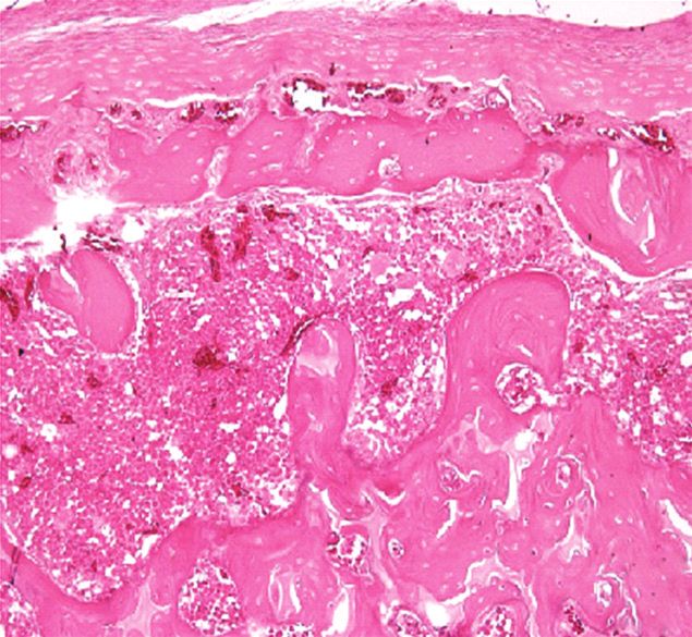
2                                                                 Evidence-Based Complementary and Alternative Medicine

joint and bone in ovariectomized rats [9]. Acanthopanacis        2.3. Animals. Male Sprague-Dawley rats (200–220 g) and
Cortex is known to show antiarthritic activity [10], and         male New Zealand white rabbits (2.0–2.2 kg) were obtained
Cibotii Rhizoma is known for its analgesic property [11]         from Dae Han Biolink Ltd. (Eumseong, Korea) and housed
along with osteoclast formation inhibition [12]. Glycine         in solid bottom cages with pellet food and water available ad
Semen is effective in reducing swelling [13] and genistin,        libitum. All animal procedures were approved by the Sung-
an active compound from Glycine Semen, shows beneficial          kyunkwan University Animal Care Committee and were per-
effect on bone loss [14]. Eucommiae Cortex exhibits strong        formed in accordance with the guidelines of the National
analgesic effect [15] and geniposide from its extract shows       Institutes of Health.
anti-inflammatory effect on rheumatoid arthritis rats [16]
and enhances the osteoblast-like cell proliferation and inhib-   2.4. Cartilage Glycosaminoglycan Assay. Rabbit knee articu-
ited osteoclast [17]. We reported strong antinociceptive and     lar cartilage explants were obtained according to the method
anti-inflammatory properties of GCSB-5 [11, 13]. Recently,       described by Sandy et al. [19]. Briefly, 200–220 mg articu-
GCSB-5 reduces the development of acute and chronic in-          lar surfaces per joint were dissected and submerged into
flammation, and its anti-inflammatory property is likely due     complete medium of DMEM supplemented with heat-inac-
to inhibition of inducible nitric oxide synthase (iNOS) and      tivated 5% FBS, penicillin (100 U/mL), and streptomycin
cyclooxygenase (COX)-2 expression via downregulation of          (100 μg/mL). After stabilization in incubator, the medium
the Akt signal pathway and inhibition of nuclear factor-κB       was replaced with basal medium made of DMEM supple-
(NF-κB) activation [18]. In phase III clinical study, GCSB-5     mented with heat-inactivated 1% FBS, 10 mM HEPES, peni-
was shown to exert therapeutic effects and acted to reduce        cillin (100 U/mL), and streptomycin (100 μg/mL). Cartilage
OA severity and improved functional recovery without             pieces (50–60 mg; 2 × 3 × 0.35 mm/piece) were placed in
apparent hepatic or renal toxicity (unpublished data).           24-well cell culture plates and treated with GCSB-5 at 1 ×
    In this study, we examined the chondroprotective and         10−3 , 1 × 10⁻² , and 1 × 10⁻¹ mg/mL or 30 μM diclofenac
anti-inflammatory effects of GCSB-5 on monoiodoacetate            (Sigma-Aldrich, St. Louis, MO, USA). After 1 h of GCSB-5 or
(MIA)-induced OA animal model, both in vitro and in vivo.        diclofenac pretreatment, 5 ng/mL of rhIL-1α (R&D Systems,
                                                                 Minneapolis, MN, USA) was added and further incubated
2. Materials and Methods                                         at 37◦ C in a humidified 5% CO2 /95% air incubator. The
                                                                 amount of glycosaminoglycan (GAG) in the medium was de-
2.1. Preparation and Composition of GCSB-5. GCSB-5 was           termined by the 1,9-dimethyl-methylene blue method using
prepared by the Hanpoong Pharmaceutical Co., Ltd., Jeonju,       the Blyscan Sulfated GAG Assay kit (Biocolor Ltd., County
Republic of Korea. The mixture of six crude drugs (Lede-         Antrim, UK) according to the manufacturer’s instructions.
bouriellae Radix (4.444 g), Achyranthis Radix (4.444 g),
Acanthopanacis Cortex (4.444 g), Cibotii Rhizoma (2.778 g),
                                                                 2.5. MIA-Induced OA. Rats were anesthetized with diethyl
Glycine Semen (2.778 g), and Eucommiae Cortex (1.389 g))
                                                                 ether and given a single intra-articular injection of 3 mg MIA
was powdered and boiled for 3 h in distilled water (1 L).
                                                                 (Sigma-Aldrich, St. Louis, MO, USA) through the infrap-
The resulting extract was subjected to ultrafiltration, and
                                                                 atellar ligament of the left knee [20]. MIA was dissolved in
the components with molecular weight over 10,000 were
                                                                 physiological saline and administered in a 50 μL volume. Rats
excluded. The filtrate was lyophilized as powder and kept
                                                                 were treated with saline, with 300 or 600 mg/kg of GCSB-5
at 4◦ C until use. GCSB-5 was administered orally at a dose
                                                                 or with 5 mg/kg of diclofenac by oral administration once
of 300 and 600 mg/kg in saline (1 kg/10 mL), and the same
                                                                 daily, for 2, 7, and 28 days since MIA injection. These GCSB-
volume of saline was used as a vehicle control group. The
                                                                 5 doses and MIA injection volume were selected based on
validation of GSCB-5 was performed by high-performance
                                                                 previous evaluations [21].
liquid chromatography analysis of each ingredient ex-
tract using six indicator biological components: cimifu-
gin for Ledebouriellae Radix, 20-hydroxyecdysone (0.311-         2.6. Gross Observation. After MIA injection, all experimental
0.312 mg/g) for Achyranthis Radix, acanthoside D (0.577-         rats were weighed and carefully inspected every 2 days to as-
0.578 mg/g) for Acanthopanacis Cortex, onitin-4-O-β-D-           sess knee joint swelling and gait disturbances under natural
glucopyranoside for Cibotii Rhizoma, genistin (0.0426-           conditions in the cages, where they moved freely. Swelling
0.0427 mg/g) for Glycine Semen, and geniposide (0.431-           and limping were classified as no change, mild, and severe on
0.432 mg/g) for Eucommiae Cortex. GCSB-5 was further             the basis of severity [22], and inspection was conducted by an
standardized for quality control according to the regulations    inspector blinded to treatment details throughout the study.
imposed by Korea Food and Drug Administration (KFDA).
                                                                 2.7. Roentgenographic Examination and Histopathological
2.2.Chemicals. Dulbecco’s modified Eagle’s medium(DMEM),         Analysis. Seven and 28 days following MIA injection, rats
penicillin/streptomycin (10,000 U/mL, 10,000 μg/mL, resp.),      were checked with roentgenography to assess chronic mor-
and fetal bovine serum (FBS) were obtained from Gibco            phological changes of knee articular bones for narrowing,
BRL, Life Technologies (Grand Island, NY, USA). All the          loss of joint region, cartilage erosion, and osteophyte forma-
other materials required for culturing of tissue were pur-       tion [23]. For histological analysis, knee joints were removed
chased from Sigma Chemical Company (St. Louis, MO,               and fixed in 10% neutral buffered formalin, decalcified with
USA).                                                            10% formic acid, and embedded in paraffin. Five micrometer
Evidence-Based Complementary and Alternative Medicine                                                                            3

                                             Table 1: RT-PCR primers used in study.

Gene             Accession number                           Primer sequences (5 → 3 )                        Product length (bp)
                                                    Sense: GTA GCC CAC GTC GTA GCA AA
TNF-α                 X66539                                                                                           346
                                                  Antisense: CCC TTC TCC AGC TGG AAG AC
                                                 Sense: TTC TTT GCT TCT GTG CTT AAT GCG
iNOS                  D44591                                                                                          1061
                                               Antisense: GTT GTT GCT GAA CTT CCA ATC GT
                                                    Sense: CTG CAT GTG GCT GAT GTC ATC
COX-2                 U03389                                                                                          1061
                                              Antisense: AGG ACC CGT CAT CTC CAG GGT AAT C
                                                     Sense: TGA TGT TCC CAT TAG ACA GC
IL-1β                 M98820                                                                                           378
                                                  Antisense: GAG GTG CTG ATG TAC CAG TT
                                                     Sense: CAG TCA GCC AGA CCC ACA T
IL-10                 X60675                                                                                           322
                                                    Antisense: GCT CCA CTG CCT TGC TTT
                                                Sense: TTG TAA CCA ACT GGG ACG ATA TGG
β-Actin               V01217                                                                                           764
                                               Antisense: GAT CTT GAT CTT CAT GGT GCT AG

(5 μm) sections were stained with hematoxylin and eosin           2.11. Total RNA Extraction and Reverse Transcription-
(H and E) or safranin-O fast green (SOFG) and observed.           Polymerase Chain Reaction (RT-PCR). Articular cartilage
Histopathological changes in each animal were quantitatively      samples collected 2 and 28 days after MIA injection were
expressed by three grades for each finding [24]. Grading was      pulverized in TRI Reagent (Molecular Research Center Inc.,
done under the authority of Medplan Pathology Laborato-           Cincinnati, OH, USA) for RNA extraction. Equal amounts
ries, Seoul, Korea.                                               of RNA from articular cartilages were subjected to reverse
                                                                  transcription using iNtRON RNA PCR kit (iNtRON Biotech-
                                                                  nology Co., Seongnam, Korea) to generate cDNA for RT-
2.8. Gelatinase Assay. Rat articular cartilage samples of MIA-
                                                                  PCR analysis. RT-PCR analysis was performed with the
induced OA were harvested 7 and 28 days after MIA injec-
                                                                  GeneAmp PCR system 2700 (Applied Biosystems Co., Foster
tion. Gelatinase activities were measured by the gelatin zy-
                                                                  City, CA, USA). The primers used in the RT-PCR are listed
mography method described by Dumond et al. [25]. Proteins
                                                                  in Table 1. All PCR reactions included an initial denaturation
were extracted from pulverized cartilage tissues and elec-
                                                                  step at 94◦ C for 5 min and a final extension at 72◦ C for 7 min.
trophoresed on 10% zymogram precast gels. The cleared gels
                                                                  The PCR amplification cycling conditions were as follows:
were captured, and the area of each band was quantified with
                                                                  32 cycles of 94◦ C (30 s), 58◦ C (30 s), and 72◦ C (30 s) for
densitometric scanning analysis program (Science Lab 98
                                                                  TNF-α; 32 cycles of 94◦ C (45 s), 65◦ C (45 s), and 72◦ C
Image Gauge, version 3.12, Fuji Photo Film Co. Ltd., Tokyo,
                                                                  (60 s) for iNOS; 40 cycles of 94◦ C (45 s), 65◦ C (45 s), and
Japan).
                                                                  73◦ C (60 s) for COX-2; 36 cycles of 94◦ C (30 s), 60◦ C
                                                                  (30 s), and 72◦ C (45 s) for IL-1β; 40 cycles of 94◦ C (30 s),
2.9. Serum Cytokine Levels. Commercial tumor necrosis             66◦ C (45 s), and 72◦ C (45 s) for IL-10; 30 cycles of 94◦ C
factor (TNF)-α, interleukin (IL)-1β, and IL-10 enzyme-            (30 s), 56◦ C (30 s), and 72◦ C (60 s) for β-actin. After RT-
linked immunosorbent assay (ELISA) kits (BD Biosciences           PCR, 10 μL samples of the amplified products were resolved
Co., CA, USA) were used for quantification of the serum           by electrophoresis on 1.5% agarose gels and stained with
levels of TNF-α, IL-1β, and IL-10, respectively.                  ethidium bromide. The intensity of each PCR product was
                                                                  evaluated semiquantitatively using a digital camera (DC120;
2.10. Western Blot Immunoassay. 15 μg of whole protein was        Eastman Kodak, Rochester, NY, USA) and a densitometric
used for determination of the content of COX-2 and iNOS.          scanning analysis program (ID Main; Advanced American
20 μg of nuclear protein was used for determination of the        Biotechnology, Fullerton, CA, USA).
content of the NF-κB/p65 subunit. 20 μg of the cytosolic
protein was used for determination of the content of the          2.12. Statistics. All results are presented as mean ± S.E.M.
inhibitor of κB (IκB)-α. ImageQuantTM TL software (Amer-          The overall significance of the experimental results was
sham Biosciences/GE Healthcare, Piscataway, NJ, USA) was          examined by one-way analysis of variance and the two-tail
used for densitometric evaluation of visualized immunore-         Dunnet’s t-test. Differences between groups were considered
active bands. The following primary antibodies were used:         significant at P < 0.05 with the appropriate Bonferroni
COX-2 (Abcam, Cambridge, UK; 1 : 1000), iNOS (Trans-              correction for multiple comparisons.
duction Lab., CA, USA; 1 : 1000), phosphoryl NF-κB/p65
(Santa Cruz Biotechnology, Santa Cruz, CA, USA; 1 : 1000),        3. Results
and IκB-α (Santa Cruz Biotechnology; 1 : 5000) were used,
and the signals were normalized to that of β-actin (Sigma         3.1. Cartilage Glycosaminoglycan Release. In the control
Chemical Co.; 1 : 1000) or lamin B1 (Abcam; 1 : 2500).            group, the level of GAG in the culture medium remained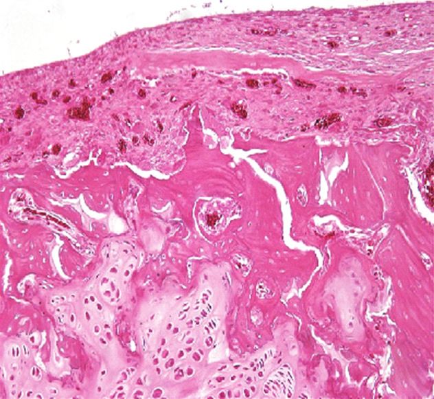
4                                                                                            Evidence-Based Complementary and Alternative Medicine

                                Table 2: Quantitative summary of gross observations in MIA-induced osteoarthritic rats treated with GCSB-5.

                                                                                                                     MIA
                                                           Control                                              GCSB-5 (mg/kg)                   Diclofenac
                                                                                Vehicle
                                                                                                          300                    600              5 mg/kg
Swelling
  No change                                  0               10/10                0/10                    1/10                 1/10                 0/10
  Mild                                       1               0/10                 3/10                    8/10                 6/10                 8/10
  Severe                                     2               0/10                 7/10                    1/10                 3/10                 2/10
  Average score                                            0.0 ± 0.0           1.7 ± 0.2a             1.0 ± 0.2a,b          1.2 ± 0.2a           1.2 ± 0.1a
Limping
  No change                                  0               10/10                0/10                    6/10                 5/10                 5/10
  Mild                                       1               0/10                 7/10                    4/10                 5/10                 5/10
  Severe                                     2               0/10                 3/10                    0/10                 0/10                 0/10
  Average score                                            0.0 ± 0.0           1.3 ± 0.2a              0.4 ± 0.2c           0.5 ± 0.2c           0.5 ± 0.2c
GCSB-5 or diclofenac was treated daily for 14 days after 2 weeks of OA induction by intra-articular injection of MIA.
a Denotes significant differences (P < 0.01) versus the control group.
b,c Denote significant differences (P < 0.05, P < 0.01) versus the vehicle-treated MIA group.

                            9                                                               These symptoms gradually aggravated at 21 days (data not
                                                                                            shown) and were the most severe at 28 days. Twenty-
                                                                          ∗∗
                                                                                            eight days after MIA injection, swelling and limping were
                                                      ∗∗                                    attenuated by both 300 and 600 mg/kg GCSB-5 treatment
    GAG (μg/mg cartilage)

                                                                          ∗∗
                            6      ∗∗                 ∗∗                  +
                                                                          ∗∗
                                                                                            (Table 2).
                                                      ∗∗                   +
                                                      ∗∗                  ∗∗
                                   ∗∗
                                   ∗∗
                                   ∗∗
                                                      ++
                                                      ∗∗
                                                                          ++
                                                                          ∗∗                3.3. Roentgenographic and Histopathological Analysis. Seven
                                   ++
                                   ∗∗
                                                                                            days after MIA injection, rats underwent the first roentgeno-
                            3                                                               graphic examination. Their roentgenographic examinations
                                                                                            revealed degenerative changes, such as irregularity or osteo-
                                                                                            phytes on the surface of the cartilage and subchondral bone
                                                                                            (data not shown). At 28 days, rats underwent the second
                            0                                                               roentgenographic examination (Figures 2(a)–2(d)). Mor-
                                 24                  48                  72                 phological changes were more significant, showing rough
                                                  Time (h)                                  edges of cartilage and the tendency of patellar displace-
                                                                                            ment. These changes were attenuated by GCSB-5 600 mg/kg
Figure 1: GAG release in rabbit articular cartilage explant cultures                        treatment. Twenty-eight days after MIA injection, H and E
at 24, 48, and 72 h. Rabbit articular cartilage explants were
                                                                                            staining revealed irregular surface accompanied by ulcera-
stimulated with rhIL-1α (5 ng/mL). The amount of GAG release
stimulated by rhIL-1α (◦) increased approximately 3.6 times
                                                                                            tion, fibrillation, and loss of cartilage tissue (Figures 2(e)–
compared to control (•) at 72 h. GCSB-5 (1.0 × 10⁻³ () and                                 2(h)). However, these cartilage damages were attenuated by
1.0 × 10⁻² () mg/mL) and diclofenac (30 μM ()) efficiently                                  GCSB-5 600 mg/kg treatment. SOFG staining also revealed
inhibited the GAG release. However, a high concentration of GCSB-                           clearly diffused PG depletion in joint cartilage tissues of
5 (1.0 × 10⁻¹ () mg/mL) slightly inhibited it. Each value represents                       MIA-injected rats (Figures 2(i)–2(l)). This loss of PG was
the mean ± S.E.M. from 6 articular cartilage explants cultures per                          attenuated by GCSB-5 600 mg/kg treatment. Summation of
group. ∗∗ Significantly different (P < 0.01) from control. + and                             all histopathologic finding scores in vehicle-treated MIA
++ Significantly different (P < 0.05, P < 0.01) from rhIL-1α.
                                                                                            group and in 300 and 600 mg/kg GCSB-5-treated MIA
                                                                                            groups were 24.5 ± 1.3, 16.1 ± 1.4, and 12.5 ± 1.1, respectively
                                                                                            (Table 3).
constant at approximately 1.5 μg/mg cartilage throughout
the experiment. In the rhIL-1α-treated group, on the other                                  3.4. Gelatinase Assay. Seven days after MIA injection, the
hand, the level of GAG in the culture medium dramatically                                   activities of matrix metalloproteinase (MMP)-2 and -9 in-
increased to approximately 4 times the control values.                                      creased to 2.7 and 2.4 times that in the control group,
GCSB-51 × 10−3 and 1 × 10⁻² mg/mL treatments attenuated                                     respectively. Similarly, 28 days after MIA injection, the
the elevation in GAG release at 72 h (Figure 1).                                            activities of MMP-2 and -9 increased to 2.3- and 2.8-fold
                                                                                            higher than the control level, respectively. On day 7, GCSB-5
3.2. Gross Observation. In the MIA-injected groups, swelling                                and diclofenac treatment showed no significant modulation
and limping were first observed 7 days after MIA injection.                                 on MMP activities (data not shown). However, on day 28,
They subsided transiently and then reappeared at 14 days.                                   GCSB-5 300 mg/kg treatment exhibited significant MMP-2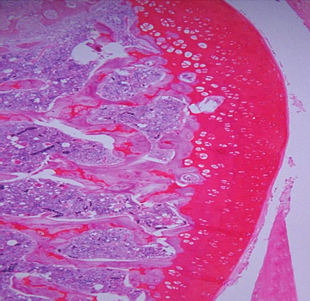
Evidence-Based Complementary and Alternative Medicine 5
Control MIA MIA + GCSB-5 MIA + diclofenac
Roentgenic
picture
(a) (b) (c) (d)
H and E
staining
(e) (f) (g) (h)
SOFG
staining
(i) (j) (k) (l)
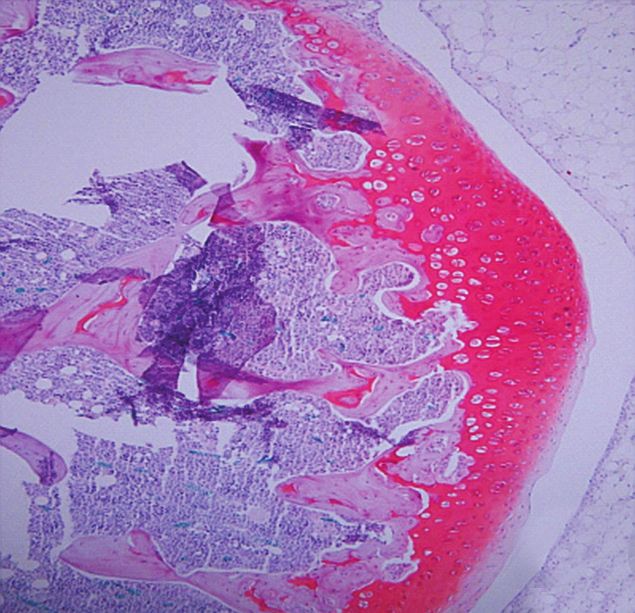
Figure 2: Roentgenography and histopathological features of osteoarthritic lesion in the knee joint of rats 28 days after intra-articular
injection of MIA (H and E staining, ×100; SOFG staining, ×100). Control (a) represents intact normal joint feature. Vehicle-treated MIA
(b) shows a severely damaged joint with rough edges around the tibia and femur, indicative of bone lysis, swelling, and tendency of patellar
displacement. However, these damages were reduced significantly by treatment with 600 mg/kg GCSB-5 (c) and 5 mg/kg diclofenac (d).
SOGF-stained control (e) represents normal cartilage PG staining, whereas vehicle-treated MIA (f) represents severely damaged cartilage
showing marked fibrillation and the depletion of SOFG staining with separation of cartilage from subchondral bone. 600 mg/kg GCSB-5 (g)
and 5 mg/kg diclofenac (h) treatments significantly reduced cartilage damage. H and E stained control (i) represents the normal status of
joint cartilage, whereas vehicle-treated MIA (j) represents severely damaged cartilage showing widespread cell necrosis and inflammation.
However, treatment with 600 mg/kg GCSB-5 (k) and 5 mg/kg diclofenac (l) treatments significantly reduced joint cartilage damage.
and -9 activities attenuation (79.6%, P < 0.01 and 81.2%, The levels of TNF-α, IL-1β, IL-10, COX-2, and iNOS
P < 0.01, resp.), while GCSB-5 600 mg/kg treatment did not mRNA expression increased 5.3, 2.1, 1.3, 7.8 and 8.8 times
affect the MMP-2 and -9 activities (91.0% and 91.6%, resp.) in the vehicle-treated MIA groups, compared to those in
(Figure 3). the control group 2 days after MIA injection, respectively
(Figures 4 and 6). Increase in TNF-α, COX-2, and iNOS
3.5. Inflammatory Mediators. The serum levels of TNF-α, IL- mRNA expression was significantly suppressed by treatment
1β, and IL-10 were 30.0 ± 4.5 pg/mL, 29.1 ± 3.7 pg/mL, and with GCSB-5. However, GCSB-5 did not affect the level of IL-
27.8 ± 0.6 pg/mL in the control. 2 days after MIA injection, 1β mRNA expression. Interestingly, increase of IL-10 mRNA
the serum levels of TNF-α, IL-1β, and IL-10 increased to 2.8-, expression was significantly potentiated by GCSB-5. At 7 and
3.4- and 2.2-fold higher than the control level, respectively. 28 days, there were no significant differences in the level of
Increase in TNF-α level was significantly suppressed by inflammatory mediators mRNA expression among any of the
treatment with GCSB-5, while increase in IL-10 level was experimental groups (data not shown).
significantly potentiated by GCSB-5. However, GCSB-5 did
not affect the serum level of IL-1β (Table 4). The levels of 3.6. Nuclear NF-κB and Cytosolic IκB-α Immunoblot Assay.
COX-2 and iNOS protein expression increased 3.3 and 12 The nuclear localization of NF-κB was measured by the pro-
times in the vehicle-treated MIA groups, compared to those tein level of NF-κB p65 subunit in the nucleus. Cytosolic IκB-
in the control group 2 days after MIA injection, respectively α was also examined from cytosol fraction as an endogenous
(Figure 5). Increase in COX-2 and iNOS protein expression NF-κB inhibitor. The level of nuclear NF-κB p65 protein
was significantly suppressed by treatment with GCSB-5. expression increased 2.5 times, whereas the level of cytosolic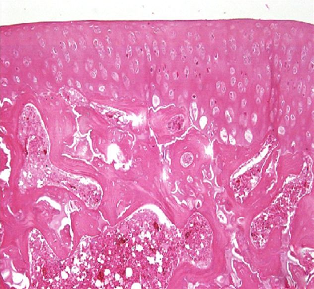
6                                                                                    Evidence-Based Complementary and Alternative Medicine

                                                      Table 3: Summary of microscopic findings.

                                                                                                                 MIA
                                                                                                            GCSB-5 (mg/kg)           Diclofenac
                                                                           Vehicle
                                                                                                      300                    600      5 mg/kg
Structural changes in the joint
   Surface irregularities                                +                   0/4                      2/4                    1/4        2/4
                                                        ++                   0/4                      1/4                    2/4        2/4
                                                        +++                  4/4                      1/4                    1/4        0/4
    Average pathology score                                                   3                       1.8                     2         1.5
Ulceration                                               +                   0/4                      1/4                    3/4        1/4
                                                        ++                   1/4                      1/4                    1/4        2/4
                                                        +++                  3/4                      2/4                    0/4        1/4
    Average pathology score                                                  2.8                      2.3                    1.3         2
Fibrillation of cartilage surface                        +                   0/4                      1/4                    2/4        3/4
                                                        ++                   3/4                      3/4                    1/4        1/4
                                                        +++                  1/4                      0/4                    1/4        0/4
    Average pathology score                                                  2.3                      1.8                    1.8        1.3
Disorganization of chondrocytes                          +                   0/4                      1/4                    3/4        3/4
                                                        ++                   2/4                      3/4                    1/4        0/4
                                                        +++                  2/4                      0/4                    0/4        1/4
    Average pathology score                                                  2.5                      1.8                    1.3        1.5
Exposure of subchondral bone                             +                   2/4                      1/4                    0/4        0/4
                                                        ++                   1/4                      0/4                    1/4        0/4
                                                        +++                  1/4                      0/4                    0/4        0/4
    Average pathology score                                                  1.8                      0.3                    0.5         0
Cellular changes of chondrocyte
  hypertrophy                                            +                   1/4                      3/4                    1/4        0/4
                                                        ++                   2/4                      1/4                    3/4        4/4
                                                        +++                  1/4                      0/4                    0/4        0/4
    Average pathology score                                                   2                       1.3                    1.8         2
Degeneration/necrosis                                    +                   0/4                      1/4                    4/4        2/4
                                                        ++                   1/4                      0/4                    0/4        2/4
                                                        +++                  3/4                      3/4                    0/4        0/4
    Average pathology score                                                  2.8                      2.5                     1         1.5
Inflammatory cell infiltration                            +                  1/4                      3/4                    2/4        3/4
in synovial tissue                                       ++                  1/4                      1/4                    1/4        0/4
                                                        +++                  2/4                      0/4                    0/4        1/4
    Average pathology score                                                  2.3                      1.3                     1         1.5
Synovial cell proliferation                              +                   1/4                      3/4                    3/4        3/4
                                                        ++                   2/4                      0/4                    0/4        1/4
                                                        +++                  1/4                      1/4                    1/4        0/4
    Average pathology score                                                   2                       1.5                    1.5        1.3
Safranin-O staining
   Reduction of staining in cartilage                    +                   0/4                      3/4                    1/4        1/4
                                                        ++                   0/4                      0/4                    0/4        1/4
                                                        +++                  4/4                      1/4                    0/4        0/4
    Average pathology score                                                   3                       1.5                    0.3        0.8
Total pathology score (average ± S.E.M)                                  24.5 ± 1.3             16.1 ± 1.4a            12.5 ± 1.1b   13.4 ± 2.2b
+: mild, ++: moderate, and +++: severe.
a,b Denote significant differences (P < 0.05, P < 0.01) versus the vehicle-treated MIA group. N = 4.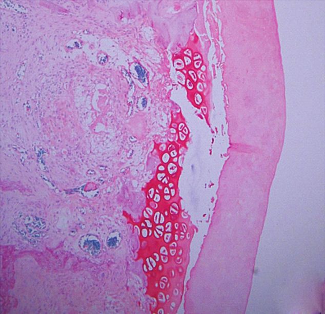
Evidence-Based Complementary and Alternative Medicine                                                                                                                                                  7

Table 4: Effect of GCSB-5 on serum TNF-α, IL-1β, and IL-10 levels                                                                                        MIA
                                                                                                                                                    GCSB-5 (mg/kg)
                                                                                                                     Control   Vehicle                                 Diclofenac
in MIA-induced osteoarthritic rats.                                                                                                                 300          600
                                                                                                  95 kD                                                                                 MMP-9 latent
                                                                                                  85 kD
                              TNF-α            IL-1β            IL-10                                                                                                                   MMP-9 active
Group                                                                                             72 kD                                                                                 MMP-2 latent
                             (pg/mL)          (pg/mL)          (pg/mL)                            62 kD                                                                                 MMP-2 active

Control                     30.0 ± 4.5       29.1 ± 3.7       27.8 ± 0.6                                       400
MIA
  Vehicle                  85.4 ± 6.6b     99.5 ± 11.2b      60.4 ± 7.2b

                                                                                  MMP activity (control (%))
                                                                                                                                                                       a
                                                                                                               300
  GCSB-5                                                                                                                                                                          a

     300 mg/kg            60.0 ± 4.6a,c    102.6 ± 10.4b 91.4 ± 8.8b,d                                                                   a
                                                                                                                                                    a
                                                                                                                                                                           a, b
                                                                                                                                                                                      a, b

     600 mg/kg            55.3 ± 6.2c       93.9 ± 9.0b 84.5 ± 8.5b,c                                          200
                                                                                                                                             a, b         a, b

  Diclofenac 5 mg/kg      50.1 ± 4.0d       49.5 ± 7.5d 86.1 ± 9.2b,c
The serum concentration of TNF-α, IL-1β, and IL-10 was determined using
enzyme-linked immunosorbent assay. The results are presented as mean ±                                         100
S.E.M. of 6 rats per group.
a,b Denote significant differences (P < 0.05, P < 0.01) compared with control

group.
c,d Denote significant differences (P < 0.05, P < 0.01) compared with                                             0
                                                                                                                                         MMP-2                         MMP-9
vehicle-treated MIA group.

                                                                                                                          Control
                                                                                                                          MIA
IκB-α protein expression decreased 2.0-fold in the vehicle-                                                               MIA + GCSB-5 300 mg/kg
treated MIA group, compared to that in the control group                                                                  MIA + GCSB-5 600 mg/kg
2 days after MIA injection. These changes were significantly                                                              MIA + diclofenac 5 mg/kg
inhibited by GCSB-5 treatment (Figure 7).
                                                                               Figure 3: Activities of MMP-2 (Gelatinase A) and MMP-9 (Gelati-
                                                                               nase B) assessed by zymography in knee joint cartilages obtained
4. Discussion                                                                  28 days after MIA injection. The latent and active amounts of
                                                                               gelatinase were combined to give a total value for each gelatinase.
Today, cure for OA remains elusive. The management of OA                       Each value represents the mean ± S.E.M. from 6 rats per group.
                                                                               a Significantly different (P < 0.01) from control. b Significantly
is largely palliative focusing on the alleviation of symptoms.
Current recommendations for the management of OA                               different (P < 0.05) from vehicle-treated MIA.
include a combination of nonpharmacological (weight loss,
education programs, and exercise) and pharmacological
interventions (paracetamol, NSAIDs, etc.) [26]. Of the phar-                   model in which lesions resembling some aspects of human
macological intervention available, analgesics and NSAIDs                      OA produced quickly and has been suggested as a model
have been proven to be highly effective in controlling the                      for the study of chondroprotective drugs [30]. In the present
symptoms and signs of OA. However, they have potential                         study, we investigated GCSB-5 on the clinical and behavioral
gastrointestinal (GI) adverse effects. Herbal medicinal prod-                   changes associated with MIA-induced OA. Swelling and
ucts (HMPs) are not yet among the recommended treatment                        limping were apparent as early as 7 days after MIA injection,
options, although they are used in a variety of oral and top-                  after which they became transiently subsided. At 14 days,
ical forms in the treatment of OA. The mechanism of action                     there was a second period of knee joint swelling and limping
of HMPs is broader than that of NSAIDs and/or analgesics                       that was progressively aggravated until day 28. Administra-
in current use for symptomatic OA. Although the exact                          tion of GCSB-5 once daily for 28 days significantly reduced
mechanisms of action have not yet been elucidated, there is                    the severity of swelling and limping. These results suggest
no doubt that all herbal medicines act via several pathways,                   that GCSB-5 may have potential as a treatment for OA.
including inhibition of COX and/or lipoxygenase, inhibition                    Roentgenographic and histological observations strongly
of cytokine release, inhibition of elastase or hyaluronidase,                  supported the behavioral changes following MIA injection
and induce antioxidative activity [27]. On the basis of this                   as well as the protective effect of GCSB-5.
knowledge, our experimental herbal extract, GCSB-5, con-                           Cartilage comprises an extracellular matrix consisting
sisted of various herbs known to exhibit antiarthritic, anti-                  of PGs, collagens (types II, IX, XI, and others), and water.
inflammatory and analgesic effects, is expected to show ther-                   Cartilage PGs consist of a protein core with GAG side chains
apeutic activity against OA.                                                   [31]. When cartilage is damaged by inflammatory mediators
     Articular cartilage destruction is a key pathological char-               such as rhIL-1α, PGs degrade and consequently release GAG,
acteristic of OA. MIA is an inhibitor of glyceraldehyde-3-                     which is a typical clinical symptom of OA. GCSB-5 did not
phosphate dehydrogenase activity, and therefore an inhibitor                   inhibit GAG release at low concentrations, but showed an
of glycolysis shown to induce chondrocyte death in vitro [28].                 inhibitory effect at moderate-to-high concentrations. This
Intra-articular injection of MIA induces chondrocyte death                     analysis reflected the histochemical appearance of cartilage.
in the articular cartilage of rodent and nonrodent species                     SOFG staining showed significant PG loss and lesion devel-
[29]. Injection of MIA into the knees of rats provides a                       opment in subchondral bone which were induced by MIA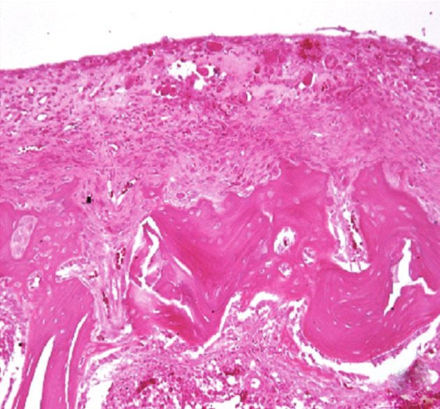
8 Evidence-Based Complementary and Alternative Medicine
MIA MIA
GCSB-5 (mg/kg) GCSB-5 (mg/kg)
Control Vehicle Diclofenac Control Vehicle Diclofenac
300 600 300 600
COX-2
TNF-α β-actin
β-actin
iNOS
IL-1β β-actin
β-actin
IL-10 2
β-actin
1 b, d
Protein expression (a.u.)
1.5
b, d b
b b, d
0.8 b
mRNA expression (a.u.)
a
1
b a, c
0.6 b b c
a, d
b, c
d
d
b, d 0.5 d
0.4 b
b
0.2 0
COX-2 iNOS
0
TNF-α IL-1β IL-10 Control
Control MIA
MIA MIA + GCSB-5 300 mg/kg
MIA + GCSB-5 300 mg/kg MIA + GCSB-5 600 mg/kg
MIA + GCSB-5 600 mg/kg MIA + diclofenac 5 mg/kg
MIA + diclofenac 5 mg/kg
Figure 5: COX-2 and iNOS protein expressions in cartilage from
Figure 4: TNF-α, IL-1β, and IL-10 mRNA expressions in cartilage knee joints of rats at day 2 after MIA injection. Each value represents
from knee joints of rats at day 2 after MIA injection. Each value the mean ± S.E.M. from 6 rats per group. a,b Significantly different
represents the mean ± S.E.M. from 6 rats per group. a,b Significantly (P < 0.01, P < 0.05) from control. c,d Significantly different (P <
different (P < 0.01, P < 0.05) from control. c,d Significantly different 0.01, P < 0.05) from vehicle-treated MIA.
(P < 0.01, P < 0.05) from vehicle-treated MIA.
MIA injection. Our results indicate that GCSB-5 inhibits
injection. These cartilage damages were attenuated by GCSB- collagen degradation through inhibition of MMP-2 and -9
5 treatment. Our data suggest that GCSB-5 may protect activities in late stage of OA.
articular cartilage from degradation. Matrix turnover is solely dependent on chondrocytes,
MMPs are a family of proteinases that together can de- which are believed to be the main site of inflammatory
grade all extracellular matrix components. Type IV colla- mediators production in human OA [36]. Overexpression
genases (gelatinases) are members of the MMP family and of MMPs is induced by several cytokines, such as TNF-
are thought to play an important role in the degradation of α, IL-1, IL-17, and IL-10. TNF-α and IL-1β drive the
extracellular components. The gelatinase subclass can be di- catabolic processes in OA, leading to cartilage degradation.
vided into gelatinase-A (MMP-2) and gelatinase-B (MMP- In this study, the levels of TNF-α, COX-2, and iNOS mRNA
9), which is capable of degrading types IV and V collagens, expression were significantly increased 2 days after MIA
elastin, and gelatin [32]. MMP-2 is known to be produced by injection and returned to control level at 28 days (data
osteoblasts and tissue structural cells, including fibroblasts not shown). These increases were attenuated by GCSB-5
and endothelial cells, whereas MMP-9 is produced by in- treatment. On the other hand, a critical function of IL-
flammatory cells such as macrophages, neutrophils, and eos- 10 is to limit inflammatory responses [37]. This cytokine
inophils [33, 34]. These MMPs are secreted as latent pre- inhibits IL-1β and TNF-α expression and is present in
cursors and can be activated by limited proteolysis. The in- OA chondrocytes, where it may counteract their catabolic
creased expression of MMP-2 and -9 in the synovium of effects [38]. Interestingly, GCSB-5 treatment significantly
patients with arthritic effusions superiorly reflects the potentiated this increase. Our results indicate that GCSB-
inflammatory condition of the joints, and a positive corre- 5 shows a significant anti-inflammatory action in the early
lation between MMP-9 production and rapid destruction of stage of OA.
the hip joint has been described in OA [35]. Although GCSB- Inappropriate regulation of NF-κB activity has been
5 did not affect MMP-2 and -9 activities 7 days after MIA implicated in the pathogenesis of inflammatory diseases,
injection (data not shown), GCSB-5 especially at a dose of such as rheumatoid arthritis and OA [39]. NF-κB signal-
300 mg/kg suppressed MMP-2 and -9 activities 28 days after ing pathways mediate critical events in the inflammatory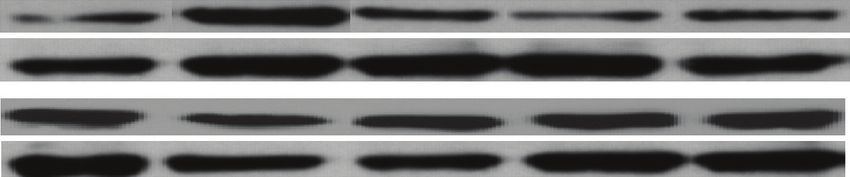
Evidence-Based Complementary and Alternative Medicine 9
MIA MIA
GCSB-5 (mg/kg) GCSB-5(mg/kg)
Control Vehicle Diclofenac Control Vehicle Diclofenac
300 600
300 600
COX-2
NF-κB p65
β-actin
Lamin B1
iNOS IκB-α
β-actin β-actin
1.2 a
1.5
b
mRNA expression (a.u.)
0.9 1.2
Protein expression(a.u.)
d c
0.9 d
0.6
c
a 0.6
b
0.3 a
b
0.3
b b
0
COX-2 iNOS 0
NF-κB p65 IκB-α
Control Control
MIA MIA
MIA + GCSB-5 300 mg/kg MIA + GCSB-5 300 mg/kg
MIA + GCSB-5 600 mg/kg MIA + GCSB-5 600 mg/kg
MIA + diclofenac 5 mg/kg MIA + diclofenac 5 mg/kg
Figure 7: Nuclear NF-κB p65 and cytosolic IκB-α protein expres-
Figure 6: COX-2 and iNOS mRNA expressions in cartilage from sions in cartilage from knee joints of rats at day 2 after MIA
knee joints of rats at day 2 after MIA injection. Each value represents injection. Each value represents the mean ± S.E.M. from 6 rats per
the mean ± S.E.M. from 6 rats per group. a Significantly different group. a,b Significantly different (P < 0.05, P < 0.01) from control.
(P < 0.05) from control. b Significantly different (P < 0.05) from c,d Significantly different (P < 0.05, P < 0.01) from vehicle-treated
vehicle-treated MIA. MIA.
Acknowledgments
response by chondrocytes, leading to progressive extracellu-
lar matrix damage and cartilage destruction. NF-κB mediates This paper was supported by Green Cross Corporation,
fibronectin fragment-induced chondrocyte activation and Korea, and Jaseng Hospital, Korea.
increased expression of proinflammatory cytokines, che-
mokines as well as MMPs such as IL-6, IL-8, MCP-1, growth-
related oncogene-α, -β, -γ, and MMP-13 by human articular References
chondrocytes [40, 41]. In this study, we showed that GCSB- [1] M. B. Goldring and S. R. Goldring, “Osteoarthritis,” Journal of
5 inhibits nuclear translocation of NF-κB/p65 subunit and Cellular Physiology, vol. 213, no. 3, pp. 626–634, 2007.
degradation of IκB-α. [2] J. Saklatvala, “Inflammatory signaling in cartilage: MAPK and
Although the results of present study provided clues for NF-κB pathways in chondrocytes and the use of inhibitors
further studies on the pharmacological mechanisms of for research into pathogenesis and therapy of osteoarthritis,”
GCSB-5, the relationship between the effects and its active Current Drug Targets, vol. 8, no. 2, pp. 305–313, 2007.
components remains to be clarified. Therefore, the detailed [3] F. E. Silverstein, G. Faich, J. L. Goldstein et al., “Gastrointesti-
nal toxicity with Celecoxib vs nonsteroidal anti-inflammatory
molecular mechanisms of GCSB-5 and further studies of
drugs for osteoarthritis and reumatoid arthritis: the CLASS
anti-inflammatory properties of the active ingredients
study: a randomized controlled trial,” Journal of the American
should be elucidated. Medical Association, vol. 284, no. 10, pp. 1247–1255, 2000.
[4] L. X. Chen, L. Lin, H. J. Wang et al., “Suppression of early
experimental osteoarthritis by in vivo delivery of the adenovi-
5. Conclusions ral vector-mediated NF-κBp65-specific siRNA,” Osteoarthritis
and Cartilage, vol. 16, no. 2, pp. 174–184, 2008.
These results indicate that GCSB-5 improves OA-induced [5] S. W. Park, C. H. Lee, S. H. Kim et al., “General pharmaco-
cartilage damage, which inhibits MMP activities, down- logical study of GCSB-5, a herbal formulation,” The Journal of
regulates the expression of inflammatory mediators, and Applied Pharmacology, vol. 14, pp. 194–201, 2006.
suppresses NF-κB activity, suggesting that GCSB-5 may be [6] H. W. Kim, Y. B. Kwon, T. W. Ham et al., “The antinociceptive
a potential therapeutic agent for OA. and anti-inflammatory effect of ethylacetate extracts from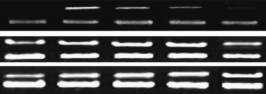
10                                                                            Evidence-Based Complementary and Alternative Medicine

       Bang-Poong (Radix ledebouriellae) on the Freund’s adjuvant-                  explant culture and collagenase-induced rabbit osteoarthritis
       induced arthritis in rats,” Journal of Veterinary Science, vol. 3,           model,” Osteoarthritis and Cartilage, vol. 10, no. 6, pp. 471–
       no. 4, pp. 343–349, 2002.                                                    478, 2002.
 [7]   B. Zhao, X.-B. Yang, X.-W. Yang et al., “Intestinal permeability      [23]   Y. Sakano, N. Terada, H. Ueda et al., “Histological study of
       of the constituents from the roots of saposhnikovia divaricata               articular cartilage in experimental rat knee arthritis induced
       in the human caco-2 cell monolayer model,” Planta Medica,                    by intracapsular injection of cationic polyethyleneimine,”
       vol. 77, no. 13, pp. 1531–1535, 2011.                                        Medical Electron Microscopy, vol. 33, no. 4, pp. 246–257, 2000.
 [8]   Y. Ida, Y. Satoh, M. Katsumata et al., “Two novel oleanolic acid      [24]   K. Kobayashi, R. Imaizumi, H. Sumichika et al., “Sodium io-
       saponins having a sialyl Lewis X mimetic structure from                      doacetate-induced experimental osteoarthritis and associated
       Achyranthesfauriei root,” Bioorganic and Medicinal Chemistry                 pain model in rats,” Journal of Veterinary Medical Science, vol.
       Letters, vol. 8, no. 18, pp. 2555–2558, 1998.                                65, no. 11, pp. 1195–1199, 2003.
 [9]   D. Seidlova-Wuttke, D. Christel, P. Kapur, B. T. Nguyen, H.           [25]   H. Dumond, N. Presle, P. Pottie et al., “Site specific changes in
       Jarry, and W. Wuttke, “β-Ecdysone has bone protective but no                 gene expression and cartilage metabolism during early experi-
       estrogenic effects in ovariectomized rats,” Phytomedicine, vol.               mental osteoarthritis,” Osteoarthritis and Cartilage, vol. 12, no.
       17, no. 11, pp. 884–889, 2010.                                               4, pp. 284–295, 2004.
[10]   H. C. Kim, S. I. Lee, and D. K. Ahn, “Effect of acanthopanacis
                                                                             [26]   L. G. Ameye and W. S. S. Chee, “Osteoarthritis and nutrition.
       cortex on the IL-8 production in human monocyte as a
                                                                                    From nutraceuticals to functional foods: a systematic review
       rheumatoid arthritis remedy,” Journal of Herbalogy, vol. 10, no.
                                                                                    of the scientific evidence,” Arthritis Research and Therapy, vol.
       1, pp. 49–59, 1995.
                                                                                    8, no. 4, article R127, 2006.
[11]   C. H. Lee, S. H. Kim, J. S. Lee et al., “Evaluation of the antino-
       ciceptive properties of GCSB-5, a herbal formulation,” Korean         [27]   M. Cameron, J. J. Gagnier, C. V. Little, T. J. Parsons, A. Blümle,
       Journal of Pharmacognosy, vol. 36, no. 4, pp. 299–304, 2005.                 and S. Chrubasik, “Evidence of effectiveness of herbal medici-
[12]   X. C. Nguyen, V. M. Chau, V. K. Phan et al., “Inhibitors of os-              nal products in the treatment of arthritis—Part 1: osteoarthri-
       teoclast formation from rhizomes of Cibotium barometz,”                      tis,” Phytotherapy Research, vol. 23, no. 11, pp. 1497–1515,
       Journal of Natural Products, vol. 72, no. 9, pp. 1673–1677, 2009.            2009.
[13]   S. H. Kim, C. H. Lee, J. S. Lee et al., “Anti-inflammatory ac-        [28]   C. Cournil, B. Liagre, L. Grossin et al., “Overexpression and
       tivities of a herbal preparation GCSB-5 on acute and chronic                 induction of heat shock protein (Hsp) 70 protects in vitro and
       inflammation,” Korean Journal of Pharmacognosy, vol. 36, no.                 in vivo from mono-iodoacetate (MIA)-induced chondrocytes
       4, pp. 311–317, 2005.                                                        death,” Arthritis Research and Therapy, vol. 3, supplement 1, p.
[14]   R. W. Wong and A. B. Rabie, “Effect of genistin on bone for-                  41, 2001.
       mation,” Frontiers in Bioscience, vol. 2, pp. 764–770, 2010.          [29]   J. Dunham, S. Hoedt-Schmidt, and D. A. Kalbhen, “Prolonged
[15]   N. D. Hong, Y. S. Rho, J. W. Kim, D. H. Won, N. J. Kim, and                  effect of iodoacetate on articular cartilage and its modification
       B. S. Cho, “Studies on the general pharmacological activities                by an anti-rheumatic drug,” International Journal of Experi-
       of Eucommia ulmoides Oliver,” Korean Journal of Pharmacog-                   mental Pathology, vol. 74, no. 3, pp. 283–289, 1993.
       nosy, vol. 19, pp. 102–110, 1998.                                     [30]   R. A. Barve, J. C. Minnerly, D. J. Weiss et al., “Transcriptional
[16]   J. Zhu, X. Gao, W. L. Xie, Y. Z. Jin, and W. J. Sun, “Effect of gen-          profiling and pathway analysis of monosodium iodoacetate-
       iposide on serum IL-1β and TNF-α of rheumatoid arthritis                     induced experimental osteoarthritis in rats: relevance to
       rats,” Zhongguo Zhongyao Zazhi, vol. 30, no. 9, pp. 708–711,                 human disease,” Osteoarthritis and Cartilage, vol. 15, no. 10,
       2005.                                                                        pp. 1190–1198, 2007.
[17]   H. Ha, J. Ho, S. Shin et al., “Effects of Eucommiae Cortex on          [31]   A. L. Stevens, C. A. Wheeler, S. R. Tannenbaum, and A. J.
       osteoblast-like cell proliferation and osteoclast inhibition,”               Grodzinsky, “Nitric oxide enhances aggrecan degradation by
       Archives of Pharmacal Research, vol. 26, no. 11, pp. 929–936,                aggrecanase in response to TNF-α but not IL-1β treatment
       2003.                                                                        at a post-transcriptional level in bovine cartilage explants,”
[18]   H. J. Chung, H. S. Lee, J. S. Shin et al., “Modulation of acute              Osteoarthritis and Cartilage, vol. 16, no. 4, pp. 489–497, 2008.
       and chronic inflammatory processes by a traditional medicine          [32]   R. Trelstad and P. Kemp, Matrix Glycoproteins and Proteogly-
       preparation GCSB-5 both in vitro and in vivo animal models,”                 cans, WB Saunders, Philadelphia, Pa, USA, 1993.
       Journal of Ethnopharmacology, vol. 130, no. 3, pp. 450–459,
                                                                             [33]   K. Kusano, C. Miyaura, M. Inada et al., “Regulation of matrix
       2010.
                                                                                    metalloproteinases (MMP-2,-3,-9, and-13) by interleukin-1
[19]   J. D. Sandy, H. L. G. Brown, and D. A. Lowther, “Degradation
                                                                                    and interleukin-6 in mouse calvaria: association of MMP
       of proteoglycan in articular cartilage,” Biochimica et Biophysica
                                                                                    induction with bone resorption,” Endocrinology, vol. 139, no.
       Acta, vol. 543, no. 4, pp. 536–544, 1978.
                                                                                    3, pp. 1338–1345, 1998.
[20]   S. E. Bove, S. L. Calcaterra, R. M. Brooker et al., “Weight
       bearing as a measure of disease progression and efficacy of             [34]   G. Murphy and A. J. Docherty, “The matrix metalloprotein-
       anti-inflammatory compounds in a model of monosodium                         ases and their inhibitors,” American Journal of Respiratory Cell
       iodoacetate-induced osteoarthritis,” Osteoarthritis and Carti-               and Molecular Biology, vol. 7, no. 2, pp. 120–125, 1992.
       lage, vol. 11, no. 11, pp. 821–830, 2003.                             [35]   S. C. Chu, S. F. Yang, K. H. Lue, Y. S. Hsieh, T. Y. Hsiao, and
[21]   M. J. Yeom, H. C. Lee, G. H. Kim, I. Shim, H. J. Lee, and D.                 K. H. Lu, “The clinical significance of gelatinase B in gouty
       H. Hahm, “Therapeutic effects of Hominis placenta injection                   arthritis of the knee,” Clinica Chimica Acta, vol. 339, no. 1-2,
       into an acupuncture point on the inflammatory responses in                   pp. 77–83, 2004.
       subchondral bone region of adjuvant-induced polyarthritic             [36]   C. Melchiorri, R. Meliconi, L. Frizziero et al., “Enhanced and
       rat,” Biological and Pharmaceutical Bulletin, vol. 26, no. 10, pp.           coordinated in vivo expression of inflammatory cytokines
       1472–1477, 2003.                                                             and nitric oxide synthase by chondrocytes from patients with
[22]   J. H. Choi, J. H. Choi, D. Y. Kim et al., “Effects of SKI 306X,               osteoarthritis,” Arthritis and Rheumatism, vol. 41, no. 12, pp.
       a new herbal agent, on proteoglycan degradation in cartilage                 2165–2174, 1998.
Evidence-Based Complementary and Alternative Medicine 11
[37] K. W. Moore, R. De Waal Malefyt, R. L. Coffman, and A.
O’Garra, “Interleukin-10 and the interleukin-10 receptor,”
Annual Review of Immunology, vol. 19, pp. 683–765, 2001.
[38] F. Iannone, C. De Bari, F. Dell’ Accio et al., “Interleukin-10 and
interleukin-10 receptor in human osteoarthritic and healthy
chondrocytes,” Clinical and Experimental Rheumatology, vol.
19, no. 2, pp. 139–145, 2001.
[39] A. S. Baldwin, “The transcription factor NF-κB and human
disease,” Journal of Clinical Investigation, vol. 107, no. 1, pp.
3–6, 2001.
[40] C. B. Forsyth, A. Cole, G. Murphy, J. L. Bienias, H. J. Im, and
R. F. Loeser Jr., “Increased matrix metalloproteinase-13 pro-
duction with aging by human articular chondrocytes in
response to catabolic stimuli,” Journals of Gerontology A, vol.
60, no. 9, pp. 1118–1124, 2005.
[41] J. I. Pulai, H. Chen, H. J. Im et al., “NF-κB mediates the stim-
ulation of cytokine and chemokine expression by human ar-
ticular chondrocytes in response to fibronectin fragments,”
Journal of Immunology, vol. 174, no. 9, pp. 5781–5788, 2005.You can also read