The size and morphology of sella turcica: A lateral cephalometric study
←
→
Page content transcription
If your browser does not render page correctly, please read the page content below
Journal of Medicine, Radiology, Pathology & Surgery (2015), 1, 3–7

ORIGINAL ARTICLE

The size and morphology of sella turcica: A lateral
cephalometric study
Tejavathi Nagaraj1, R. Shruthi2, Leena James3, I. Keerthi2, Lakshmi Balraj2, Rahul Dev Goswami2
1
 Professor and Head, Department of Oral Medicine and Radiology, Sri Rajiv Gandhi College of Dental Sciences & Hospital, Bengaluru, Karnataka, India,
2
 Post‑graduate Student, Department of Oral Medicine and Radiology, Sri Rajiv Gandhi College of Dental Sciences & Hospital, Bengaluru, Karnataka, India,
3
 Professor, Department of Oral Medicine and Radiology, Sri Rajiv Gandhi College of Dental Sciences & Hospital, Bengaluru, Karnataka, India

Keywords                                             Abstract
Lateral cephalogram, sella turcica, sella turcica    Background: Sella turcica is a saddle-shaped concavity in the body of sphenoid bone
morphology and size
                                                     situated in the middle cranial fossa of skull, clearly seen on lateral cephalometric radiograph.
                                                     Aim: The purpose of the study was to measure the size and describe the morphology of
Correspondence:
Dr. R. Shruthi, Department of Oral
                                                     sella turcica in different age groups and gender.
Medicine and Radiology, Sri Rajiv                    Materials and Methods: Lateral cephalometric radiographs of 200 subjects of which
Gandhi Dental College and Hospital,                  100 males and 100 females in the age group of 8-30 years were included in the study
Cholanagar, Bengaluru - 560 032,                     population. Linear dimensions which include the length, depth, and anteroposterior
Karnataka, India. Phone: +91-9686666902,             diameter were measured and the shape of sella turcica was analyzed and skeletal class of
Email: drshruthir@gmail.com                          malocclusion was noted. Chi-square test and ANOVA test were used for statistical analysis.
                                                     Results: In the present study, morphology of sella turcica appeared to be normal shape
Received 15 February 2015;                           (upper contour of anterior wall of sella turcica appears to be perpendicular to floor)
Accepted 28 March 2015                               in 46.5% of the study population and morphological variations in shape were seen in
                                                     53.5% of study population. Whereas size was considered there was statistically significant
doi: 10.15713/ins.jmrps.14
                                                     increase in the depth and anteroposterior diameter of sella turcica as age advanced.
                                                     There was no significant difference in the linear measurements of sella turcica between
                                                     males and females.
                                                     Conclusion: The most common shape of sella turcica in the study population was
                                                     normal shape. There is a gradual increase in the size of sella turcica as age advances.

Introduction                                                                     acromegaly, empty Sella syndrome, and Nelson syndrome.
                                                                                 A small size may lead to decreased pituitary function causing
Sella turcica is a saddle-shaped concavity in the body of sphenoid
                                                                                 symptoms such as short stature and retarded skeletal growth.[4]
bone situated in the middle cranial fossa of the skull. Sella turcica
                                                                                 Research concerning the sella turcica has focused on both size
gets its name from Turkish language because of its similarity to
                                                                                 and morphology. A normal morphological variation of sella
the Turkish saddle. The depression in saddle is noted as pituitary
                                                                                 turcica vary greatly from individual to individual.[5] The aim
fossa or hypophyseal fossa. The pituitary gland is situated in
the hypophyseal fossa. It is limited by bony constituents of the                 of this study was to determine the average dimensions and
sella turcica, anteriorly by tuberculum sellae, posteriorly by                   morphological variations of the sella turcica in different age
dorsum sellae and inferiorly by the bony roof of sphenoid air                    groups and to evaluate any difference in size between males and
sinus.[1,2] Sella turcica on lateral cephalometric radiograph can be             females in the study population.
observed clearly and consecutively traced during cephalometric
analysis.[3] A larger size may be an indication of pituitary tumor               Materials and Methods
over producing hormones such as an adrenocorticotropic
hormone, prolactin, growth hormone, thyroid stimulating                          This radiographic study was conducted during a time period
hormone, antidiuretic hormone. The enlarged sella turcica on                     of 6 months, from July 2014 to December 2014. The study
a radiograph has been found to be associated with adenomas,                      included a total of 200 digital lateral cephalometric radiographs
meningioma, primary hypothyroidism, prolactinoma, gigantism,                     of 100 males and 100 females (between 8 and 30 years of age)

Journal of Medicine, Radiology, Pathology & Surgery ● Vol. 1:3 ● May-Jun 20153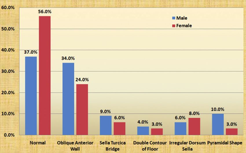
Size and morphology of sella turcica                                                                                                Nagaraj, et al.

who attended the Department of Oral Medicine and Radiology              contour of the floor, irregular surface (notch like depression) in
and were grouped into five categories based on age i.e., 4° belongs to Class II
                                                                        normal type of 46.5%.
skeletal base. ANB angle of
Nagaraj, et al. Size and morphology of sella turcica
b c
a
d e f
Figure 2: Different morphological shapes of sella turcica: (a) Normal, (b) Oblique anterior wall, (c) sella turcica bridging, (d) double contour
of floor, (e) irregular dorsum sella, (f) pyramidal shape
anterior wall, bridging of sella turcica, double contour of the
floor, irregular surface (notch like depression) in the posterior
aspect of the dorsum sellae, pyramid-like shape of the dorsum
sellae were recognized.[6]
Alkofide conducted a study to evaluate the morphological
shapes of sella turcica in cleft lip and palate patients in 2008,
according to study he arrived at result majority of cleft subjects
had morphological aberrations such as a double contour of the
floor, an irregular posterior wall found more commonly than the
normally shaped sella turcica. Contrary to individuals with clefts,
in most non-cleft subjects the morphology of the sella turcica
appears to be normal.[7]
The morphological variations of sella turcica with greater
Graph 1: Distribution of different shapes of sella turcica among severity are more commonly seen in syndromic patients such as
genders
Down’s syndrome, William’s syndrome, Seckel syndrome, and
Axenfeld-Rieger syndrome.[8-11]
Shape of sella turcica A study was done by Sathyanarayana et al. in 2012 to assess the
Gorden and Bell in 1922 examined radiographs of normal size and morphology of sella turcica in south Indian population
children in between 1 and 12 years of age and categorized sella having Class I, Class II, and Class III skeletal patterns. In this
turcica into three shapes, circular, oval, flat/saucer shaped. study, 61% of the subjects had normal morphology whereas the
Circular or oval shaped sella turcica were observed in majority of remaining 39% had variations in the shape, lowest being oblique
subjects, and they arrived at a conclusion that not all cases could anterior wall in 5%, double contour of the floor in 5.5%, pyramid-
easily be put into such a broad three-way classification.[5] like shape of the dorsum sellae in 5.5%, bridging of sella turcica in
Axelsson et al. conducted a study in Norway using lateral 8% of subjects, irregularity (notch like depression) in the posterior
cephalometric radiographs of males and females in age range surface of the dorsum sellae in 15% of study population.[12]
of 6-21 year in 2004 to determine variations in size and shape In contrast to above, study done by Chauhan et al. in 2014
of sella turcica. The sella turcica morphology was analyzed and showed morphology of sella turcica to be typical in just 28% of
five types of different morphological aberration like oblique cases. Within the atypical sellae most had oblique anterior wall
Journal of Medicine, Radiology, Pathology & Surgery ● Vol. 1:3 ● May-Jun 20155Size and morphology of sella turcica Nagaraj, et al.
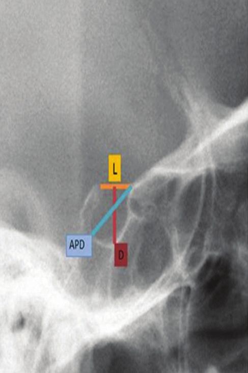
Table 1: Age‑wise distribution of sella turcica parameters The presence of a sella turcica bridge in normal individuals
Parameters N Mean SD Minimum Maximum F value P value has been shown to occur in 5.5-22% of subjects, with an increased
Length incidence in patients with craniofacial deviations.[14-16] Najim
≤10 years 13 9.12 1.464 7.07 13.00 0.638 0.636 and Al-Nakib conducted a cephalometric study in 2011 to assess
morphology of sella turcica in patients with maxillary malposed
11‑15 years 93 9.45 1.716 5.93 14.83
canine and normal population and found that prevalence of
16‑20 years 39 9.86 1.945 5.62 14.91 sella turcica bridging was comparatively greater in subjects with
21‑25 years 39 9.39 1.586 6.76 12.07 abnormally placed canine as compared to control group.[17] In
26‑30 years 16 9.67 2.216 5.26 15.32 our study, 7.5% of the population had bridging of sella turcica.
Total 200 9.52 1.762 5.26 15.32
Size of sella turcica
Depth
Tetradis and Kantor conducted a study in 1999 with sample of
≤10 years 13 7.65 1.238 5.47 10.81 3.919 0.004
325 orthodontic patients in which 134 patients were males and
11‑15 years 93 7.94 1.545 4.37 13.69 191 were female patients, varying from 6 to 49 years with mean
16‑20 years 39 8.31 1.470 5.66 11.73 age of 14.8 years. They measured linear dimensions of sella
21‑25 years 39 8.53 1.312 6.03 11.65 turcica on the lateral cephalogram, the anteroposterior diameter
ranged from 6.0 to 17.0 mm, mean value was found to be 10.9 ±
26‑30 years 16 9.26 1.160 6.34 10.62
1.8 mm, while the depth varied from 2.5 to 12.5 mm with a mean
Total 200 8.21 1.484 4.37 13.69 of 7.6 ± 1.7 mm.[18]
Diameter The size of sella turcica was studied by Axelsson et al.
≤10 years 13 11.19 1.171 9.61 13.31 7.899Nagaraj, et al. Size and morphology of sella turcica
seen more commonly in females than males. The results can be four siblings with Seckel syndrome. Cleft Palate Craniofac J
used as a reference in future studies with larger study population. 2001;38:645-51.
11. Meyer-Marcotty P, Weisschuh N, Dressler P, Hartmann J,
Clinical significance Stellzig-Eisenhauer A. Morphology of the sella turcica in
Axenfeld-Rieger syndrome with PITX2 mutation. J Oral Pathol
The normal anatomy and variations in the morphology and size Med 2008;37:504-10.
of sella turcica on a lateral cephalometric radiograph should be 12. Sathyanarayana HP, Kailasam V, Chitharanjan AB. The size and
acquainted by clinicians, in order to analyze deviations that may morphology of sella turcica in different skeletal patterns among
reflect pathological situations. Growth of the individual can be South Indian population: A lateral cephalometric study. J Indian
assessed based on the size of the sella turcica at different age Orthod Soc 2013;47:266-71.
period. 13. Chauhan P, Kalra S, Mongia SM, Ali S, Anurag A. Morphometric
analysis of sella turcica in North Indian population:
A radiological study. Int J Res Med Sci 2014;2:521-6.
References 14. Becktor JP, Einersen S, Kjaer I. A sella turcica bridge in subjects
with severe craniofacial deviations. Eur J Orthod 2000;22:69-74.
1. Subhadra Devi V, Baburao S. Age and sex related morphology 15. Sathyanarayana HP, Kailasam V, Chitharanjan AB. Sella
and morphometry of sellar region of sphenoid in prenatal and turcica - Its importance in orthodontics and craniofacial
postnatal human cadavers. Int J Res Dev Health 2013;1:141-8. morphology. Dent Res J (Isfahan) 2013;10:571-5.
2. Chaurasia BD. BD Chaurasia’s Human Anatomy. Head Neck 16. Andredaki M, Koumantanou A, Dorotheou D, Halazonetis DJ.
and Brain. 4th ed., Vol. 3. New Delhi: CBS Publishers; 2004. p. 22. A cephalometric morphometric study of the sella turcica. Eur J
3. Leonardi R, Barbato E, Vichi M, Caltabiano M. A sella turcica Orthod 2007;29:449-56.
bridge in subjects with dental anomalies. Eur J Orthod 17. Najim AA, Al-Nakib L. A cephalometric study of sella turcica
2006;28:580-5. size and morphologyamong young Iraqi normal population in
4. Meyer-Marcotty P, Reuther T, Stellzig-Eisenhauer A. Bridging comparison to patients with maxillary malposed canine. J Bagh
of the sella turcica in skeletal Class III subjects. Eur J Orthod College Dent 2011;23:53-8.
2010;32:148-53. 18. Tetradis S, Kantor ML. Prevalence of skeletal and dental
5. Shah AM, Bashir U, Ilyas T. The shape and size of the sella anomalies and normal variants seen in cephalometric and other
turcica in skeletal class I, II, III in patients presenting at Islamic radiographs of orthodontic patients. Am J Orthod Dentofac
International Dental Hospital, Islamabad. Pak Oral Dent J Orthop 1999;116:572-7.
2011;31:104-10. 19. Yassir AY, Nahidh M, Yousif HA. Size and morphology of sella
6. Axelsson S, Storhaug K, Kjaer I. Post-natal size and morphology turcica in Iraqi adults. Mustansiria Dent J 2010;7:23-30.
of the sella turcica. Longitudinal cephalometric standards 20. Chavan SR, Kathole MA, Katti AS, Herekar NG. Radiological
for Norwegians between 6 and 21 years of age. Eur J Orthod analysis of sella turcica. Int J Recent Trends Sci Technol
2004;26:597-604. 2012;4:36-40.
7. Alkofide EA. Sella turcica morphology and dimensions in cleft 21. Osunwoke EA, Mokwe CR, Amah-Tariah FS. Radiologic
subjects. Cleft Palate Craniofac J 2008;45:647-53. measurements of the sella turcica in an adult Nigerian
8. Axelsson S, Storhaug K, Kjaer I. Post-natal size and morphology population. Int J Pharm Res 2014;4:115-7.
of the sella turcica in Williams syndrome. Eur J Orthod
2004;26:613-21.
9. Korayem M, AlKofide E. Size and shape of the sella turcica How to cite this article: Nagaraj T, Shruthi R, James L,
in subjects with Down syndrome. Orthod Craniofac Res Keerthi I, Balraj L, Goswami RD. The size and morphology of
2015;18:43-50.
sella turcica: A lateral cephalometric study. J Med Radiol Pathol
10. Kjaer I, Hansen N, Becktor KB, Birkebaek N, Balslev T.
Craniofacial morphology, dentition, and skeletal maturity in
Surg 2015;1:3-7.
Journal of Medicine, Radiology, Pathology & Surgery ● Vol. 1:3 ● May-Jun 20157You can also read