Theme 3 Physiological Medicine 2019/2020 - MRC DTP
←
→
Page content transcription
If your browser does not render page correctly, please read the page content below

Theme 3 Physiological Medicine 2019/2020
Theme 3 Physiological Medicine

This theme explores synergies between organ-based physiology disciplines and has a
translational research emphasis, focusing on cardiovascular and respiratory
disease, foetal and maternal health, and diabetes/obesity. These areas are the core of
our “Clinical Medicine” research area, and link strongly into the Guy’s and St. Thomas’
Biomedical Research Centre. Links to other foci of scientific excellence (e.g. in vivo
imaging, bioinformatics, computational modelling) underpin an interdisciplinary ethos.

Lead: Professor Cathy Shanahan

When choosing a project from this catalogue in the funding section of the online
application form please enter MRCDTP2018_Theme3

Application Deadline: Sunday 25th November
Shortlisted candidates will be contacted in mid-January.

Interviews: 30th and 31st January 2019
The 2019/20 studentships will commence in September 2019.

For further Information or queries relating to the application process please contact mrc-
dtp@kcl.ac.uk

  Projects listed in this catalogue are subject to amendments, candidates
invited to interview will have the opportunity to discuss projects in further
                                     detail.

                                                                                             2
Contents
1.3 Gestational diabetes and depression: A role for islet serotonin ...............................................4
2.3 Defining soluble Nogo-B/NgBR binding site: towards novel treatment for endothelial
dysfunction and chronic vascular complications in diabetes. ..........................................................5
3.3 Targeting senescence to rejuvenate the aged heart ......................................................................6
4.3 A gut Feeling: Investigating Gut epithelial adaptations to metabolic disease. .....................7
5.3 Using supramolecular drug-polyamine assemblies to target cancer cells through the
polyamine transport system. ........................................................................................................................8
6.3 Strategies to improve islet transplantation outcomes .................................................................9
7.3 The role of the breast milk microbiome in infant growth and development .................... 10
8.3 Effects of air pollution exposure in pregnancy women on infant health; a clinical data
linkage study in South London. ................................................................................................................. 11
9.3 A novel mechanism underlying GnRH pulse generation by KNDy neurones..................... 13
10.3 Prevention of Gestational Diabetes in Obese Pregnant Women; Targeting Early
Pregnancy Intervention to Women at Risk ........................................................................................... 14
11.3 Identifying adipocyte-derived peptides that regulate islet function: implications for
diabetes therapy ............................................................................................................................................. 15
12.3 Impact of critical care on skeletal muscle strength .................................................................. 16
13.3 Linking epigenetic dysregulation to vascular calcification in diabetes ............................ 17
14.3 Quantitative in-vivo biomarkers of in-utero inflammation .................................................. 17

                                                                                                                                                             3
1.3 Gestational diabetes and depression: A role for islet serotonin
Co-Supervisor 1: Dr James Bowe
Research Division or CAG: School of Life Course Sciences
E-mail: james.bowe@kcl.ac.uk
Website: https://kclpure.kcl.ac.uk/portal/james.bowe.html
Co-Supervisor 2: Dr Paul Taylor
Research Division or CAG: School of Life Course Sciences
Email: paul.taylor@kcl.ac.uk
Website: https://kclpure.kcl.ac.uk/portal/paul.taylor.html
Project description:
During pregnancy insulin resistance increases and maternal islets of Langerhans adapt by increasing both insulin
secretory response and β-cell mass. Gestational diabetes (GDM) occurs when the islets are unable to adapt
sufficiently. The signals underlying this islet adaptation are poorly understood, however recent studies show islet
serotonin to be a key local mediator. Normally β-cell serotonin expression is negligible, but levels increase during
pregnancy due to placental signals such as lactogens and kisspeptin. Reduced islet serotonin signalling during
pregnancy leads to impaired glucose tolerance and GDM.
GDM is also associated with depression, though the mechanism is unclear. The most common therapeutics for
treatment of depression are serotonin reuptake inhibitors (SSRIs), which increase availability of endogenous
serotonin. SSRIs have also been shown to stimulate islet function and are likely to influence the action of
endogenous islet serotonin during pregnancy.
This project will use a combination of in vivo and primary tissue studies to address two aims:
• How does is endogenous β-cell serotonin involved in the islet adaptation to pregnancy?
• Do SSRIs improve the islet adaptation to pregnancy?
Initially the student will use tissues from mouse models to assess the effects of serotonin on β-cells. Techniques
will include RNAscope, immunohistochemistry and hormone assays. From year 2 onwards the student will use in
vivo models to examine the effects of SSRIs on glucose homeostasis in pregnant animals and models of
depression. Subsequent studies may examine the longer-term effects of SSRIs in pregnancy on the metabolic
health of both mother and offspring.
Two representative publications:
1. Drynda, R., Peters, C. J., Jones, P. M. & Bowe, J. E. (2015) The role of non-placental signals in the
adaptation of islets to pregnancy. Hormone and Metabolic Research. 47; 64-71
2. Taylor, P. D., Matthews, P. A., Khan, I. Y., Rees, D., Itani, N. & Poston, L. (2018) Generation of Maternal
Obesity Models in Studies of Developmental Programming in Rodents. Methods in molecular biology
1735; 167-199
42.3 Defining soluble Nogo-B/NgBR binding site: towards novel treatment for
endothelial dysfunction and chronic vascular complications in diabetes.
Co-Supervisor 1: Prof Maria R (Sasi) Conte
Research Division or CAG: School of Basic & Medical Biosciences
E-mail: sasi.conte@kcl.ac.uk
Website: https://www.kcl.ac.uk/nms/depts/chemistry/people/shadowaffiliate/contesasi.aspx
Co-Supervisor 2: Prof Luigi Gnudi
Research Division or CAG: School of Cardiovascular Medicine & Sciences
Email: luigi.gnudi@kcl.ac.uk
Website: https://www.kcl.ac.uk/lsm/research/divisions/cardio/about/people/gnudil.aspx
Project description:
Endothelial dysfunction is one of the major determinants of diabetes micro and macrovascular complications.
We have been working on a novel pathway, the sNogo-B/NgBR, and demonstrated, in animal experimental
model of diabetes, that targeting this pathway results in amelioration of endothelial dysfunction.
Data in the literature and in our laboratory (immunoprecipitation, proximity ligation assay) strongly support the
idea that the soluble Nogo-B N-terminus (sNogo-B) interacts with NgBR.
In this proposal, we aim to investigate the structure and biophysics of the sNogo-B/NgBR receptor interaction,
with the aim of defining specific receptor features that will allow the development of future molecules to
modulate NgBR receptor activity in the clinical setting.
Specifically, we will prepare recombinant proteins and purify them to high grade in order to test their binary
interaction in vitro using biophysical techniques, taking advantage of the wide range of available methodologies
in the Randall Centre and in the centre for Biomolecular Spectroscopy (e.g. Isothermal titration calorimetry,
fluorescence, circular dichroism, surface plasmon resonance, nuclear magnetic resonance - NMR).
The plan is also to purse structural investigations of the isolated components as well as any sNogo-B/NgBR
complex using X-ray crystallography, NMR and small angle X-ray scattering (SAXS).
The detailed knowledge of the binding surface and the biophysical characterisation of the interaction will guide
the design of specific ligand/receptor mutants that will then be tested both in vitro and in endothelial cells for
their ability to block or potentiate the protein-receptor interaction.
Two representative publications:
1. Martino, L., Pennell, S., Kelly, G., Busi, B., Brown, P., Atkinson, R.A., Salisbury, N.J.H., Ooi, Z-H., See, K-W.,
Smerdon, S.J., Alfano, C., Bui, T.T., Conte, M.R.* Synergic interplay of the La motif, RRM1, and the
interdomain linker of LARP6 in the recognition of collagen mRNA expands the RNA binding repertoire of
the La module. Nucleic Acids Res, 2015, 43, 645-60.
2. Dessapt-Baradez C, Woolf AS, White KE, Pan J, Huang JL, Hayward A, Price KL, Kolatsi-Joannou M,
Locatelli M, Diennet M, Webster Z, Smillie SJ, Nair V, Kretzler M, Cohen CD, Long DA, Gnudi L. Targeted
glomerular angiopoietin-1 therapy for early diabetic kidney disease. J Am Soc Nephrol, 2014, 25(1):33-
42
53.3 Targeting senescence to rejuvenate the aged heart
Co-Supervisor 1: Dr Georgina Ellison-Hughes
Research Division or CAG: School of Basic & Medical Biosciences
E-mail: georgina.ellison@kcl.ac.uk
Website: https://kclpure.kcl.ac.uk/portal/georgina.ellison.html
Co-Supervisor 2: Dr Julien Ochala
Research Division or CAG: School of Basic & Medical Biosciences
Email: julien.ochala@kcl.ac.uk
Website: https://kclpure.kcl.ac.uk/portal/julien.ochala.html
Project description:
Aging leads to increased cellular senescence and is associated with impaired tissue regeneration. Recent work
from the Ellison-Hughes lab and her collaborators at the Mayo Clinic (USA) shows cellular senescence is causally
implicated in generating age-related phenotypes and that removal of senescent cells (senolysis) can prevent or
delay tissue dysfunction, physical dysfunction and extend health- and lifespan.
This novel project will determine how senescence affects the regenerative capacity and function of the aged
mouse heart. We will also eliminate senescent cells using senolytic drugs, Dasatinib and Quercetin, in aged mice
and determine cellular and physiological changes.
AIM 1: TO DETERMINE HOW AGEING AND SENESCENCE AFFECTS THE REPLENISHMENT OF CARDIOMYOCYTES
(DURATION: 2.5 YEARS)
Hypothesis - ageing and senescence negatively affects the heart to regenerate new cardiomyocytes. We will
determine the source of new cardiomyocytes using double transgenic mhy6-mER-Cre-mER/R26RmT-mG aged
mice (young 3m vs. old 24m) subjected to diffuse cardiac injury by acute Isoproternol injection.
AIM 2: TO DETERMINE HOW SENESCENCE ALTERS CONTRACTILE FUNCTION (DURATION: 1 YEAR)
Hypothesis – senescence negatively affects the molecular contractility of cardiomyocytes isolated from aged
hearts. We will focus on the most abundant motor protein, myosin, and assess its mechanical behaviour using in
vitro motility assays.
WHAT THIS PROJECT BRINGS TO THE PHD STUDENT
The student will benefit from the pluridisciplinary nature of this project and the experience and technical
expertise of the two supervisors. They will receive a unique training experience combining cardiac muscle,
senolysis, in vivo animal models, molecular and cellular biology, advanced microscopy and biophysics. This
combination is likely to be highly sought after by future employers and funders.
Two representative publications:
1. Li M, Ogilvie H, Ochala J, Artemenko K, Iwamoto H, Yagi N, Bergquist J, Larsson L. Aberrant post-
translational modifications compromise human myosin motor function in old age. Aging Cell. 2015.
2. Ellison GM, Vicinanza C, Smith AJ, Aquila I, Leone A, Waring CD, Henning BJ, Stirparo GG, Papait R, Scarfo
M, Agosti V, Viglietto G, Condorelli G, Indolfi C, Ottolenghi S, Torella D & Nadal-Ginard B (2013). Adult c-
kitpos Cardiac Stem Cells Are Necessary and Sufficient for Functional Cardiac Regeneration and Repair.
Cell, 154: 827-842. doi: 10.1016/j.cell.2013.07.039.
64.3 A gut Feeling: Investigating Gut epithelial adaptations to metabolic disease.
Co-Supervisor 1: Dr Bu’Hussain Hayee
Research Division or CAG: Medicine, DTIMB
Email: b.hayee@kcl.ac.uk
Website:
Co-Supervisor 2: Dr Gavin Bewick
Research Division or CAG: Life Course Sciences
E-mail: gavin.bewick@kcl.ac.uk
Website:https://kclpure.kcl.ac.uk/portal/en/persons/gavin-bewick(72bb89df-4e3c-41e6-a686-
03a2af284806).html
Project description:
Obesity and Type 2 diabetes are together the greatest modern public health threat. Only a small proportion of
patients achieve glucose control using current pharmacological interventions. Novel, approaches are urgently
required.
There is one intervention which resolves diabetes and produces long-term durable weight-loss; Roux-en-Y gastric
bypass but is not practical for treating all obese patients. Diabetes remission can also be achieved with non-
surgical interventions which exclude nutrients from the duodenum. This highlights the important glucoregulatory
role of the small intestine and places it front and centre in the pathophysiology and treatment of metabolic
disease. Key questions remain:
1. What are the pathophysiological changes in the gut epithelium that occur in response to obesity and
high fat diet?
2. Are the changes stem cell driven and how is gut epithelial cell fate and function altered?
3. How does duodenal exclusion improve gut physiology and glucose homeostasis?
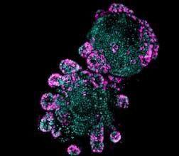
The student will explore the role of the small intestinal epithelium
in the pathology of metabolic disease by answering the above
questions. They will use whole animal physiology, endoscopy
samples from lean and obese patients, 3D organoids as a near
physiological model of the gut epithelium (mouse and human, Fig1)
and be trained in techniques ranging from confocal/lightsheet
imaging, FACS sorting, RNA-seq, and genetic engineering of
Fig 1. Examples of 3D organoids. A,
organoids.
human derived duodenal organoid. B,
mouse small intestinal organoid
showing proliferating stem cells in
Objectives pink.
Year 1: investigate the effect of obesity and diabetes on the
duodenal epithelium.
Year 2: Understand the effect of diet on the duodenal epithelium.
Year 3: Define how duodenal exclusion alters the pathology of the duodenal epithelium.
Two representative publications:
1. Elective endoscopic clipping for the treatment of symptomatic diverticular disease: a potential for
'cure'. Haji A, Plastiras A, Ortenzi M, Gulati S, Emmanuel A, Hayee B. Gut. 2018 May 5. pii: gutjnl-2017-
315509. doi: 10.1136/gutjnl-2017-315509
2. 3D intestinal organoids in metabolic research: Virtual reality in a dish. Tsakmaki A, Fonseca Pedro P.
and Bewick GA. Current Opinion in Pharmacology 37C (2017) pp. 51-58 DOI:
10.1016/j.coph.2017.09.003.
75.3 Using supramolecular drug-polyamine assemblies to target cancer cells through the
polyamine transport system.
Co-Supervisor 1: Dr Stuart Jones
Research Division or CAG: School of Cancer and Pharmaceutical Science
E-mail: stuart.jones@kcl.ac.uk
Website: https://www.kcl.ac.uk/lsm/research/divisions/ips/about/people/jonesstuart/index.aspx
Co-Supervisor 2: Dr Christopher Corpe
Research Division or CAG:
Email: christopher.corpe@kcl.ac.uk
Website: https://kclpure.kcl.ac.uk/portal/christopher.corpe.html
Project description:
Polyamines, such as putrescine, spermidine and spermine are widely distributed in the diet, but can also be
synthesised intracellularly in people. It is known that controlling the uptake and metabolism of polyamines (PAs)
by cells can have important physiological (e.g. stem cell regeneration) and therapeutic (cancer cell proliferation
inhibition) implications. However, the polyamine transport system (PTS), which is responsible for actively
sequestering these essential nutrients into cells has yet to be fully characterised.
Three putative models of PA transport have been proposed including glypican-mediated endocytosis, plasma
transport, vesicular sequestration, and caveolin-mediated endocytosis, but a gene for a polyamine-specific
transporter has not yet been isolated. Recent work at King’s has identified that rat endothelial cells and human
lung adenocarcinoma cell express high levels of the PTS, which can be used to target supramolecular drug-
polyamine assemblies into lung cells. Building on this preliminary work this PHD project aims to isolate and
functionally characterise the polyamine transporter expressed in cancer cell lines, understand how the PTS
transports drug-polyamine assemblies into cancer cells and then use this knowledge to optimise the assemblies
design in order to target and supress cancer cell growth.
In the first year of the work the objectives will be to characterise the supramolecular drug-polyamine assembly
transport in a series of cancer cell lines, varying in source and genetic modifications. In the second year the
objectives will be to isolate the PTS transporter gene, use the gene to express the PTS transporter in oocyte
membranes and then use this model, which will only display the PTS transporter, to understand better
supramolecular drug-polyamine assemblies transport mechanisms. In the third year the objective will be to
translate the knowledge of the supramolecular drug-polyamine assembly transport to modify, characterise and
optimise the uptake of the supramolecular drug-polyamine assemblies into cells in order to inhibit cancer cell
growth. In the fourth year a proof-of-concept in-vivo study will be used test the therapeutic potential of the
developed approach.
Two representative publications:
1. Benaouda, F., Jones, S.A., Chana, J., Dal Corno, B.M., Barlow, D.J., Hider, R.C., Page, C.P., Forbes, B. Ion-
Pairing with Spermine Targets Theophylline to the Lungs via the Polyamine Transport System (2018)
Molecular Pharmaceutics, 15 (3), pp. 861-870.
2. Pharmacologic ascorbic acid concentrations selectively kill cancer cells: action as a pro-drug to deliver
hydrogen peroxide to tissues. Chen Q, Espey MG, Krishna MC, Mitchell JB, Corpe CP, Buettner GR,
Shacter E, Levine M. Proc Natl Acad Sci U S A. 2005 Sep 20;102(38):13604-9.
86.3 Strategies to improve islet transplantation outcomes
Co-Supervisor 1: Dr Aileen King
Research Division or CAG: Life Course Sciences/Diabetes, Endocrinology, Nutrition, Obesity, Vision and Related
Surgeries Clinical Academic Group
E-mail: aileen.king@kcl.ac.uk
Website: https://www.kcl.ac.uk/lsm/Schools/life-course-sciences/departments/diabetes/research/Diabetes-
Research-Group-at-Guys-.aspx
Co-Supervisor 2: Dr Pratik Choudhary
Research Division: Life Course Sciences/Diabetes, Endocrinology, Nutrition, Obesity, Vision and Related Surgeries
Clinical Academic Group
Email: pratik.choudhary@kcl.ac.uk
Website: https://www.kcl.ac.uk/lsm/Schools/life-course-sciences/departments/diabetes/research/Diabetes-
Research-Group-at-Denmark-Hill-.aspx
Project description:
Islet transplantation as a treatment for Type 1 diabetes can stabilise blood glucose concentrations and protect
against hypoglycaemia. However, only half of patients undergoing the procedure become insulin independent.
This project aims to understand the dynamics of islet function after transplantation by using novel glucose
sensors in a variety of rodent transplantation models. Using this state-of-the-art continuous glucose monitoring
technology will allow treatments that have previously shown potential in rodent models to be fully optimised to
improve islet transplantation outcome in humans. Initially we will assess the effect of transplantation site on
blood glucose excursions in vascularised and non-vascularised islet transplantation models. We will then assess
at which stage of the transplantation treatments such as GLP-1 receptor agonists are most beneficial. We will
use this information to translate our findings to human islet transplantation. The two supervisors of this project
are experts in preclinical and clinical islet transplantation respectively which will allow for rapid translation of
our preclinical findings to clinical application. In the first year the student will be trained in rodent islet isolation,
culture and transplantation. In the second year the student will carry out transplantation studies in rodents with
state-of-the-art continuous glucose monitoring to allow minute-by-minute visualisation of the effect of
interventions on blood glucose concentrations. In the third year, the student will repeat the most promising
studies with human islets in a rodent model and, if appropriate, become involved in the first proof of concept
studies in humans.
Two representative publications:
1. Diabetes in rats is cured by islet transplantation… but only during daytime. King AJ, Austin AL, Nandi
M, Bowe JE. In Press, Cell Transplant. Cell Transplant. 26:171-172, 2017.
2. Clinical use of continuous glucose monitoring in adults with type 1 diabetes Slattery, D. & Choudhary,
Diabetes Technology and Therapeutics. 19: S55-S61, 2017.
97.3 The role of the breast milk microbiome in infant growth and development
Co-Supervisor 1: Dr Sophie Moore
Research Division or CAG: School of Life Course Science/Department of Women & Children’s Health
E-mail: Sophie.moore@kcl.ac.uk
Website: https://www.kcl.ac.uk/lsm/research/divisions/wh/groups/globalhealth/index.aspx
Co-Supervisor 2: Prof Rachel Tribe
Research Division or CAG: School of Life Course Science/Department of Women & Children’s Health
Email: Rachel.tribe@kcl.ac.uk
Website: https://kclpure.kcl.ac.uk/portal/rachel.tribe.html
Project description:
Maternal milk is a complex and dynamic fluid that provides nutrients, antigens, passive immunity, gut growth
factors, and other bioactive compounds that can actively shape and educate the infant immune system,
influencing infant growth and development. It is known that the immunological potential of human milk differs
between mothers; however the control and regulation of the critical immune and other bioactive components
of human milk is not well understood. A better understanding of how natural variation in these factors influences
infant development, may inform the development of therapeutic and preventative strategies.
The aim of this project is to contribute to our understanding of the regulation of human milk bioactives and how
variation in these components influences infant development. To meet this aim, the project will require access
to relevant populations and training in laboratory techniques for sample analysis. With access to samples from
an ongoing study in four contrasting settings (Gambia, Bangladesh, Brazil, Denmark), the objectives of this project
will be:
1. Develop novel methods to define the milk microbiome, and determine the influence of collection
methods on this (Year 1)
2. Using existing milk and stool samples, measure the breast milk and infant stool microbiome from
mother-infant dyads in each setting (Year 2)
3. Investigate the relationships between maternal health, breast milk microbiome, infant stool microbiome
and infant growth and development (Years 2/3)
The required training (method development, field, laboratory, analytical) will be provided through existing
collaborations both within and outside of KCL.
Two representative publications:
1. Davis JC, Lewis ZT, Krishnan S, Bernstein RM, Moore SE, Prentice AM, Mills DA, Lebrilla CB, Zivkovic AM.
Growth and morbidity of Gambian infants are influenced by maternal milk oligosaccharides and infant
gut microbiota. Sci Rep. 2017 Jan 12;7:40466. doi: 10.1038/srep40466.
2. Tribe RM, Taylor PD, Kelly NM, Rees D, Sandall J, Kennedy HP. Parturition and the perinatal period: can
mode of delivery impact on the future health of theneonate? J Physiol. 2018 Mar 13. doi:
10.1113/JP275429.
108.3 Effects of air pollution exposure in pregnancy women on infant health; a clinical
data linkage study in South London.
Co-Supervisor 1: Dr Ian S Mudway
Research Division or CAG:
E-mail: ian.mudway@kcl.ac.uk
Website: https://kclpure.kcl.ac.uk/portal/ian.mudway.html
Co-Supervisor 2: Prof Lucilla Poston
Research Division or CAG: School of Population Health & Environmental Sciences / Department of Analytical,
Environmental & Forensic Sciences
Email: lucilla.poston@kcl.ac.uk
Website: https://kclpure.kcl.ac.uk/portal/lucilla.poston.html
Collaborating Academic: Professor Anne Greenough
School/Division or CAG: Asthma, Allergy & Lung Biology
Email: anne.greenough@kcl.ac.uk
Website: https://kclpure.kcl.ac.uk/portal/anne.greenough.html
Summary of role: Professor Greenough, CO-I MRC eLIXIR parternship and Professor of Neonatalogy and Clinical
Respiratory Physiology with a major interest in respiratory physiology will advise on neonatal and infant health
in general and respiratory function in particular.
Project description:
Adverse in utero environmental exposures are implicated in disorders of childhood and adult health. In this study,
the student will investigate relationships between critical periods of environmental air pollution exposure during
pregnancy and the health of the child in later life. Influences on birthweight, neonatal health and infant health,
especially lung function will be studied. Data from a new data linkage (eLIXIR) will provide information at the
population level, on pregnancy, neonatal and primary care health from women and their children living in the
London Boroughs of Southwark and Lambeth. Monthly air pollution exposures (PM 2.5, PM10, NO2 and O3) will be
produced for each individual within the cohort to allow exposure estimation based on residential address for
each trimester and then annually post partition. Where possible detailed compositional information on metals
and PAHs will also be ascribed at a city-wide level based on continuous moinoring networks within London.
Statistical modelling will be used to address relationships with adjustment for relevant confounders, including
noise and measures of urban deprivation.
Skills training; statistical modelling, maternal and infant health outcomes, air pollution modelling, lung function.
Year 1. Generation of air pollution data, idenfication of data fields, maternal, infant and primary care data
linkage, data cleaning, literature review and PhD Upgrade
Year 2. Data linkage of all data sets. Statistical training course, preliminary analyses.
Year 3. Interrogation of data sets to address relationships between maternal air pollution exposures and infant
health outcomes including but not confined to birthweight centile, neonatal health outcomes, infant infection,
respiratory disorders and allergy.
Two representative publications:
1. Samoli E, Atkinson RW, Analitis A, Fuller GW, Green DC, Mudway I, Anderson HR, Kelly FJ. Associations
of short-term exposure to traffic-related air pollution with cardiovascular and respiratory hospital
admissions in London, UK. Occup Environ Med. 2016;73(5):300-7. doi: 10.1136/oemed-2015-103136
2. Patel N, Godfrey KM, Pasupathy D, Levin J, Flynn AC, Hayes L, Briley AL, Bell R, Lawlor DA, Oteng-Ntim E,
Nelson SM, Robson SC, Sattar N, Singh C, Wardle J, White S, Seed PT, Poston L. Infant adiposity following
a randomised controlled trial of a behavioural intervention in obese pregnancy.Int J Obes (Lond).
2017;;41:1018-1026. doi: 10.1038/ijo.2017.44.
113. Rossor T, Ali K, Bhat R, Trenear R, Rafferty GF, Greenough A. The effects of sleeping position, maternal
smoking and substance misuse on the ventilator response to hypoxia in the newborn period. Pediatr Res
2018 doi: 10.1038/s41390-018-0090-0 [Epub ahead of print].
129.3 A novel mechanism underlying GnRH pulse generation by KNDy neurones
Co-Supervisor 1: Prof Kevin O’Byrne
Research Division or CAG: Department of Women and Children’s Health, School of Life Course Sciences
E-mail: kevin.o'byrne@kcl.ac.uk
Website: https://kclpure.kcl.ac.uk/portal/kevin.o'byrne.html
Co-Supervisor 2: ProfHelen Cox
Research Division or CAG: Wolfson Centre for Age Related Diseases
Email: helen.cox@kcl.ac.uk
Website: https://kclpure.kcl.ac.uk/portal/helen.cox.html
Project description:
The hypothalamic gonadotrophin-releasing hormone (GnRH) pulse generator that drives the pulsatile secretion
of the gonadotrophic hormones, LH and FSH, is critical for reproduction. The KNDy neurones (derive their
acronym from the neuropeptides they co-express: Kisspeptin, Neurokinin B and Dynorphin) of the hypothalamus
directly stimulate GnRH neurones. It’s speculated that the KNDy neuronal network generates synchronized
oscillatory patterns of activity through shared excitatory and inhibitory inputs, and comprise the GnRH pulse
generator. The most pressing question now is, what initiates and maintains the rhythmic activation of the
KNDy neural network to drive pulsatile secretion of GnRH?
Combining mathematical modelling (Tsaneva-Atanasova, Exeter) with cutting edge in-vivo experimentation,
including optogenetics (O’Byrne) and neuropharmacology (Cox) will provide unprecedented access to the
function of the KNDy network and a paradigm shift in our understanding of the key mechanisms underpinning
the oscillatory activity of the GnRH pulse generator.
The Rotation Project will expose students to sophisticated robotic-stereotaxic surgery for injection of viral
vectors containing channelrhodopsins for selective optogenetic stimulation of KNDy neurones in freely behaving
mice. Serial blood sampling will allow detection of LH pulse frequency as a proxy for GnRH pulse frequency. In-
vivo optogenetics will determine the stimulation parameters underlying LH pulsatility.
The PhD programme will utilize in-vivo optogenetics, electrophysiological recordings and neuropharmacological
techniques to investigate how the KNDy network integrates different neuropeptides to generate and modulate
the GnRH pulse generator. In-silico investigations will facilitate a better understanding of this complex dynamic
system through generation of new testable hypotheses which will be interrogated experimentally.
Two representative publications:
1. Voliotis M, Li XF, De Burgh R, Lass G, Lightman SL, O'Byrne KT, Tsaneva-Atanasova K. (2018)
Mathematical modelling elucidates core mechanisms underpinning GnRH pulse generation.
doi: https://doi.org/10.1101/245548
2. Ghamari-Langroudi M, Digby GJ, Sebag JA, Millhauser GL, Palomino R, Matthews R, Gillyard T, Panaro BL,
Tough IR, Cox HM, Denton JS, Cone RD. (2015) G-protein-independent coupling of MC4R to Kir7.1 in
hypothalamic neurons. Nature. 520(7545):94-8.
1310.3 Prevention of Gestational Diabetes in Obese Pregnant Women; Targeting Early
Pregnancy Intervention to Women at Risk
Co-Supervisor 1: Mr Dharmintra Pasupathy
Research Division or CAG: School of Life Course Sciences / Department of Women and Children’s Health/
Women's Health Clinical Academic Group
E-mail: dharmintra.pasupathy@kcl.ac.uk
Website: https://kclpure.kcl.ac.uk/portal/en/persons/dharmintra-pasupathy.html
Co-Supervisor 2: Prof Catherine Williamson
Research Division or CAG: School of Life Course Sciences / Department of Women and Children’s Health/
Women's Health Clinical Academic Group
Email: catherine.williamson@kcl.ac.uk
Website: https://kclpure.kcl.ac.uk/portal/catherine.williamson.html
Project description:
Obesity in pregnancy increases the risk of gestational diabetes (GDM) and associated adverse outcomes. UK NICE
guidelines recommend that all obese women have an oral glucose tolerance test at 24-28 weeks’ gestation for
detection of GDM. However, excessive fetal growth in obese women is evident before diagnosis of GDM,
accompanied by an abnormal metabolome. Targeted early pregnancy intervention is therefore required to
prevent GDM and improve clinical outcomes in obese women. The KCL GDM research group have recently
developed a novel early pregnancy GDM prediction tool. This project will address the hypothesis that early
pregnancy dietary advice and/or metformin, the two ‘first line’ treatments for women with established GDM,
will prevent gestational diabetes by improving glucose tolerance and metabolic function in obese pregnant
women identified as 'at risk' by this prediction tool.
Skills:
Maternal metabolic profiling
Nutritional epidemiology
Use of novel glucose monitoring methodologies to assess glycaemic status in obese pregnancy
Large scale data analysis and curation
Study design
Objectives:
Year One: Trial site set up, participant recruitment and follow up, intervention delivery
Year Two: Participant recruitment and follow up, intervention delivery, data analysis plan
Year Three: Data and statistical analysis and interpretation, research dissemination
Two representative publications:
1. White SL, Lawlor D, Seed PT, Vieira M, Sattar N, Nelson S, Briley A, Robson R, Whitworth M, Oteng-Ntim
E, Poston L, Pasupathy D. Early antenatal prediction of Gestational diabetes mellitus (GDM) in obese
women. PLOS One 2016 Dec 8;11(12):e0167846. doi:10.1371/journal.pone. 0167846.
2. Martineau MG, Raker C, Dixon PH, Chambers J, Machirori M, King NM, Hooks ML, Manoharan R, Chen K,
Powrie R, Williamson C. The Metabolic Profile of Intrahepatic Cholestasis of Pregnancy is Associated
with Impaired Glucose Tolerance, Dyslipidemia, and Increased Fetal Growth. Diabetes Care 2015
Feb;38(2):243-248 PMID: 25504029.DOI: 10.2337/dc14-2143
1411.3 Identifying adipocyte-derived peptides that regulate islet function: implications for
diabetes therapy
Co-Supervisor 1: Prof Shanta Persaud
Research Division or CAG: Department of Diabetes, School of Life Course Sciences
E-mail: shanta.persaud@kcl.ac.uk
Website: https://www.kcl.ac.uk/lsm/research/divisions/dns/about/people/Profiles/shantapersaud.aspx
Co-Supervisor 2: Prof Peter Jones
Research Division or CAG: Department of Diabetes, School of Life Course Sciences
Email: peter.jones@kcl.ac.uk
Website: https://www.kcl.ac.uk/lsm/research/divisions/dns/about/people/Profiles/peterjones.aspx
Project description:
Around 400 million people worldwide currently have type 2 diabetes (T2D), in which peripheral cells show
reduced sensitivity to insulin and islet beta-cells do not secrete sufficient insulin to maintain low blood glucose
levels. Several pharmacotherapies for T2D are available, but they all have side-effects associated with their use
and there is a need to identify safe, effective drugs that maintain beta-cell mass and improve insulin secretory
function. We have identified that islets express nearly 300 G-protein-coupled receptors (GPCRs), but only one of
them (GLP-1 receptor) is targeted for treating T2D. There is also evidence that peptides secreted from insulin
target tissues, such as adipocytes, can act at beta-cells to regulate their function. This PhD project will identify
GPCR-activating peptides that are secreted from adipocytes under insulin-sensitive and insulin-resistant
conditions, and provide data underpinning the development of adipocyte-derived GPCR ligands as novel
therapeutics to increase beta-cell functional mass.
Overarching Objectives and Skills Training:
This project has 4 main objectives
1) Identify the expression profile of genes encoding islet GPCR ligands in adipocytes under insulin-sensitive and
insulin-resistant conditions (adipocyte isolation from mice; cell culture; induction of insulin resistance; PAGE and
western blotting; RNA extraction, cDNA synthesis and quantitative PCR: year 1).
2) Detect secreted GPCR-activating bioactive peptides in adipocyte supernatants (peptide immunoassays; beta-
arrestin reporter assays: year 2).
3) Identify the effects of key adipocyte-derived peptide ligands that interact with islet GPCRs to improve beta-
cell function in vitro and identify signaling pathways (mouse islet isolation, static and dynamic insulin secretion;
islet hormone immunoassays; second messenger quantification; PAGE and western blotting; apoptosis and
viability assays: years 2 and 3).
4) Identify the effects of key adipocyte-derived peptide ligands that interact with islet GPCRs to improve beta-
cell function in vivo. (GTTs and ITTs, plasma insulin quantification, BrdU incorporation, islet isolation, insulin
secretion and gene expression quantification: year 4).
Two representative publications:
1. Atanes P, Ruz-Maldonado I, Hawkes R, Liu B, Zhao M, Huang GC, Al-Amily IM, Salehi A, Amisten S and
Persaud SJ (2018) Defining G-protein coupled receptor peptide ligand expressomes and signalomes in
human and mouse islets. Cell. Mol. Life Sci. 75, 3039-3050
2. Rackham CL, Vargas AE, Hawkes RG, Amisten S, Persaud SJ, Austin AL, King AJ and Jones PM (2016)
Annexin A1 is a key modulator of mesenchymal stromal cell-mediated improvements in islet function.
Diabetes 65, 129-39
1512.3 Impact of critical care on skeletal muscle strength
Co-Supervisor 1: Dr Gerrard Rafferty
Research Division or CAG: Asthma Allergy & Lung Biology
E-mail: gerrard.rafferty@kcl.ac.uk
Website: https://kclpure.kcl.ac.uk/portal/gerrard.rafferty.html
Co-Supervisor 2: Prof Nicholas Hart
Research Division or CAG: Asthma Allergy & Lung Biology
Email: Nicholas.hart@gstt.nhs.uk
Website:http://www.guysandstthomas.nhs.uk/our-services/consultant-profiles/lane-fox/nicholas-hart.aspx#na
Project description:
Skeletal muscle wasting and weakness occurs in up to 65% of ICU patients and is a major complication of critical
illness. Intensive care unit-acquired weakness (ICU-AW) influences not only short term but also long-term clinical
outcomes, contributing to ‘post intensive care syndrome’ a collection of common health disorders of which
muscle weakness is a significant component. Muscle force generation is influenced by multiple factors and while
the anatomical and physiological characteristics of muscle itself are significant determinants of strength, the
central nervous system also plays an important role.
Research examining ICU-AW has focused primarily on peripheral neuromuscular function while much less is
known regarding potentially important neurological changes within the central nervous system (CNS). Studies
in healthy subjects employing short periods of limb immobilisation have described decrements in muscle
strength greater than that expected from the degree of muscle atrophy observed. Such reductions in strength
are, therefore, potentially due to reduced neural drive to the muscle from the CNS.
The proposed study examines the impact of critical illness on muscle strength and central nervous system and
motor cortex function in patients following critical illness in relation to ICU-AW. Training in a broad range of
human physiological technique to assess muscle strength and architecture and physical function will be provided
including electrical and magnetic motor nerve stimulation and force assessment as well as transcranial magnetic
stimulation. Year 1 training, study setup and commencement of data acquisition in controls. Year 2 - 3 patient
data acquisition. Year 3-4 completion of data acquisition and PhD thesis preparation
Two representative publications:
1. Connolly B, Maddocks M, MacBean V, Bernal W, Hart N, Hopkins P & Rafferty GF. (2018). Nonvolitional
assessment of tibialis anterior force and architecture during critical illness. Muscle Nerve 57, 964-972.
2. Puthucheary ZA, Rawal J, McPhail M, Connolly B, Ratnayake G, Chan P, Hopkinson NS, Phadke R, Dew T,
Sidhu PS, Velloso C, Seymour J, Agley CC, Selby A, Limb M, Edwards LM, Smith K, Rowlerson A, Rennie
MJ, Moxham J, Harridge SD, Hart N & Montgomery HE. (2013). Acute skeletal muscle wasting in critical
illness. JAMA 310, 1591-1600. Joint Senior Authorship: Harridge-Hart-Montgomery
1613.3 Linking epigenetic dysregulation to vascular calcification in diabetes
Co-Supervisor 1: Prof Catherine Shanahan
Research Division or CAG: School of Cardiovascular Medicine and Sciences
E-mail: cathy.shanahan@kcl.ac.uk
Website: http://www.kcl.ac.uk/lsm/research/divisions/cardio/about/people/shanahanc.aspx
Co-Supervisor 2: Dr Alison Brewer
Research Division or CAG: School of Cardiovascular Medicine and Sciences
Email: Alison.brewer@kcl.ac.uk
Website: https://www.kcl.ac.uk/lsm/research/divisions/cardio/about/people/brewera.aspx
Project description:
Vascular calcification is a life-threatening pathology which is accelerated markedly in diabetes. Calcification is
driven by vascular smooth muscle cells (VSMCs), that in response to metabolic stressors convert to an
osteo/chondrogenic mineralizing cell phenotype. Epigenetic change is a key driver of vascular dysfunction in
diabetes. DNA methylation is regulated by the opposing actions of DNA-Methyl-Transferases, and Ten-Eleven-
Translocation Enzymes (TETs), which convert 5-Methyl-Cytosine (5mC) to successive oxidised variants, to
facilitate its replacement by unmethylated cytosine. TET2 has been shown to be a master regulator of the
epigenetic landscape of VSMCs. Its loss has profound effects on VSMC phenotype and its function is
compromised in response to hyperglycaemia. This project aims to determine whether and how hyperglycaemia
mediates vascular calcification via dysregulation of TET2 to identify new therapeutic targets for intervention.
The effect of genetic ablation of TET2 upon the transcriptome and genomic DNA methylation status of VSMCs
in vitro will be determined by RNA-seq and 5hMeDIP-seq analyses to identify TET2 targets (year 1).
The effect of high glucose on the expression and methylation status of these targets will be determined in
VSMCs from control and diabetic patients and in vessels from a hyperglycaemic rat model of vascular
calcification (Year 2-3).
The effects of TET2 ablation and/or high glucose exposure on the development of vascular calcification will be
assessed in VSMCs in vitro and in rat vessel rings ex vivo (Year 2-3).
The student will be trained in molecular and cellular biology, tissue culture, bioinformatics, redox metabolism
and epigenetics.
Two representative publications:
1. Burr S, Caldwell A, Chong M, Beretta M, Metcalf S, Hancock M, Arno M, Balu S, Kropf VL, Mistry RK,
Shah AM, Mann GE, Brewer AC. Oxygen gradients can determine epigenetic asymmetry and cellular
differentiation via differential regulation of tet activity in embryonic stem cells. Nucleic Acids Res.
2018;46:1210-1226.
2. Liu Y, Drozdov I, Shroff R, Beltran LE, Shanahan CM. (2013). Prelamin A accelerates vascular
calcification via activation of the DNA damage response and senescence-associated secretory
phenotype in vascular smooth muscle cells. Circ Res 10;112(10):e99-109.
14.3 Quantitative in-vivo biomarkers of in-utero inflammation
17Co-Supervisor 1: Dr Lisa Story
Research Division or CAG: Division of Women’s and Children’s Health
E-mail: lisa.story@kcl.ac.uk
Website:https://kclpure.kcl.ac.uk/portal/en/persons/lisa-story(672f0408-d780-4af0-8e9d-d16a70c9dc23).html
Co-Supervisor 2: Prof Joseph V Hajnal
Research Division or CAG: Biomedical Engineering and Imaging Science/Biomedical Engineering
Email: jo.hajnal@kcl.ac.uk
Website: https://kclpure.kcl.ac.uk/portal/jo.hajnal.html
Project description:
Preterm premature rupture of the membranes (PPROM) affects 3% of pregnancies. In the absence of signs of
infection in the mother, current practice is to continue the pregnancy until term to reduce complications
associated with prematurity.
Novel techniques to assess the presence of infection has established in the fetal compartment are urgently
required. The current approach of prolonging the pregnancy may actually be detrimental for long-term health
outcomes (neurological and respiratory) of the child in the presence of infection.
This PhD proposal will utilise magnetic resonance (MR) relaxometry, a technique already used to assess
infection/inflammation in other organs including bowel and heart, to assess the placenta for chorioamnionitis
(placental infection) in vivo. Findings will be correlated with neonatal outcome data and inflammatory
biomarkers from umbilical cord blood and placental histology.
If fetal infection/inflammation can be accurately predicted this could prove invaluable in informing the timing
of delivery to minimise the morbidity/mortality associated with PPROM.
Training:
• Clinical data collection
• Pulse programming on clinical MRI scanners
• Image reconstruction and development of processing pipelines
• Analysis of medical imaging data
• Correlation of imaging data with inflammatory biomarkers and placental histology
Objectives:
• Develop in utero imaging protocols for the investigation of infection/inflammation in the fetal
compartment.
• Correlation of imaging parameters with umbilical cord biomarkers of infection/inflammation and
placental histology.
Dr Story will supervise clinical aspects of the project and correlation of outcomes with biomarkers and
placental histology and Professor Hajnal will provide guidance on MR aspects.
Two representative publications:
1. Story, L., Hutter, J., Zhang, T., Shennan, A. H. & Rutherford, M. 1 Mar 2018 The use of antenatal fetal
magnetic resonance imaging in the assessment of patients at high risk of preterm birth. Eur J Obstet
Gynecol Reprod Biol. 222, p. 134-141
2. Hutter, J., Christiaens, D. J., Schneider, T., Cordero-Grande, L., Slator, P. J., Deprez, M., … Hajnal, J. V.
(2018). Slice-level diffusion encoding for motion and distortion correction. Med Image Anal, 48, p. 214–
229.
18You can also read