Transformer Convolutional Neural Networks for Automated Artifact Detection in Scalp EEG
←
→
Page content transcription
If your browser does not render page correctly, please read the page content below
Transformer Convolutional Neural Networks for Automated Artifact
                                                                 Detection in Scalp EEG
                                                                           Wei Yan Peh1,2∗ , Yuanyuan Yao3 , and Justin Dauwels3 †

                                             Abstract— It is well known that electroencephalograms
                                          (EEGs) often contain artifacts due to muscle activity, eye blinks,
                                          and various other causes. Detecting such artifacts is an essential
                                          first step toward a correct interpretation of EEGs. Although
                                          much effort has been devoted to semi-automated and automated
arXiv:2208.02405v1 [eess.SP] 4 Aug 2022

                                          artifact detection in EEG, the problem of artifact detection
                                          remains challenging. In this paper, we propose a convolutional
                                          neural network (CNN) enhanced by transformers using belief
                                          matching (BM) loss for automated detection of five types of
                                          artifacts: chewing, electrode pop, eye movement, muscle, and
                                          shiver. Specifically, we apply these five detectors at individual
                                          EEG channels to distinguish artifacts from background EEG.                            (a) Background.                        (b) Chewing.
                                          Next, for each of these five types of artifacts, we combine the
                                          output of these channel-wise detectors to detect artifacts in
                                          multi-channel EEG segments. These segment-level classifiers
                                          can detect specific artifacts with a balanced accuracy (BAC) of
                                          0.947, 0.735, 0.826, 0.857, and 0.655 for chewing, electrode pop,
                                          eye movement, muscle, and shiver artifacts, respectively. Finally,
                                          we combine the outputs of the five segment-level detectors to
                                          perform a combined binary classification (any artifact vs. back-
                                          ground). The resulting detector achieves a sensitivity (SEN) of
                                          60.4%, 51.8%, and 35.5%, at a specificity (SPE) of 95%, 97%,
                                          and 99%, respectively. This artifact detection module can reject
                                          artifact segments while only removing a small fraction of the
                                          background EEG, leading to a cleaner EEG for further analysis.                    (c) Electrode pop.                     (d) Eye movement.
                                             Keywords — Artifact detection, electroencephalogram, deep

                                          learning, belief matching, multi-class classification, multi-class
                                          multi-label classification.

                                                                I. INTRODUCTION
                                             Electroencephalography (EEG) is a widely used tech-
                                          nology in neurology, e.g., helpful for the diagnosis of
                                          epilepsy [1]. However, EEG recordings often contain ar-
                                          tifacts, which can be due to eyeblinks, head movements,
                                          chewing, interference from electronic equipment, and other                              (e) Muscle.                          (f) Shiver.
                                          causes [2]. These artifacts may resemble epileptiform abnor-
                                          malities or other transient waveforms, resulting in mistakes                Fig. 1.    Examples of clean EEG and EEG with various types of artifacts.
                                          during annotation [3]. Knowledge of the plausible scalp
                                          distribution of EEG abnormalities is essential to distinguish                  When reading EEGs, one must distinguish artifacts from
                                          artifacts from brain waves [4]. For instance, muscle artifacts              brain waves. An automatic artifact detection system can
                                          usually appear in multiple channels, whereas artifacts such                 improve the readability of an EEG [2]. Common artifact
                                          as electrode pop may only be visible in a single channel.                   rejection and detection methods includes high amplitude
                                             This is an extension to a paper presented at the 2022 44th Annual
                                                                                                                      rejection [5], common average referencing (CAR) [6], in-
                                          International Conference of the IEEE Engineering in Medicine & Biology      dependent component analysis (ICA) [4], [3], wavelet trans-
                                          Society (EMBC) Scottish Event Campus, Glasgow, UK, July 11-15, 2022.        forms (WT) [4], machine learning [7], convolutional neural
                                             1 Nanyang Technological University (NTU), Interdisciplinary Graduate
                                                                                                                      networks (CNN) [8], [3], and generative adversarial networks
                                          Programme (IGP), Singapore 639798.
                                             2 NTU, School of Computer Science and Engineering (SCSE), Singapore      (GAN) [3]. The majority of the studies are not validated
                                          639798.                                                                     on large datasets (more than 100 patients), but instead on
                                             3 Department of Microelectronics, Delft University of Technology, 2628
                                                                                                                      small datasets (less than 100 patients) or semi-simulated
                                          CD Delft, Netherlands
                                             * corresponding author email: pehw0012@e.ntu.edu.sg                      datasets [4], [8] by injecting noise into regular EEGs. Ad-
                                             † corresponding author email: j.h.g.dauwels@tudelft.nl                   ditionally, most studies detect artifacts directly from multi-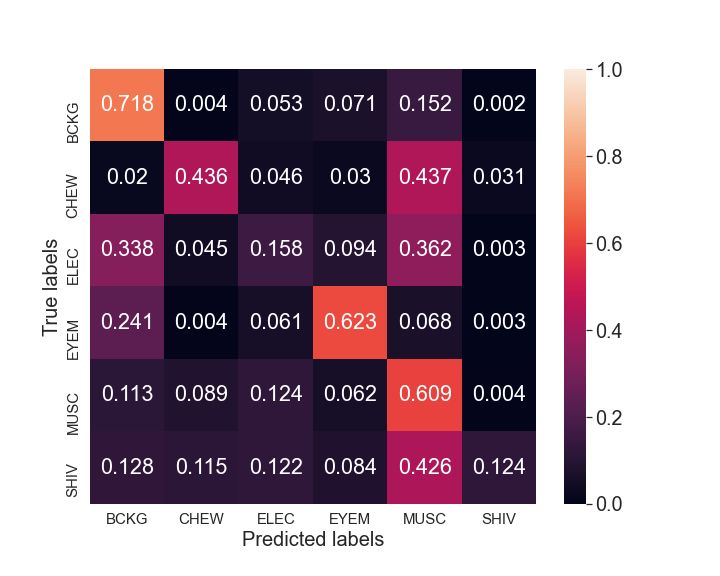
TABLE I
                    Channel-level    Segment-level
                                                                                S UMMARY OF THE TUH ART EEG DATASET.

                                                                          Background/Artifacts   Duration (hr)   Number of Events
                                                                              Background            62.533              -
                                                                               Chewing               0.995             804
                                                                             Electrode Pop           5.554            6090
                                                                             Eye Movement            8.04             9480
                                                                                Muscle              16.303           11267
                                                                                 Shiver              0.047             31

                                                                    channel-wise detector on the TUH-ART dataset for each of
                                                                    the five artifact types. The channel-level artifact detector
                                                                    is a CNN cascaded with a transformer, while the learning
                                                                    objective function is a BM loss [11] (see Figure 3).
      Fig. 2.   Channel-level and segment-level analysis of EEG.
                                                                       A CNN is not adequate for modelling correlations between
                                                                    distant data points. This inherent limitation makes CNN
                                                                    less suitable for time series, especially when correlations
channel segments [7]; as a result, many of those methods            over relatively long periods are expected, such as long
are only applicable to a fixed number of channels, whereas          artifacts patterns (e.g., eye movement artifacts). Therefore,
the proposed method can be applied to EEG with any                  we augment the CNN with a transformer to compensate
number of channels. Ultimately, most studies failed to deploy       for this limitation since transformers can extract long-range
proper evaluation metrics to measure the effectiveness of           patterns in the features extracted by the CNN.
their methods [5], [6]. Consequently, it is challenging to             In addition, it is essential to have a reliable measure of the
compare the existing artifact detectors.                            uncertainty associated with a detection (output of the neural
   In this paper, we propose a CNN equipped with a trans-           network) such that we can be confident in the detections
former (CNN-TRF) trained through a belief matching loss             with low uncertainty. To this end, we deploy a BM loss
(BM) to detect five different types of artifacts (see Figure 1)     instead of the traditional softmax (SM) loss, as it yields more
from the TUH Artifact (TUH-ART) dataset. The proposed               reliable uncertainty estimates [11]. The BM framework is a
system detects artifacts in individual EEG channels and also        Bayesian approach that views the binary classification from a
in multi-channel EEG segments (see Figure 2) [9]. The               distribution matching perspective, making it a more reliable
artifact detector can detect specific artifacts at segment-level    detector. Moreover, Joo et al. observed improvements in
with a balanced accuracy (BAC) of 0.947, 0.735, 0.826,              generalization, a desirable property for the application at
0.857, and 0.655 for chewing, electrode pop, eye movement,          hand [11]. The BM loss is defined as:
muscle, and shiver artifacts, respectively. When combined                                        m
                                                                                             1 X
to perform binary artifact classification (any artifact type                   L(W) ≈ −              lEB y (i) , αW (x(i) ) ,     (1)
vs. background EEG), the binary artifact detector achieves                                   m i=1
a sensitivity (SEN) of 42.0%, 32.0%, and 13.3% at 95%,
                                                                    where x(i) and y (i) are the i-th training data and its label,
97%, and 99% specificity (SPE), respectively. This artifact
                                                                    respectively, m is the total number of training data, lEB is
detector can detect specific artifacts and reject them from
                                                                    the evidence lower bound (ELBO) [11], and αW = exp(W),
EEGs, resulting in a cleaner EEG for a better reviewing
                                                                    where W are the weights of the neural network classifier.
experience.
                                                                       The input of the CNN-TRF is the L-second single-channel
                         II. M ETHODS                               EEG window that is split into 0.5s local segments with
A. Scalp EEG recordings and preprocessing                           25% overlap (see Figure 3). We trained the model with
    In this study, we analyzed the public TUH Artifact Corpus       different window lengths L, i.e., 1, 3, and 5s. We varied
(TUH-ART), containing EEGs with artifact annotations [10].          the window lengths to determine the best window length
The dataset consists of five artifacts types: chewing, electrode    to detect artifacts. For instance, a short window length of
pop, eye movement, muscle, and shiver (see Table I). On             1s is adequate to remove short-duration eye blinks, while a
each EEG, we applied a Butterworth notch filter (4th order)         long window length of 5s is suitable to remove long-duration
at 60Hz (USA) to remove interference and a 1Hz high-pass            muscle artifacts.
filter (4th order) to remove noise [9]. We downsampled all             The CNN architecture consists of 5 convolution layers, all
EEGs to 128Hz. We trained the artifact detectors via 5-             with a filter size of 3 and a ReLU activation function. The
fold cross-validation (CV), where each fold contains different      number of filters is 8, 16, 32, 64, and 128 in layers 1, 2, 3, 4,
patients and similar distribution across all five artifact types.   and 5, respectively. After each convolution layer, we apply
                                                                    max-pooling with stride 2. Next, we deploy a transformer
B. Channel-level Artifact Detection                                 encoder to identify patterns in the features extracted by the
  First, we develop a system to detect artifacts at individual      CNN [12]. The encoder relies on an activation function that
EEG channels (channel-level analysis). We train a separate          maps the query and a set of key-value pairs to an output.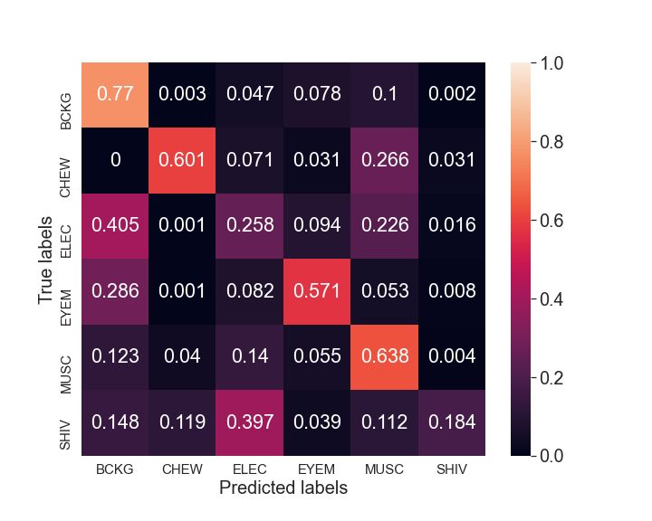
L-second single-channel EEG window
                                                                                   account for eye blink features in each feature set. Eventually,
                                                                                   we obtain 74 features from each multi-channel segment for
                                                                          Local    the training and testing of each artifact class. We performed
                                                    …
                                                                        Segments   segment-level binary classification (specific artifact vs. back-
                                        CNN                                        ground) for each artifact type. We classify the features with
                                                                                   CatBoost [14], and optimize the hyperparameters by grid
        Features             Features               …        Features              search.
                                                                                      Lastly, we concatenate the probability outputs from the
                                Transformer Encoder                                five segment-level classifiers. With all the features, we
                                                                                   trained a CatBoost classifier to detect any of the five artifacts
        Refined              Refined                         Refined
                                                    …                              types; in other words, this system is designed to determine
        Features             Features                        Features

                                            Concatenation
                                                                                   whether a multi-channel EEG segment is clean or contains
                                                                                   artifact(s). Finally, we evaluate the systems with the follow-
                                  Window Features
                                                                                   ing metrics: area under the receiver operator characteristic
                                                                                   (AUC), area under the precision-recall curve (AUPRC), ac-
                                Fully Connected Layers
                                                                                   curacy (ACC), balanced accuracy (BAC), sensitivity (SEN),
                                                                                   and specificity (SPE) [9]. NVIDIA GeForce GTX1080 GPU
                                    Output: [0,1]
                                                                                   machines, Keras 2.2.0 and TensorFlow 2.6.0 were adopted
                                                                                   in this study.
                   Fig. 3.    CNN with transformer encoder.
                                                                                                               III. R ESULTS
                                                                                   A. Channel-level Artifact Detection Results
Here, the local features extracted by the CNN are the query,
                                                                                      We displayed the results for the channel-level artifact
key, and value simultaneously. We set the number of heads in
                                                                                   detector in Table II. The chewing artifact achieved the best
the transformer to the commonly chosen value of 8 [12] and
                                                                                   BAC (0.911), while the electrode pop artifact achieved the
the number of neurons in the hidden layer of the feed-forward
                                                                                   poorest BAC (0.716). For most artifacts, the best BAC is
network (FNN) module to 1024. Two fully connected (FC)
                                                                                   achieved at a window length of 5s. However, the best BAC
layers containing 100 and 2 neurons follow the CNN-TRF
                                                                                   for each artifact class is obtained at a different window
module. Before the final FC layer, we include a dropout layer
                                                                                   length.
with a probability of 0.5. The output of the second FC is
the prediction for that particular window of EEG. Finally,                                                      TABLE II
we applied the Adam optimizer [13] with an initial learning                                   C HANNEL - LEVEL ARTIFACT DETECTION RESULTS .
rate equal to 10−4 to minimize the BM loss. The batch
                                                                                        Artifact      Window                      Binary
size for training is 1000. We applied balanced training to                               Type         Length   AUC     AUPRC    ACC     BAC     SEN     SPE
avoid overfitting during training by applying weights to each                                            1     0.961    0.950   0.901 0.901     0.894   0.907
                                                                                        Chewing          3     0.966    0.904   0.933   0.911   0.856   0.966
class. We optimized the hyperparameters of the CNN-TRF                                                   5     0.967    0.864   0.950   0.906   0.835   0.978
via nested CV on the training data with an 80%:20% split                                                 1     0.792    0.926   0.802 0.663     0.914   0.412
                                                                                      Electrode pop      3     0.802    0.802   0.709   0.716   0.718   0.713
for training and validation.                                                                             5     0.783    0.681   0.711   0.684   0.559   0.809
                                                                                                         1     0.856    0.921   0.795   0.734   0.905   0.564
                                                                                     Eye movement        3     0.866    0.799   0.807   0.788   0.723   0.853
C. Segment-level Artifact Detection                                                                      5     0.895    0.758   0.877   0.792   0.644   0.940
                                                                                                         1     0.794    0.949   0.936 0.760     0.990   0.529
   Next, we wish to detect artifacts in multi-channel segments                           Muscle          3     0.931    0.973   0.907 0.836     0.961   0.711
                                                                                                         5     0.934    0.957   0.888   0.861   0.942   0.780
(see Figure 4). To perform binary classification of a specific                                           1     0.657    0.138   0.948 0.527     0.091   0.994
artifact type, we performed the following:                                               Shiver          3     0.610    0.056   0.986 0.563     0.182   0.943
                                                                                                         5     0.756    0.066   0.994   0.621   0.364   0.878
   1) Perform channel-level predictions on all channels in a
       multi-channel segment.
   2) With the set of probabilities outputs and knowledge                          B. Segment-level Artifact Detection Results: Binary
       of their location, we distribute probability outputs                           We displayed the results for the binary artifact classifi-
       accordingly to seven regions: frontal, frontal-temporal,                    cation for each artifact class in Table III. We performed
       non-frontal (all non-frontal channels), central, parietal,                  classification using two feature sets. The first feature set
       occipital, and the entire scalp.                                            (specific features) deployed only the features extracted from
   3) From each region, we extract statistical features: mean,                     its respective channel-level artifact detector. The second fea-
       median, standard deviation, maximum values, mini-                           ture set (all features) utilized the features extracted from all
       mum values, and the histogram features (5 bins, range:                      five channel-level artifact detectors. Similarly, the chewing
       [0,1]). This corresponds to 10 features per region.                         artifact achieved the best BAC, while the electrode pop
   This results in 70 features for each artifact type. Addition-                   artifact achieved the poorest BAC. We noticed that the best
ally, we include the cross-correlation and auto-correlation of                     BAC is achieved at different window lengths with different
the signals between channel FP1/FP2 and channel F7/F8 to                           feature sets for each artifact type. Generally, the best results
Multi-channel
Segment
⋮
⋮
Single-channel
Segment
Channel-level Electrode Eye
Chew Muscle Shiver
Detector Pop Movement
Compute
Correlation
Features
Channel Probabilities Electrode Eye
For
Chew Muscle Shiver FP1/FP2
Outputs Pop Movement
and
F7/F8
Regional Features Electrode Eye
Chew Muscle Shiver
Extraction Pop Movement
354 Features
Segment-level
Prediction Probability
[0, 1]
Fig. 4. Artifact detection pipeline for segment-level classification.
for the electrode pop and eye movement artifacts are obtained results into individual artifacts to determine performance for
at shorter window lengths. In contrast, chewing, muscle, each artifact class. Here, the chewing artifact achieved the
and shiver artifacts achieve better results at longer window best BAC, while the shiver artifact achieved the poorest
lengths. BAC. This is because the shiver artifact is the minority
class, with only a small amount of events. Consequently,
TABLE III the performance of the shiver artifact would be poor; in fact,
S EGMENT- LEVEL BINARY ARTIFACT DETECTION RESULTS . the system rarely predicts any shiver class. Here, the best
Artifact Window Binary
results for eye movement artifacts are achieved at shorter
Type Length AUC AUPRC ACC BAC SEN SPE window lengths, while chewing, electrode pop, muscle, and
1 0.953 0.730 0.971 0.922 0.868 0.976
Chewing 3 0.963 0.742 0.983 0.941 0.895 0.987
shiver artifacts achieved better results at longer window
5 0.936 0.734 0.986 0.929 0.866 0.992 lengths. The results achieved here are much poorer than those
1 0.815 0.362 0.814 0.756 0.675 0.837
Electrode pop 3 0.824 0.264 0.850 0.739 0.601 0.877 from the binary classification, as it is more complicated to
5 0.813 0.218 0.878 0.725 0.551 0.899
1 0.895 0.609 0.823 0.819 0.816 0.823
distinguish multiple labels simultaneously.
Eye movement 3 0.881 0.424 0.870 0.814 0.745 0.883
5 0.876 0.374 0.881 0.824 0.759 0.889
1 0.894 0.736 0.796 0.832 0.939 0.725 D. Segment-level Artifact Detection Results: Multi-Class
Muscle 3 0.903 0.659 0.815 0.858 0.940 0.775
5 0.902 0.579 0.814 0.856 0.922 0.791
Multi-Label (MCML)
1 0.691 0.027 0.993 0.516 0.034 0.997
Shiver 3 0.770 0.068 0.994 0.530 0.062 0.998 We displayed the results for the multi-class multi-label
5 0.661 0.308 0.996 0.655 0.311 0.997
(MCML) segment-level artifact detector in Table V. While
the outputs are multi-labels, we split the classification results
into individual artifacts to determine the most prominent
C. Segment-level Artifact Detection Results: Multi-Class artifact. The chewing artifact achieved the best BAC, while
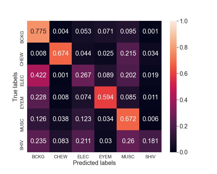
We displayed the results for the multi-class artifact de- the electrode pop artifact attained the poorest BAC. Overall,
tector in Table IV. Additionally, we illustrated the multi- better results for eye movement artifacts are obtained at
class confusion matrix in Figure 5. We split the classification shorter window lengths, while the other artifacts achieved(a) Window length of 1s. (b) Window length of 3s. (c) Window length of 5s.
Fig. 5. Confusion matrix of the multi-class segment-level classification at a window length of (a) 1s, (b) 3s, and (c) 5s. The background, chewing,
electrode pop, eye movement, muscle, and shiver classes are denoted as BCKG, CHEW, ELEC, EYEM, MUSC, and SHIV, respectively. Note that the
confusion matrix is computed by taking the sum of all the predictions, while the results reported in Table IV are computed by taking the average of the
results obtained from each fold (5-fold CV).
TABLE IV TABLE V
S EGMENT- LEVEL MULTI - CLASS ARTIFACT DETECTION RESULTS . S EGMENT- LEVEL MULTI - CLASS MULTI - LABEL (MCML) ARTIFACT
DETECTION RESULTS .
Artifact Window Multi-Class
Type Length AUC AUPRC ACC BAC SEN SPE
Artifact Window Multi-Class Multi-Label
1 0.799 0.264 0.632 0.710 0.447 0.972 Type Length AUC AUPRC ACC BAC SEN SPE
Chewing 3 0.911 0.470 0.698 0.813 0.636 0.990
5 0.939 0.629 0.725 0.879 0.768 0.990 1 0.909 0.342 0.941 0.799 0.647 0.951
1 0.600 0.098 0.632 0.561 0.203 0.920 Chewing 3 0.905 0.480 0.967 0.846 0.715 0.978
Electrode pop 3 0.645 0.098 0.698 0.586 0.248 0.924 5 0.932 0.612 0.980 0.879 0.770 0.988
5 0.649 0.087 0.725 0.615 0.301 0.929 1 0.690 0.168 0.843 0.610 0.327 0.893
1 0.809 0.428 0.632 0.774 0.614 0.933 Electrode pop 3 0.747 0.199 0.859 0.652 0.410 0.894
Eye movement 3 0.771 0.299 0.698 0.754 0.584 0.924 5 0.732 0.155 0.887 0.632 0.344 0.919
5 0.775 0.258 0.725 0.764 0.596 0.931 1 0.865 0.560 0.803 0.795 0.783 0.808
1 0.779 0.505 0.632 0.722 0.625 0.820 Eye movement 3 0.856 0.398 0.850 0.769 0.666 0.872
Muscle 3 0.745 0.476 0.698 0.770 0.651 0.889 5 0.853 0.345 0.852 0.782 0.698 0.866
5 0.755 0.448 0.725 0.789 0.682 0.895 1 0.833 0.597 0.727 0.780 0.911 0.648
1 0.866 0.020 0.632 0.562 0.124 0.999 Muscle 3 0.859 0.564 0.777 0.811 0.880 0.743
Shiver 3 0.870 0.258 0.698 0.592 0.184 1.000 5 0.870 0.535 0.770 0.811 0.881 0.741
5 0.575 0.007 0.725 0.590 0.181 1.000 1 0.498 0.009 0.997 0.700 0.000 0.999
Shiver 3 0.769 0.021 0.995 0.798 0.000 0.998
5 0.831 0.030 0.998 0.799 0.000 0.999
better results at longer window lengths. TABLE VI
S EGMENT- LEVEL COMBINED BINARY ARTIFACT DETECTION RESULTS .
E. Segment-level Artifact Detection Results: Combined Bi-
nary Window SPE @95% SPE @97% SPE @99%
AUC AUPRC
Length SEN Th SEN Th SEN Th
Finally, we combined the five segment-level artifact de- 1 0.852 0.979 0.436 0.784 0.342 0.834 0.191 0.900
tectors and obtained the results for the combined binary 3 0.897 0.962 0.571 0.780 0.488 0.845 0.300 0.921
5 0.905 0.946 0.604 0.776 0.518 0.852 0.353 0.929
segment-level artifact detector in Table VI. We combined
all the artifact classes into a single class to distinguish
artifacts against backgrounds. At an SPE of 95%, 97%, and
99%, the highest SEN achieved is 0.604, 0.518, and 0.353, measured the effectiveness of their artifact detector indirectly
respectively, at a window length of 3, 3, and 5, respectively. by performing brain-computer interface (BCI) classification
However, we note that this evaluation approach no longer and achieved an ACC improvement from 63% to 72.5%
accounts for the artifact class imbalance; there are much with the artifact rejection module [4]. Mashhadi et al. reject
more muscle artifacts than eye movement artifacts. Hence ocular artifacts by means of U-NET, and reported a mean
the combined binary segment-level detector will be overfitted square error (MSE) of 0.00712 [8]. Meanwhile, Dhindsa
to muscle artifacts. performed artifact classification on an EEG dataset with
four channels and achieved an ACC of 93.3% and AUC of
IV. D ISCUSSION 0.923 [15]. Finally, Pion-Tonachini et al. deployed ICA and
In the following, we compare our results to the literature. CNN/GAN to classify EEG independent components (IC),
Roy performed multi-class artifact classification and reported and achieved BAC of 0.855, 0.623, and 0.597, for 2, 5, and
a SEN of 72.39% for the background class [7]. Abdi et al. de- 7-class classification, respectively [3]. For all scenarios, they
ployed wavelet transform to detect and reject artifacts. They reported SEN of 73% for the background class.Compared to these studies, our system performs better in [10] A. Hamid, K. Gagliano, S. Rahman, N. Tulin, V. Tchiong, I. Obeid,
terms of SPE, as our system reports a high SPE of 95% J. Picone, The temple university artifact corpus: An annotated corpus
of eeg artifacts, in: 2020 IEEE Signal Processing in Medicine and
while achieving decent SEN of 57.1% for the artifact class, Biology Symposium (SPMB), IEEE, 2020, pp. 1–4.
making it suitable for real-world application. In contrast, [11] T. Joo, U. Chung, M.-G. Seo, Being bayesian about categorical
the studies by [7], [4], [8], [3] might be less suitable for probability, in: International Conference on Machine Learning, PMLR,
2020, pp. 4950–4961.
real-world applications which require high SPE to avoid [12] A. Vaswani, N. Shazeer, N. Parmar, J. Uszkoreit, L. Jones, A. N.
rejecting too much clean EEG. The majority of existing Gomez, Ł. Kaiser, I. Polosukhin, Attention is all you need, in:
studies reported low SPE for the clean EEG (less than 75%), Advances in neural information processing systems, 2017, pp. 5998–
6008.
which is unacceptable as it can lead to a significant loss of [13] D. P. Kingma, J. Ba, Adam: A method for stochastic optimization,
valuable EEG information. arXiv preprint arXiv:1412.6980 (2014).
[14] A. V. Dorogush, V. Ershov, A. Gulin, Catboost: gradient boosting with
V. C ONCLUSION categorical features support, arXiv preprint arXiv:1810.11363 (2018).
[15] K. Dhindsa, Filter-bank artifact rejection: High performance real-time
We have proposed a neural system for automated detection single-channel artifact detection for eeg, Biomedical Signal Processing
and Control 38 (2017) 224–235.
of five artifact classes: chewing, electrode pop, eye move-
ment, muscle, and shiver artifacts. The channel-wise detector
consists of a CNN followed by a transformer optimized via a
BM loss. The outputs of the CNN-TRF at multiple channels
are then combined via another classifier for artifact detection
in multi-channel EEG segments. We evaluated the segment-
level detector via binary, multi-class, and multi-class multi-
label classification. Finally, we implemented a non-artifact
versus artifact combined binary classification for a general
artifact rejection system. The proposed system can reject
a substantial fraction of artifacts while only removing a
small fraction of clean EEG, thus potentially improving the
readability of EEG recordings. In future work, the artifact
rejection can be implemented with other EEG abnormality
detectors, such as seizure or slowing detectors, to enhance
the performance of those abnormalities detectors.
R EFERENCES
[1] C.-Y. Chang, S.-H. Hsu, L. Pion-Tonachini, T.-P. Jung, Evaluation of
artifact subspace reconstruction for automatic eeg artifact removal, in:
2018 40th Annual International Conference of the IEEE Engineering
in Medicine and Biology Society (EMBC), IEEE, 2018, pp. 1242–
1245.
[2] X. Jiang, G.-B. Bian, Z. Tian, Removal of artifacts from eeg signals:
a review, Sensors 19 (5) (2019) 987.
[3] L. Pion-Tonachini, K. Kreutz-Delgado, S. Makeig, Iclabel: An au-
tomated electroencephalographic independent component classifier,
dataset, and website, NeuroImage 198 (2019) 181–197.
[4] B. Abdi-Sargezeh, R. Foodeh, V. Shalchyan, M. R. Daliri, Eeg
artifact rejection by extracting spatial and spatio-spectral common
components, Journal of Neuroscience Methods 358 (2021) 109182.
[5] J. Thomas, P. Thangavel, W. Y. Peh, J. Jing, R. Yuvaraj, S. S.
Cash, R. Chaudhari, S. Karia, R. Rathakrishnan, V. Saini, et al.,
Automated adult epilepsy diagnostic tool based on interictal scalp
electroencephalogram characteristics: A six-center study, International
Journal of Neural Systems (2021) 2050074.
[6] P. Thangavel, J. Thomas, W. Y. Peh, J. Jing, R. Yuvaraj, S. S. Cash,
R. Chaudhari, S. Karia, R. Rathakrishnan, V. Saini, et al., Time–
frequency decomposition of scalp electroencephalograms improves
deep learning-based epilepsy diagnosis, International Journal of Neural
Systems (2021) 2150032.
[7] S. Roy, Machine learning for removing eeg artifacts: Setting the
benchmark, arXiv preprint arXiv:1903.07825 (2019).
[8] N. Mashhadi, A. Z. Khuzani, M. Heidari, D. Khaledyan, Deep learning
denoising for eog artifacts removal from eeg signals, in: 2020 IEEE
Global Humanitarian Technology Conference (GHTC), IEEE, 2020,
pp. 1–6.
[9] W. Y. Peh, J. Thomas, E. Bagheri, R. Chaudhari, S. Karia, R. Rathakr-
ishnan, V. Saini, N. Shah, R. Srivastava, Y.-L. Tan, et al., Multi-center
validation study of automated classification of pathological slowing in
adult scalp electroencephalograms via frequency features, International
Journal of Neural Systems (2021) 2150016.You can also read