TUBERCULOSIS DIAGNOSTICS - DIGITAL CHEST RADIOGRAPHY AND COMPUTER-AIDED DETECTION (CAD) SOLUTIONS FOR - FindDx.org
←
→
Page content transcription
If your browser does not render page correctly, please read the page content below
D I G I TAL C HE ST RADIOGRAPH Y
A N D C OMPUTE R- AIDED DETECTION
( CA D) SOLUTI ON S F O R

TUBERCULOSIS
DIAGNOSTICS
TEC HN OLOGY LAN D S CA P E A N A LYS I S

                                            FI ND 2021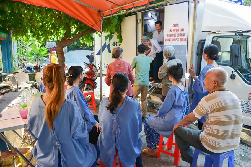
TA B L E O F
CONTENTS
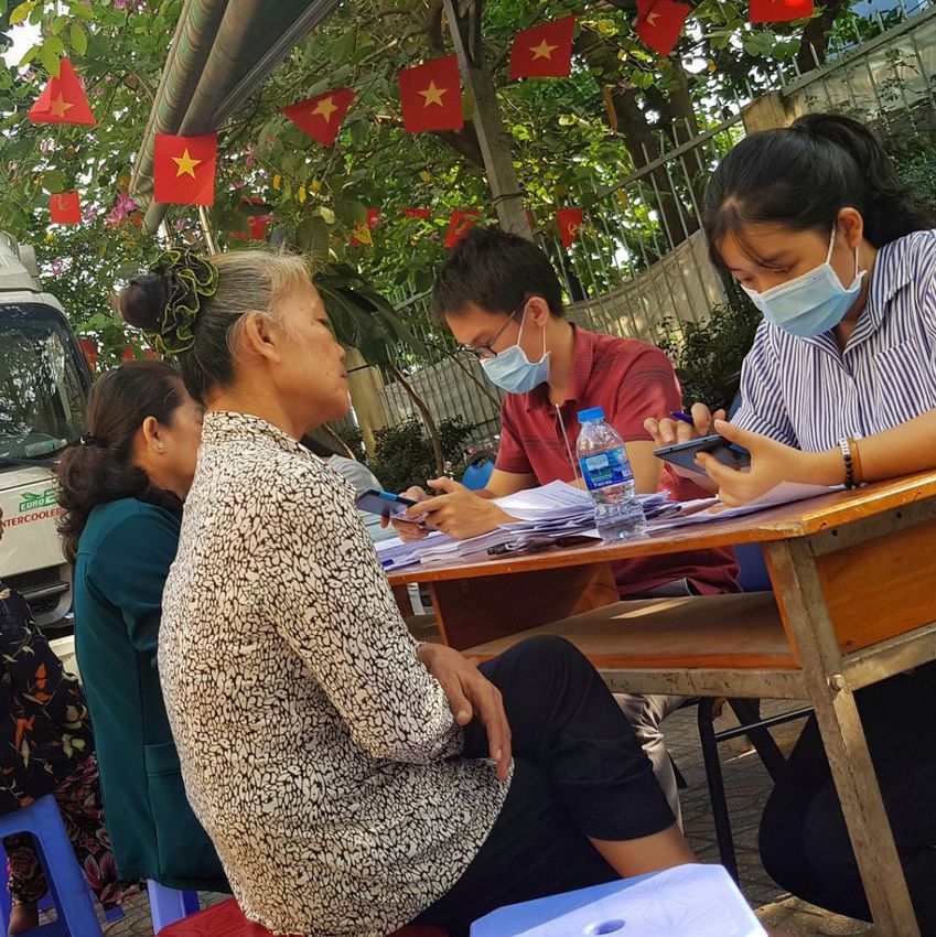
Patient registration at screening
campaign in Viet Nam.
(Image credit: Friends for
International TB Relief
(FIT), Viet Nam)
2
ACKNOWLEDGEMENT, CONFLICT OF INTEREST DECLARATION, DISCLAIMER AND COPYRIGHT                   04
ACRONYMS                                                                                      06
EXECUTIVE SUMMARY                                                                             07

CHAPTER 1: INTRODUCTION                                                                       08
     BACKGROUND                                                                               08
     OBJECTIVES                                                                               09
     METHODOLOGY                                                                              10

CHAPTER 2: OVERVIEW OF CXR TECHNOLOGY                                                         11
     CXR IMAGING                                                                              11
     DIGITAL RADIOGRAPHY                                                                      12
     ANALOG RETROFIT DIGITAL RADIOGRAPHY                                                      13
     COMPUTED RADIOGRAPHY                                                                     14
     ANALOG RADIOGRAPHY                                                                       15

CHAPTER 3: RECENT ADVANCES IN DIGITAL RADIOGRAPHY                                             18
     DETECTOR                                                                                 18
     INNOVATION, R&D IN DETECTOR TECHNOLOGY                                                   20
     DUAL ENERGY X-RAY DETECTORS                                                              20

CHAPTER 4: X-RAY EQUIPMENT CATEGORIZATION FOR TB PROGRAMMES                                   21
     CATEGORY 1 – STATIONARY X-RAY EQUIPMENT                                                  24
     CATEGORY 2 – PORTABLE X-RAY EQUIPMENT                                                    25
     CATEGORY 3 – ULTRA-PORTABLE X-RAY EQUIPMENT                                              26
     CONSIDERATIONS FOR X-RAY EQUIPMENT SELECTION FOR TB PROGRAMMES                           30

CHAPTER 5: OVERVIEW OF AI-POWERED CAD SOLUTIONS FOR TB                                        32
     COMMERCIALLY AVAILABLE CAD SOLUTIONS FOR TB                                              33

CHAPTER 6: GETTING SET UP FOR CAD AND DIGITAL RADIOLOGY                                       36

CHAPTER 7: REGULATORY CONSIDERATIONS FOR IMPLEMENTING CAD AND PORTABLE CXR                    40
     REGULATORY CONSIDERATIONS FOR IONIZING RADIATION EQUIPMENT                               40
     IN NON-HOSPITAL ENVIRONMENTS
     REGULATORY CONSIDERATIONS FOR USE OF CAD                                                 41

CHAPTER 8: EARLY ADOPTER EXPERIENCE WITH CXR CAD IMPLEMENTATION FOR TB DIAGNOSIS              42
     IMPLEMENTATION EXPERIENCE OF PORTABLE X-RAY SYSTEM AND CAD TECHNOLOGY FOR TB             42
     DIAGNOSTICS IN MALAWI
     IMPLEMENTATION EXPERIENCE OF ULTRA-PORTABLE X-RAY SYSTEM AND CAD TECHNOLOGY              45
     FOR TB DIAGNOSTICS IN UGANDA
     IMPLEMENTATION EXPERIENCE OF ULTRA-PORTABLE X-RAY SYSTEM AND CAD TECHNOLOGY              48
     FOR TB AND COVID-19 SCREENING IN ZAMBIA

ANNEX 1                                                                                       50
     COMPLETE RANGE OF STATIONARY (FACILITY-BASED) X-RAY EQUIPMENT FOR CXR IMAGING FOR TB     50
     PROGRAMMES (SPECIFICATIONS AS PER WHO CRITERIA FOR STATIONARY DIGITAL X-RAY EQUIPMENT)

ANNEX 2                                                                                       52
     PRODUCT COMPARISON SHEET: FULL RANGE OF AVAILABLE PRODUCTS IN THE MARKET                 52
     FOR PORTABLE AND ULTRA-PORTABLE X-RAY EQUIPMENT

REFERENCES                                                                                    54

                                                    3
ACKNOWLEDGEMENT, CONFLICT OF INTEREST
DECLARATION, DISCLAIMER AND COPYRIGHT

       This report has been prepared by Yogesh Jha, Consultant for the
       Foundation for Innovative New Diagnostics (FIND), with contributions
       from Dr Morten Ruhwald, Head of Tuberculosis at FIND and Dr Sandra
       Kik, Consultant and Project manager of CAD-related projects at FIND.

       The information covered in this report is based on work done by FIND, the
       World Health Organization and Stop TB Partnership. It is also based on
       extensive research and analysis of publicly available information provided
       by the X-ray equipment and CAD industries; published and unpublished
       literature; interviews with manufacturers and CAD developers; and the
       experience of TB programme implementers around the world. None of
       the members of the FIND team or consultants involved in the preparation
       of this report have any commercial links or receive incentives from any of
       the radiology equipment manufacturers or CAD developers mentioned in
       the report.

       The authors express their gratitude to all the companies and product
       developers who contributed to this report.

       We are also grateful to the TB programme implementers in Malawi,
       Zambia, and Uganda (Peter MacPherson, Rachael Burke, Dr Aldo Burua,
       Moses Eyaru, Dr Turyahabwe Stavia, Tila T.M. Mainga and Christabel
       Nkonde) for sharing their experiences using the technology described in
       this report.

       And finally, we would like to thank WHO and the Stop TB Partnership
       for participating in early discussions on this topic, and in particular the
       following individuals for their review of the prefinal version of the report:
       Arlene Chua (Médecins Sans Frontières), Andrew Codlin (Friends for
       International TB Relief), Jacob Creswell (Stop TB Partnership), Iona
       Crumley (Médecins Sans Frontières), ThuVan Dinh (Clinton Health Access
       Initiative), Sifrash Gelaw (International Organization for Migration), Wayne
       Van Gemert and Zhi Zhen Qin (Stop TB Partnership), Beatrice Gordis
       (FIND), Kekeletso Kao (FIND), Fransesco Ribolzi and Dennis Falzon
       (WHO), Peter MacPherson (Liverpool School of Tropical Medicine and
       Malawi-Liverpool-Wellcome Trust Clinical Research Programme), Claudia
       Denkinger (Heidelberg University Hospital), Kinz-ul-Eman (DOPASI
       Foundation), Madhukar Pai (McGill International TB Center), and Lindsay
       Palazuelos (Partners in Health).

                                             FIND Radiology Landscape Project Team

                                            4
Chest X-ray screening
in Viet Nam.
(Image credit: Friends for
International TB Relief
(FIT), Viet Nam)

        COPYRIGHT NOTICE: This work is subject to a Creative Commons Public License BY-SA 4.0 (https://
        creativecommons.org/licenses/by/4.0/ August 2019).

        DISCLAIMER: The published material is being distributed without warranty of any kind, either expressed
        or implied. The responsibility for the interpretation and use of the material lies with the reader. In no event
        shall FIND be liable for damage arising from its use by a third party.

        NO ENDORSEMENT OR RECOMMENDATION: The mention of specific companies or of certain
        manufacturers’ products does not imply that they are endorsed or recommended by FIND in preference to
        others of a similar nature that are not mentioned.

        FUNDING: This work was supported by the Netherlands Enterprise Agency.

                                                              5
AC:         Alternating current

               AEC:        Automatic exposure control

               AI:         Artificial intelligence

               AUC:        Area under curve

               AP:         Anterior posterior

AC RON Y M S   API:        Application programming interface

               APR:        Anatomically programmed radiography

               CAD:        Computer-aided design

               CE:         Conformité européenne (EU; standard)

               CR:         Computed radiography

               CXR:        Chest X-ray

               DC:         Direct current

               DICOM: Digital Imaging and Communications in Medicine

               DR:         Digital radiography

               DQE:        Detective quantum efficiency
               FDA:        Food and Drug Administration (USA; standard)

               FIND:       Foundation for Innovative New Diagnostics

               FPD:        Flat panel detector

               FS:         Focal spot

               GDF:        Global Drug Facility

               HF:         High frequency (X-ray generator)

               HU:         Heat unit (Anode)

               IAEA:       International Atomic Energy Agency

               ICU:        Intensive care unit

               ICT:        Information and communications technology

               Kg:         Kilogram

               kV:         Kilo Volt

               kW:         Kilo Watt

               LMIC:       Low- and middle-income country

               mA:         milli Ampere (filament current)

               mAs:        milli Ampere seconds

               mm AL: millimetre equivalent Aluminium (filtration of X-ray beam)

               PACS:       Picture archiving and communication system

               PTB:        Pulmonary tuberculosis
               R&D:        Research and development

               ROC:        Receiver operating characteristic

               TB:         Tuberculosis

               WHO:        World Health Organization

               VAC:        Volt AC (Input power supply)

                       6
EXECUTIVE SUMMARY

Tuberculosis (TB) remains one of the leading causes of mortality
worldwide 139 years after the discovery of Mycobacterium
tuberculosis, the bacteria that causes TB. Although TB is
a treatable disease, effective screening and diagnosis
remain a challenge due to varied presentations of the
disease and its long incubation period. An estimated 2.9
million of the 10 million people who were infected with
TB in 2019 were either not diagnosed or not reported                      solutions to interpret CXRs for signs of TB. Although
to the World Health Organization (WHO) (1). There is                      several studies have reported equivalent performance
an urgent need to effectively deploy evidence-based,                      by CAD compared to human readers, only recently has
innovative, and highly effective screening strategies                     WHO recommended the use of CAD as an alternative to
for TB to reach the ambitious target of diagnosing and                    human readers to interpret CXR for screening and triage
treating 40 million people from 2018 to 2022, as per                      of PTB in individuals aged 15 or over (3). Consequently,
the Political Declaration adopted at the United Nations                   there is growing interest from TB programmes across
General Assembly in September 2018 (1).                                   the world to use a combination of portable digital X-ray
                                                                          systems together with CAD solutions for effective TB
WHO recommends the use of chest X-ray radiography (CXR) as                screening.
an effective screening test for pulmonary tuberculosis (PTB),
as well as an aid in the diagnostic pathway to complement                 There are numerous challenges in implementing TB screening
bacteriological tests (2). However, there are two main                    using digital CXR and CAD technology in rural and urban
barriers to the uptake of CXR technology for TB: limited                  communities in high TB burden countries. TB programme
access to high diagnostic quality digital CXR imaging                     implementers in low- and middle-income countries
in low resource settings and a shortage of trained                        (LMICs) frequently encounter problems with selecting
readers (usually physician radiologists) to interpret                     the right type of X-ray equipment and CAD solution, and
the images. Recent advances in digital imaging and                        often face practical hurdles with set-up and workflow.
software technologies have led to the development                         Also, the market of X-ray equipment technology and
of highly compact, portable, battery-operated digital                     CAD solution for TB is evolving dynamically, with a
radiography equipment and artificial intelligence (AI)-                   significant number of equipment manufacturers and
based computer-aided detection (CAD) software                             CAD developers now entering the market.

This report

FIND published the first technology landscape report titled Digital       manufacturers interested in producing highly compact, portable
radiology solutions for TB diagnostics in low- and middle-income          battery-operated digital X-ray products. Similarly, there has been a
countries in 2015 (4) to explore the market status of radiology           significant increase in the number of CAD solutions with applicability
equipment and CAD solutions for TB screening applicability. The           for TB screening, of which many have been CE certified.
report found very limited choice for facility-based, dedicated CXR
imaging equipment and only one commercially available CAD                 This updated report by FIND aims to provide a comprehensive
solution.                                                                 overview of digital X-ray and CAD technologies for TB
                                                                          diagnosis that are currently available in market. Additionally,
Coming up to 2021, the market of digital X-ray technology has             the report captures the implementation experiences of some early
witnessed radical transformations in design and features, with            adopters of these technologies in high TB burden countries.

                                                                      7
CHAPTER 1 – INTRODUCTION

BAC K GROU ND                                                         Although CXR is the most commonly performed radiography
                                                                      worldwide (11), it can be challenging to produce a high-quality
Chest X-ray radiography (CXR) is one of the most sensitive            diagnostic image because of the technical limitations of
tests for detecting pulmonary tuberculosis (PTB) (5-7) and            the equipment and image processing, and the lack
is often used as a screening test or diagnostic tool                  of qualified radiographers across LMICs. Maintaining
where PTB cannot be ruled out microbiologically.                      diagnostic quality of CXR images over time (crucial for
This includes patients for whom bacteriological tests                 detecting subtle chest pathologies) is also challenging
tend to have lower sensitivity, such as people living                 when having to use traditional X-ray equipment with
with HIV or people with other immune-compromising                     poor service and maintenance. With increasing access
conditions, as well as patients for whom it is difficult to           to digital X-ray imaging technologies, it is gradually
obtain samples for bacteriological confirmation, such                 becoming easier to produce better diagnostic quality
as children or patients whose immune systems are                      CXRs. The introduction of AI-based computer-aided
severly compromised. Sometimes bacteriological tests                  detection (CAD) solutions for interpreting CXRs has
are not available, are contaminated, or even negative                 addressed not only the intra and inter reader variability
yet CXR findings are nevertheless suggestive of TB.                   among radiologists but also the challenges faced when
                                                                      they are not available.
The role of CXR in improving TB notification in high prevalence
settings has been established (8). While CXR is usually               Most of the global market for digital X-ray equipment is
conducted in presumptive or symptomatic TB cases, a                   targeted at industrialized countries and privatized health
subsequent analysis of TB prevalence surveys showed                   service providers in emerging economies, whereas in
that between 36-80% of bacteriologically-confirmed TB                 LMICs this equipment is used for general radiography
cases have a negative symptom screen. CXR may detect                  purposes, due to the high cost of digital radiography.
89% of individuals with bacteriologically-confirmed TB                With innovations in design and technology, several
disease and therefore should be considered to be done                 equipment manufacturers have been able to
in parallel to symptoms screening in order to find more               significantly reduce the size of X-ray equipment, some
of the missing TB patients (7). Recent studies in TB high             of which even weighs less than 5 kg and fitting easily
burden countries have revalidated the role of CXRs in                 in a backpack. These highly portable X-ray generators
active case finding and present a strong argument why                 are powered by rechargeable batteries and when used
TB programmes need to focus more on community-                        in combination with modern, ultra-sensitive detectors
based TB screening programmes (9, 10). This is even                   can produce decent diagnostic-quality CXR images at
more important due to the significant number of people                improved radiation-dose efficiency. Resultant digital
who aren’t screened and therefore missed, not to                      images can immediately be run through highly sensitive
mention the additional complications arising from the                 and reasonably specific, dedicated CAD solutions to
COVID-19 pandemic.                                                    screen for signs of TB or other radiographic findings
                                                                      and results can be obtained at point of care in under
                                                                      a minute.

                                                                      With a package combining portable or ultra-portable digital
                                                                      X-ray and an offline CAD solution, it has been possible to
                                                                      bring TB screening into communities in remote locations
                                                                      where healthcare facilities are either nonexistent or
                                                                      often have very limited essential infrastructure, like
                                                                      reliable electricity and internet.

                                                                  8
Although CAD technology has been around for more than
a decade, the uptake by TB programmes in LMICs remains
limited (12). This has been partly due to technical,
operational, industry and policy-related factors but
also to the lack of independent evidence on the
performance of CAD solutions in clinical settings (2,
13, 14). However, in recent years, several independent
evaluations of CE-certified commercially available CAD
solutions have demonstrated that the accuracy of these
products is comparable or sometimes even superior to
experienced, certified physician radiologist readings
(15, 16). Based on the evidence, WHO released new
guidelines which stipulate that CAD may be used as
an alternative to human reader interpretation of digital
CXR for TB screening and triage, but that its use should
be limited to interpreting CXR for pulmonary TB in
individuals aged 15 years or more (17, 18). As a result,
uptake of CAD is expected to increase, bringing new
opportunities for industry to market digital CXR with
CAD technology for TB, and for TB programmes across
the world to explore the added value of this innovative
diagnostic approach in the fight against this highly
infectious disease.

     OBJ E C T I V E S
     This report aims to provide readers with a compre-                 The report also attempts to summarize existing
     hensive understanding of various technologies used to              commercially available, certified CAD solutions for
     produce CXRs, commercially available CAD solutions aren’t          TB screening using CXR and highlights important
     screened and therefore missed, not to mention the additional       considerations for choosing a particular product, which are
     complications arising from the COVID-19 pandemic for               discussed in Chapter 5 and Chapter 6, respectively.
     TB screening and practical considerations that may apply
     during the implementation of CXR and CAD technology in the         Lastly, the report briefly discusses regulatory
     context of TB programmes.                                          considerations for implementing CXR-CAD technology
                                                                        (Chapter 7) and aims to provide end-user information for
     In Chapter 2, we give an overview of the different X-ray           X-ray equipment manufacturers and CAD developers by
     technologies that exist, whereas in Chapter 3 we describe          showcasing several early adopter experiences (Chapter 8).
     the latest developments in diagnostic X-ray imaging. In
     Chapter 4, we categorize digital X-ray equipment to support        The intended audience for this report includes
     CXR imaging for TB programmes based on the setting and             governmental as well as non-governmental organizations,
     intended use-case. Given the role CXR plays in detecting           and researchers in the TB field with an interest in scaling
     active TB cases in community-based screening, this report          up TB screening through the use of digital CXR and CAD
     particularly focuses on portable and ultra-portable                technology.
     digital X-ray equipment from leading manufacturers and
     outlines their standard specifications in Annex 2 to support
     decision makers in making equipment selection.

                                                                    9
ME T HOD OL OG Y
This landscape analysis is based on an extensive review of information from industry, interviews with manufacturers and TB
programme implementers in LMICs, a review of the scientific literature, and information searches on the internet.

     Digital X-ray equipment
     An internet-based search was conducted to find                      Given the wide range of X-ray equipment available in the
     manufacturers of portable and ultra-portable digital                portable and ultra-portable category, we included field-
     X-ray equipment with potential applicability for TB                 tested models within TB programmes and/or those that
     programmes in LMICs. Twenty-one X-ray manufacturers                 had been certified (CE/FDA, including those in process of
     were identified and contacted based on information from             obtaining certification). We focused the internet search on
     various sources and contacts from previous work done by             products available for sale mostly in the European Union and
     FIND after reviewing publicly available information about           in North America for inclusion in the product comparison
     the products. Manufacturers were asked to share detailed            sheet (Annex 2), as the products available in these markets
     information and specifications about their product(s) using         are more likely to have received CE/FDA certification.
     a standard specification template. Seven X-ray equipment
     manufacturers with experience or interest in CXR application
     for TB programmes were interviewed regarding their product
     specifications and market experience.

     CAD solution for TB                                                        TB programme
                                                                                implementer experience
     Information on the status of CAD solutions was
     gathered from review of previous work carried                              Interviews were conducted with implementers
     out by FIND in collaboration with the Stop TB                              in Malawi, Zambia, and Uganda to explore
     Partnership (www.ai4hlth.org), literature reviews                          and document their practical experience with
     and interactions with leading CAD developers with                          implementing digital portable or ultra-portable X-ray
     CE-certified commercial products. Eight commer-                            equipment in combination with CAD for TB screening.
     cially available CAD developers were contacted and
     asked to share qualitative information regarding
     market status and implementation challenges
     associated with CAD solutions for TB, of which four
     CE-certified CAD developers complied.

                                                                    10
CHAPTER 2 – OVERVIEW OF CXR TECHNOLOGY
CXR I M AGI NG
Applicability of X-ray equipment within the scope of TB The second step involves processing the latent image into
programmes is somewhat limited for acquiring CXRs. It is a radiograph with multiple shades of grey. The use of a
important to appreciate that all types of X-ray equipment conventional image receptor (X-ray film) requires wet
can be used to take CXRs and that no X-ray equipment processing technology and produces an analog image,
manufacturer makes X-ray equipment exclusively for whereas both the phosphor plate (CR technology-
chest images. This segment of the report attempts based) and the flat panel detector (DR technology-
to briefly explain various types of X-ray imaging and based) image receptors produce digital images.
processing technologies that are currently being used The only difference between CR and DR is that CR
to produce CXRs. technology requires an additional processing device to
scan the exposed phosphor plate (often called digitizer
CXR imaging is a two-step process. The first step involves or CR reader), whereas no additional processing device
image acquisition, in which X-ray beams (electromagnetic is needed for the DR image receptor, as the processing
radiation) produced from a source (X-ray generator) happens instantly. Final output digital images are
penetrate the patient’s thorax and sensitize the image in DICOM (Digital Imaging and Communications in
receptor. The receptor can be a digital flat panel Medicine) or JPEG file formats, allowing for electronic
detector (digital radiography (DR)), a reusable phosphor viewing, archiving and sharing using standardized
plate (computed radiography (CR)), or a conventional PACS (Picture Archiving and Communication System)
X-ray film (analog radiography), forming a so-called and teleradiology services (19).
latent image.
People waiting to get screened in the mobile X-ray truck in Viet Nam.
(Image credit: Friends for International TB Relief (FIT), Viet Nam)
11DI GI TAL R A D I O G R A P H Y
Digital radiography (DR), also known as complete digital a calibrated X-ray generator, a sensitive detector and
radiography or direct digital radiography, is the latest electronic workstation with the required software, including
development in X-ray technology that makes use of anatomically programmed radiography (APR), a series of
different types of solid-state detectors (also called flat-panel anatomy-specific predetermined settings, which reduces
detectors or simply detectors). These are made of glittering manual steps for the radiographer during the process of
materials like cesium iodide, or selenium crystals, which image acquisition. All the benefits of digital images such
act as image receptors, that are then activated by an X-ray as archiving, sharing via local PACS network for on-site
generator specifically calibrated to produce a digital image reporting and the option to include CAD screening or
with excellent contrast and resolution. Once the image is teleradiology can be efficiently performed with DR. However,
formed, it is instantaneously transmitted to the workstation considerable capital investment is needed due to the cost
monitor/device and the detector can be used for subsequent of the detector and calibrated X-ray generator. Furthermore,
exposures without the need for preparation or erasing the detectors are very fragile and need to be handled with
before a next exam. The current generation of detectors are care during radiography and/or transportation as even minor
compact, lightweight, and come in various sizes including mechanical damage can mean total malfunction, large
portable battery-operated versions able to wirelessly financial implications, and disruption in services.
transmit digital images in real time to PACS.
A less expensive model for a DR set-up can be achieved
DR is increasingly becoming the preferred modality when an already existing analogue X-ray machine (X-ray
of radiographic imaging by reducing steps involved in generator) is in place. These systems can be retrofitted
image processing, hence, delivering high throughput with with a new DR detector and image acquisition software,
excellent image characteristics (contrast and resolution) thereby transforming an analog system into a DR system.
and a better radiation-dose efficiency. A modern DR set-up This is referred to as analog retrofit digital radiography and
usually comes as an integrated package comprised of is discussed below.
A B
C
Figure 1
Complete DR X-ray Equipments. (A) MinXray Impact
portable Digital X-ray system with transportation kit,
(B) Europa AirTouch battery-operated handheld digital
X-ray from Aspen Imaging, (C) Stationary U arm digital
X-ray system from Swissray.
(Image credit: MinXray, Aspen Imaging and Swissray)
12A
C
B
Figure 2
An analog X-ray machine (A), a wireless detector (B) and a monitor with image viewing software (C)
(Image credit: Maya, Cannon and Barco)
ANA L OG RE T R O F I T D I G I TA L R A D IO G R A PH Y
Analog retrofit DR-based imaging systems make use of Usually, analog retrofit DR set-up is done by imaging service
existing analog X-ray equipment and are retrofitted with a flat providers with the intention to enhance diagnostic image
panel detector to produce high-quality digital radiographs. An quality, augment workflow, increase efficiency and throughput
analog retrofitted digital system delivers a similar image at hospitals or busy imaging facilities. There is a significant
quality as a new DR system but may be less radiation cost-saving component when making use of existing,
dose efficient since the existing X-ray generator might traditional X-ray equipment, although these systems
not be properly calibrated to the new highly sensitive are usually heavier, bulkier and require a stable power
digital detectors. Radiation dose efficiency can be source to operate. There is also a significant market for
improved by using lower exposure parameters and is portable analog retrofit DR equipment; this means that
often more efficient than computed radiography (CR) existing analog portable X-ray equipment can be used
and/or analog X-ray equipment, which are discussed with a modern flat panel detector for offsite radiography.
later in this chapter. Although image acquisition and The long-term economic considerations linked to the
post-processing software are usually provided by lifespan of the equipment, maintenance cost and type
detector manufacturers while retrofitting the existing of detectors being considered for retrofitting, cost of
equipment, it is often difficult to integrate APR for image viewing software, type of use (at facility or offsite)
CXR acquisition, hence manual selection of exposure etc. need to be considered by TB programmes before
parameters may have to be done by the radiographer. deciding to invest in an analog retrofit DR set-up.
13A C
B
Figure 3
An analog X-ray equipment (A) with
phosphor plate cassettes (B) and a CR
reader with viewing monitor (C)
(Image credit: Maya and Fujifilm)
COM PU T ED R A D I O G R A P H Y
Computed radiography (CR) systems are a combination process several cassettes at a time; the average size of a CR
of analog X-ray equipment for taking the image reader is like a moderate-sized photocopy machine which
combined with reusable photostimulable phosphor has the capacity to process one cassette at a time and there
(usually referred to as a phosphor plate) technology for are larger heavy-duty models with capacity to process up to
image processing. Once the X-ray exposure is made, the five cassettes (or more) at a time. The number of phosphor
exposed cassette containing the phosphor plate is placed plates (cassettes) required at imaging site depends on the
inside a special scanner/reader (usually referred to as CR volume of examinations, usually two would suffice for a small
reader or digitizer) that converts the latent image into a workload environment, however, multiple CR plates might be
digital image (DICOM or JPEG) as output. The image on the needed at a significantly busy radiography site where more
phosphor plate is erased during the processing phase and people need to be X-rayed in less time. Additionally, specific
therefore the phosphor plate is immediately available for operational training of the radiographer on the proper use
the next exposure after the processing is completed. The and maintenance of the CR reader, care, and maintenance of
entire cycle of image acquisition up to image processing phosphor plates (which are susceptible to creating artifacts
usually takes about 2 to 3 minutes (depending upon make on images if not cleaned periodically) is needed from quality
and model). Since the output image is in DICOM or JPEG assurance perspectives. Regarding the life of phosphor
format, all the image characteristics and benefits associated plates, there is quite a variation in terms of manufacturers
with DR-based imaging are also applicable to CR-based recommendations to replace them every 5 years, after a
imaging, only at the expense of increased radiation dose certain number of exposures (usually in thousands) or visible
and reduced throughput compared to DR-based systems degradation in image quality. CR remains the most common
(image processing time is usually < 5 sec for DR systems). form of ‘digital’ imaging system in most LMICs because of
Radiation dose efficiency can be improved to match DR- the robustness and relatively low initial investment cost
based imaging by careful selection of exposure parameters compared to a complete DR equipment or analog retrofit
depending upon different body habitus of patients. DR set-up. While in majority of circumstances CR-based
imaging is seen at hospitals and imaging clinics, it can also
The CR reader/digitizer is a separate piece of equipment be implemented for offsite imaging using analog portable
that needs to be installed in addition to the X-ray X-ray generator, however this usually presents with logistical
generator to scan the cassettes containing the phosphor challenges with transportation, more space requirement,
plate after each exposure. There is a large variation in terms steady power supply and increased susceptibility to
of size of the CR reader and cost based on the capacity to mechanical damage.
14ANA L OG RAD I O G R A P H Y
Analog radiography is the traditional method of X-ray imaging The output is an analog image (X-ray film), which in its
and is often referred to as film-based radiography. It is still original form cannot be archived digitally in a PACS or used
in use in many low resource, high TB burden settings, directly as an input file for CAD solutions, so requires a
especially at rural healthcare facilities. Analog X-ray highly experienced radiologist to interpret the image.
equipment generates X-rays combined with X-ray However, if the output image can be digitized using a
films (made of silver halide crystals and loaded inside good digital camera or smartphone, it can be transmitted
X-ray cassettes) which serve as the image receptor. electronically to a remote telemedicine service staffed
The exposed X-ray film requires a wet processing by consultant radiologists for reporting. Some CAD
mechanism (consisting of developing and fixing the developers accept non-DICOM images such as JPEG,
image using chemicals) either done manually in a PNG and even low-resolution smartphone-acquired
dark room or using an automatic processor. The images of the X-ray film placed on a lightbox. However,
dependence on a dark room-based process involving there might be concerns with the diagnostic quality
chemicals, complex accessories like X-ray cassettes, of the original image and thus far no scientific studies
and extra human resources, not to mention the difficulty have reported the performance of CAD solutions using
of maintaining quality assurance parameters are all digitized images of manual X-ray films.
factors that limit the wide-scale use of this method for
fast and effective TB screening at the population level.
A B
C
Figure 4
Equipment and steps involved in analog
radiography. (A) Analog X-ray equipment,
(B) manual processing of X-ray in dark
room, (C) a reader interpreting a CXR film
(Image credit: MAYA and Science Source Images)
15Table 1. Comparative analysis of all CXR imaging technologies
COMPLETE DR-BASED RETROFIT DR-BASED
CXR IMAGING CXR IMAGING
(NEW X-RAY GENERATOR (EXISTING ANALOG X-RAY GENERATOR
+ DETECTOR PACKAGE) + NEW DETECTOR)
Combination package of a calibrated X-ray generator Analog X-ray generator and retrofit DR panel
TECHNOLOGY and DR panel (no additional processing technology (no additional processing technology required).
required). Existing X-ray generator can be used.
Available for facility-based or offsite portable Can be used for facility-based or offsite portable
application (portable models are highly compact and application (some logistical limitations may apply in
EQUIPMENT
present with significantly less logistical limitations terms of portable applications).
MODELS compared to analog retrofit DR- and CR-based
equipment).
Digital image (usually DICOM). Digital image (usually DICOM).
OUTPUT IMAGE, PACS compatible, can run CAD. PACS compatible, can run CAD.
PACS AND CAD
IMPLEMENTATION
Image parameters (contrast, resolution) are Image parameters (contrast, resolution) are
comparable to or sometimes slightly superior to comparable to complete DR equipment and
CR-based imaging or retrofit DR-based imaging. CR-based imaging.
IMAGE Least manual steps in terms of achieving highest More manual steps compared to complete DR
CHARACTERISTICS possible image parameters. equipment to achieve highest possible image quality.
Image is less susceptible to artifacts compared to Image is less susceptible to artifacts compared to CR
CR and analog imaging. and analog imaging.
Excellent throughput, modern equipment has less Excellent throughput, slightly more image processing
THROUGHPUT than 5 seconds of image generation time. time compared to complete DR equipment.
Highly dose efficient. Modern equipment uses Better dose efficiency compared to CR-based
RADIATION-DOSE sensitive detectors and X-ray generators calibrated equipment or analog equipment, slightly less dose
EFFICIENCY to specific detector. efficient compared to complete DR equipment.
Detectors are highly susceptible to mechanical Detectors are highly susceptible to mechanical
SERVICE AND damage. damage.
MAINTENANCE Usually, replacement needed in case of damage. Usually, replacement needed in case of damage.
Initial investment: More expensive compared to CR, less expensive
(Additional insurance cost might be needed for compared to complete DR as there is cost saving in
detectors.) X-ray generator component.
Additional insurance cost might be needed for
COST Estimated price range of complete package of new detectors.
equipment set-up (X-ray generator, detector, viewing
software, essential accessories): Estimated price range of detector, viewing
Facility-based US$ 125,000 to US$ 150,000 software (excludes X-ray generator cost):
Portable US$ 40,000 to US$ 100,000 US$ 20,000 to US$ 100,000 (or higher).
Ultra-portable US$ 40,000 to US$ 70,000.
16CR-BASED CXR IMAGING ANALOG CXR IMAGING
(EXISTING ANALOG X-RAY GENERATOR (EXISTING ANALOG X-RAY GENERATOR
+ CR READER) + MANUAL IMAGE PROCESSING)
Analog X-ray generator and reusable phosphor plates Analog X-ray generator and accessories (X-ray films,
TECHNOLOGY (with CR reader / digitizer for image processing is cassettes with screens) along with darkroom-based wet
required). Existing X-ray generator can be used. processing mechanism / automatic chemical processing.
Can be used for facility-based or portable application Mostly used at facility-based imaging, very difficult to
(significant logistical limitations for portable application implement for offsite or portable application due to
EQUIPMENT
compared to DR-based systems due to requirement of manual image processing requirements.
MODELS additional processing equipment and multiple
phosphor plates depending on workload).
Digital image (usually DICOM). Analog image, X-ray film. Not compatible with PACS
PACS compatible, can run CAD. and CAD unless the original output image is digitized
OUTPUT IMAGE,
using a digital camera or smartphone (JPEG or PNG file
PACS AND CAD
formats only). Narrow choice of CAD solutions as digital
IMPLEMENTATION image is in non-DICOM format. Reduced diagnostic
quality of digital image.
Image parameters (contrast, resolution) are usually Image parameters (contrast, resolution) are
comparable to complete DR and analog retrofit DR significantly inferior as compared to digital images
equipment. produced by DR-based and CR-based equipment.
More manual steps compared to complete DR or Careful selection of exposure parameters and heavy
IMAGE
analog retrofit DR equipment to achieve highest dependency on processing conditions to maintain
CHARACTERISTICS possible Image quality. diagnostic image quality.
Image is more susceptible to artifacts as compared to Image is more susceptible to artifacts as compared
DR-based imaging. Phosphor plates need to be to DR and CR-based imaging.
cleaned periodically.
Moderate throughput, usually 2 to 3 minutes of image Low throughput, huge variation in terms of image
THROUGHPUT processing cycle by CR scanner. processing time depending upon choice of dark
room-based processing or automatic processing.
Less dose efficient compared to DR-based equipment, Less dose efficient compared to CR and DR X-ray
RADIATION-DOSE dose efficiency can be improved by careful selection of equipment.
EFFICIENCY exposure parameters because of greater sensitivity
range of CR image receptors.
Greater degree of robustness compared to detectors; Manual processing technology usually present with
SERVICE AND however, more susceptible to mechanical issues and more technical or operational issues. X-ray cassettes,
MAINTENANCE needs periodic maintenance. screens need to be maintained, replaced periodically.
Less expensive option for digital imaging Significant indirect costs (consumables – X-ray films,
compared to DR but might be expensive in long run cassettes with screens, chemicals, processing tank or
depending upon use-case (throughput, service, and automatic processor, plus HR costs) can add up in the
maintenance cost). long run.
COST Estimated price of a CR digitizer able to process Excluding the cost of existing analog X-ray
one CR cassette at a time (excludes X-ray generator equipment, significant cost on image processing
cost, additional cost for viewing software may apply): component: consumables (X-ray film, cassettes,
US$ 10,000 (approx. variation in terms of size and screens, processing chemicals) and other indirect costs
capacity) including additional HR which is often required only for
Phosphor plate and cassette US$ 600 (approx.). processing the images.
17CHAPTER 3 – RECENT ADVANCES
IN DIGITAL RADIOGRAPHY
Complete DR systems were introduced more than a decade efficiency and maintaining diagnostic image quality.
ago by leading radiology manufacturers in the form of multi- As a result, the current market of DR systems consists
purpose stationary equipment and were mostly intended for of a diverse range of compact, lightweight, alternating
general radiography applications in hospitals and clinics. The current (AC)-powered and/or battery-powered efficient
technology also required powerful X-ray generators. X-ray generators combined with highly sensitive
Use of such equipment outside typical hospital settings detectors capable of producing diagnostic quality X-ray
was out of scope due to the size of the apparatus, need images even in locations lacking electricity (disaster
for stable power for the X-ray generator and flat panel operations, ambulances, makeshift hospitals, refugee
detector, and difficulties in transporting the equipment camps, community centres, prisons). These hardware
to multiple sites. To install DR systems inside vehicles and software advancements have completely redefined
required large trucks, power supply and good road the standards of “portable radiography”, including CXR
conditions to minimize mechanical damage. Gradually, imaging. However, only a small number of this novel
with the evolution in detector technology (less radiation portable and ultra-portable X-ray gear has proven
required to generate an image, battery-powered experience in LMICs and in TB programmes.
detectors, wireless image transmission capabilities
etc.), mobile DR systems (medium- to large-size X-ray
equipment on wheels that could be rolled through
hospital corridors for intensive care unit (ICU) or ward
radiography) became available in market. DE T E C T O R
More recently, X-ray detector technology has progressed The detector is the main component of DR X-ray equipment.
significantly, allowing the production of sensitive, lightweight, Detectors are specialized devices that trap incident
wireless detectors with extended battery power supply, leading X-ray photons and electronically convert them into a
to a greater degree of portability. Due to the enhanced digital visual output. Detectors have a wired or wireless
sensitivity of these detectors and the simultaneous connection with the electronic workstation of the X-ray
use of advanced imaging/post-processing software equipment to instantaneously transmit X-ray images to
(automated noise correction, anti-scatter suppression, workstation computers. They need either AC or battery
etc.) to enhance image parameters, it is now possible power to work. Current generation wireless detectors
to produce diagnostic quality X-ray images of even usually process an image within 1 to 2 seconds of X-ray
thicker body parts, like the abdomen and thorax with exposure and can last for 1-200 exposures (depending
substantially less X-ray exposure. This means reducing on the model and specifications) when fully charged.
the power of the X-ray generator while increasing dose Usually, the charging time ranges between 4 to 8 hours.
A B
Figure 5
Wired detectors. (A) A wired CXDI Flat Panel Detector (FPD) from Canon, (B) wireless XMARU FPD from Rayence
(Image credit: Cannon and Rayence)
18There are two distinct categories of
detectors used in digital radiography
based on the method of conversion of X-ray
photons by the detectors:
Indirect conversion detectors Direct conversion detectors
Indirect conversion detectors are made up of scintillating Direct conversion detectors are based on semiconductor
crystals, which upon activation absorb the X-rays materials, which absorb X-ray photons and directly convert
produced by light photons which in turn are detected by a the absorbed X-ray into a proportionally sized electrical
photosensor and converted into a digital image. charge that is further processed into a digital image.
Commonly made of Cesium Iodide and gadolinium Commonly made of amorphous selenium.
oxysulphide.
High detective quantum efficiency (DQE), low modulation Higher DQE, MTF, and higher contrast-to-noise ratio (CNR).
transfer (MTF), low image contrast, higher quantum noise.
Suitable for imaging large anatomic structures using more Suitable for imaging small or fine anatomic structures (i.e.,
powerful X-ray beams. extremities).
Radiation dose reduction below current clinical levels is Low radiation dose compared to indirect detectors but can
difficult. only function with low energy X-ray beams.
Commonly used for CXR imaging. Not commonly used for CXR imaging.
There are other technical parameters, such as pixel pitch, X-ray detectors have high production costs and there is con-
pixel matrix, resolution, size of the detector etc., that affect siderable variation in the price among global manufacturers,
the diagnostic quality of image. The size of the detector is ranging from US$ 20,000 to US$ 100,000 or higher for a
based on the anatomical part to be imaged. For CXR detector, including workstation and software. Usually,
applications, usually 14 x 17 inches (35 x 43 cm) active the higher price is for FDA or CE approved products
pixel area detectors are used. Although X-ray detectors with features like wireless capabilities, extended
are a separate component of the DR system (and battery life, and inclusion of various image influencing
manufactured separately), they are usually sold as an software (for example, automated noise reduction,
integrated package by X-ray generator manufacturers grid-suppression, specific patented technologies). The
to match with the X-ray energy range (kV) produced current market of X-ray detectors is worth US$ 2.8
by the generator, type application and desired features billion (as of 2020) and is projected to reach US$ 3.8
(small part or large body part radiography, stationary billion by 2024, at an annual growth rate of 6.1% (20).
or portable use, wired or wireless transmission). While
procuring DR X-ray equipment, all possible detector
options recommended by the X-ray equipment
manufacturer should be investigated as this can impact
the image quality, throughput, and overall cost.
19INN OVAT I ON, R & D I N D ET E C T O R TE C H NO LO G Y
R&D in detector technology and progress in computational science are driving innovation in a new generation of flat panel
detectors. One of the promising substances to be used for production of the next generation of highly efficient flat
panel detectors is hybrid methylammonium lead iodide perovskite-based semiconductor material, which shows
excellent response to X-ray energies used in diagnostic radiography, including CXR imaging (21).
Dual energy X-ray detectors
Another recent development in X-ray detector radiography images. This technology is particularly
technology are dual energy detectors that automatically promising for detecting subtle pathologies in CXR, which
separate different energy levels of X-ray beams from a can be obscured by bony thorax or mediastinal structures,
single exposure, resulting in visualization of soft tissues and hence has potential applicability for TB diagnosis.
without the superposition of bones. The advantage of this However, at the writing of this report, the currently available
technology is that it does not require extra radiation and is CAD solutions for TB are not (yet) compatible with bone-
free of motion artifact. subtracted or soft tissue-subtracted images and therefore
require human interpreters.
A Canadian manufacturer, KA Imaging has released
a portable and retrofittable detector based on this
technology (22, 23). KA Imaging’s unique patented
technology simultaneously captures dual-energy images
and very high detective quantum efficiency (DQE) digital
A B C
CXR taken in a single exposure using the Reveal™ dual energy detector, (A) normal image, (B) bone
subtracted image and (C) soft tissue subtracted image
Figure 6 (Image credit: KA Imaging)
20CHAPTER 4 – X-RAY EQUIPMENT
CATEGORIZATION FOR TB PROGRAMMES
While it is important to know the technological It often requires one or more of the following approaches
categorization of X-ray equipment based on image for TB programmes to run effective screening campaigns or
generation and processing mechanism (complete to set up temporary clinics within the reach of these target
DR, retrofit DR, CR or analog), it is equally important to population:
understand and assess how and where the equipment will be • Ability to transport the X-ray equipment to multiple locations
used, especially for TB programmes in LMICs that are for temporary use at local healthcare facilities
considering introducing or scaling up CXR screening. • Setting up temporary imaging clinics for event-based
screening in rural/semi-urban areas
CXR imaging appropriate for TB programmes can be broadly • Transforming a truck or van into a mobile X-ray clinic for TB
divided into two use-cases: facility-based X-ray screening screening (sometimes also equipped with a a workstation
and non-facility-based screening. Both use-cases might for rapid molecular diagnostic testing)
have further applications for CXR imaging depending
on specific programme needs. The following schematic
representation explains this. Depending on the use, size, required
Although a majority of CXRs are taken at hospitals or health- infrastructure and portability across a
care facilities, some TB programmes have specific needs range of scenarios discussed above,
pertaining to CXR application. To find missing cases, TB
the X-ray equipment needed for TB
programmes are increasingly investing in screening
vulnerable populations at elevated risk of acquiring programmes can be classified into
TB infection. It is often difficult to reach out to specific three categories:
populations in low resource settings who often do not
have access to healthcare and do not visit healthcare
Category 1 - Stationary X-ray equipment
facilities. Such high-risk populations include but are not
limited to the prison population, people living with HIV,
Category 2 - Portable X-ray equipment
coal miners, tribal communities in remote settlements,
people living in densely populated urban slums, and
crowded refugee camps. However, it is not always the Category 3 - Ultra-portable X-ray equipment
case that only people from vulnerable and marginalized
sectors are hard to reach for TB screening; often people
from higher socioeconomic sectors in LMICs are also
missed by TB screening and diagnostic programmes. Although the above categorization of X-ray equipment
While this can be attributed to several reasons, the for TB programmes is applicable to all technological
major one may be a lack of trust on the quality of classifications of X-ray equipment (complete DR /
health services being provided for TB screening and analog retrofit DR / CR / analog), in the remainder of the
diagnosis. report we present a brief overview of only complete DR X-ray
equipment in each category (stationary, portable, and
It may well be that TB programmes have set targets in their ultra-portable), with the greatest focus on portable and
national strategic plans to diagnose and treat a certain ultra-portable equipment.
number of TB patients who can only be found through active
case finding/screening. To increase detection of TB, There is considerable overlap between category 2 and
screening needs to be conducted at local communities 3 equipment. Some industry experts might prefer to
in the proximity of high-risk populations. Most of these collectively put them under one umbrella. However,
settings lack essential infrastructure such as power in this report, the distinction is made between portable and
supply, internet, functioning roads, etc. ultra-portable based on the degree of portability and the
input power mode. For example, ultra-portable X-ray
equipment is completely battery-operable, weighs less
than 20 kg and can fit into a moderate-to-large size
backpack.
21Illustration 1. Different modes of CXR operation for
TB programmes to consider, dependent on the location
and use-case where screening will take place
C X R FOR TB DI AGNOSIS
F A CILI TY- BASE D I MAGI NG
URBAN CENTRES R U R A L C E N TR E S
High case volume, stable ICT Low case volume, critical ICT
and electric infrastructure and electric infrastructure
22NON-F AC I LI TY- BASE D I MAGI NG
HI G H -R I S K TA R G E T R E M O TE S E TT LE ME NTS
DENSELY P OP ULATED GROUPS ( M E D I U M TO
URBAN S LUM S ( HI G H TH R O U G H P U T) L O W TH R O U G H PUT)
Mobile radiography, van/truck Prisons, refugee camps Temporary X-ray clinics
or temporary clinic Event-based X-ray
screening clinics
Footnote: Stable ICT and electric Infrastructure – Reliable power supply and
internet bandwidth supporting trouble-free upload and download of DICOM files.
23CAT E GORY 1 – S TAT I O N A RY X -R AY E Q U IPME NT
Stationary X-ray equipment is composed of a powerful Some of the TB programmes around the world have used
X-ray generator (mostly AC powered or large rechargeable this category of equipment also for mobile community
battery for temporary backup) and detectors with a chest TB screening, where the X-ray equipment was installed
stand. It usually weighs over 200 kg and has an output in a truck. However, there are serious limitations, operational
power of 20 kW or higher. Robust, integrated equipment challenges and costs associated with mobile applications,
designed with a dedicated chest stand, detector assembly as this equipment is designed for facility-based imaging.
and excellent diagnostic images makes it an ideal choice
for facility-based chest imaging in a high workload scenario The recommended specifications for this category of
(>300 X-rays per day). X-ray equipment are based on the WHO specifications
for stationary digital X-ray equipment (24) (see Annex 1,
Several leading X-ray manufacturers produce this which includes a comprehensive list of X-ray equipment
category of equipment. There is significant variation with CE/FDA certification for this category).
in the price of products, ranging from US$ 43,000 to US$
140,000 depending upon location, brand, and regulatory
approvals. Usually, CE or FDA approved products cost over
US$ 100,000.
Figure 7
Delft Easy DR stationary X-ray equipment
(Image credit: Delft Imaging)
Product link
(Image credit: Ben Philips, FIND)
24CAT E GORY 2 – P O RTA B L E X -R AY E Q U IPME NT
It is important to understand the difference between moderately high throughput of 100−300 X-rays per day − in
‘mobile’ and ‘portable’ X-ray equipment. Transportability some cases even higher depending upon power and X-ray
is the principal criterion for a portable system. Traditional, generator components.
heavy mobile X-ray equipment on wheels used in ICUs or in
hospital radiography wards is a mobile, not a portable tool. New generation X-ray equipment in this category is
based on innovative designs with ergonomic features
Portable X-ray equipment is significantly lighter (usually and can produce high quality CXRs, with excellent image
< 100 kg) with output power in the range of 5−10 kW, can characteristics, requiring less X-ray power while still
easily be assembled and disassembled and fits in large satisfying radiation dose limits.
carry-on cases, allowing for easy transport in a mid-size car
or van for temporary use at a local facility and requires only There is a large variation in terms of cost, depending on
a household power supply or equivalent. It can be installed the make, regulatory clearance, availability and terms of
inside a large van or mini-truck for community-based procurement; FDA or CE approved models cost in the range
screening. Portable X-ray equipment can reach medium to of US$ 40,000 to US$ 100,000.
Figure 9
Fujifilm FDR NANO Portable DR X-ray System
(Image credit: Fujifilm)
Product link
Figure 8
MinXray CMDR-2S Portable DR X-ray System with wireless detector
(Image credit: MinXray)
Product link
25CAT E GORY 3 – ULT R A - P O RTA B LE X -R AY E Q U IPME NT
Ultra-portable equipment applicable for TB programmes first responders, and more recently with CXR imaging for
is the newest to join the market. These machines are COVID-19 screening and severity assessment.
fully battery operated, highly compact in design, have an
output power < 5 kW, low weight (< 20 kg, fits in a backpack), Advanced X-ray generation technology, modern
and can produce digital diagnostic-quality CXR images batteries and highly efficient flat detector panels allow
when used with a recommended highly sensitive detector. this category of equipment to last between 40 to 100
The smallest ultra-portable systems are less powerful than exposures with fully charged batteries (charging time
their larger counterparts, which impacts image quality. varies between 2−6 hours), hence, making them useful for
This category of equipment comes with the highest degree TB screening in remote communities, temporary clinics, or
of portability and is usually marketed by manufacturers for event-based screenings with a small number of people
as an integrated package with essential accessories like to be X-rayed. However, depending on the availability of a
tripod stand, detector, and workstation (laptop/tablet) in an recharging power source or replaceable batteries, they can
ergonomic carrying bag or case. deliver screening volumes comparable to larger systems.
For most equipment in this category there is limited There is large variation in terms of instrument cost,
documented experience within TB programmes, but depending on the make, regulatory clearance, availability
these systems are gaining a significant market for use and terms of procurement; FDA or CE approved models cost
across a range of applications like disaster settings, in the range of US$ 40,000 to US$ 70,000.
veterinary clinics, forensic applications, sports imaging,
A B
Product link Product link
C
Figure 10
Battery-operated lightweight ultra-portable DR systems (A)
Aspen AirTouch Europa from Aspen Imaging, (B) Fujifilm FDR
Xair from Fujifilm and (C) MINE ALNU from HDT
(Image credit: (A) Aspen Imaging; (B) Fujifilm; (C) HDT)
Product link
26CXR screening with the Delft Ultra in Ghana.
(Image credit: Delft Imaging)
For this report, FIND did extensive internet-based search, list to include 21 commercially available products
contacting X-ray manufacturers and TB programme from 13 manufacturers with CE/FDA certifications
implementers in various countries who have experience (some products were in the process of obtaining such
with community-based TB screening using portable or certification). Only 7 manufacturers provided details
ultra-portable DR X-ray equipment to identify a full range of regarding their product, including the estimated price
available products in the market. We outlined a detailed and certification status. For the rest of the models,
specifications list we collected from equipment we relied on publicly available information and were
manufactures and compared our specifications with unable to verify the CE/FDA certification status. Most
existing WHO specifications for stationary digital X-ray of the equipment considered is available in the North
equipment (24), WHO-IAEA technical specifications American and European Union market, hence more
for imaging devices applicable for COVID-19 likely to be CE or FDA certified.
screening (mobile digital radiography system) (25), and
specifications for portable/lightweight X-ray equipment Please check Annex 2 - Product Comparison Sheet: Full range
released by Stop TB Partnership – Global Drug Facility of available products in the market for portable and ultra-
(GDF) for the Invitation to Bid (26). We curated our portable DR X-ray equipment
27You can also read