Variation in Chin and Mandibular Symphysis Size and Shape in Males and Females: A CT-Based Study
←
→
Page content transcription
If your browser does not render page correctly, please read the page content below
International Journal of
           Environmental Research
           and Public Health

Article
Variation in Chin and Mandibular Symphysis Size
and Shape in Males and Females: A CT-Based Study
Tatiana Sella Tunis 1,2,3, * , Israel Hershkovitz 1,2 , Hila May 1,2 , Alexander Dan Vardimon 3 ,
Rachel Sarig 2,3,4 and Nir Shpack 3
 1    Department of Anatomy and Anthropology, Sackler Faculty of Medicine, Tel Aviv University,
      Ramat Aviv 69978, Israel; anatom2@tauex.tau.ac.il (I.H.); 2hilamay@gmail.com (H.M.)
 2    Dan David Center for Human Evolution and Biohistory Research, Shmunis Family Anthropology Institute,
      Sackler Faculty of Medicine, Tel Aviv University, Ramat Aviv 69978, Israel; rachel.sarig@gmail.com
 3    Department of Orthodontics, The Maurice and Gabriela Goldschleger School of Dental Medicine,
      Sackler Faculty of Medicine, Tel Aviv University, Ramat Aviv 69978, Israel; andyva@post.tau.ac.il (A.D.V.);
      nir@shpack.co.il (N.S.)
 4    Department of Oral Biology, The Maurice and Gabriela Goldschleger School of Dental Medicine,
      Sackler Faculty of Medicine, Tel Aviv University, Ramat Aviv 69978, Israel
 *    Correspondence: tanya.tuniss@gmail.com; Tel.: +972-3-640-7310

 Received: 12 May 2020; Accepted: 11 June 2020; Published: 14 June 2020

 Abstract: The chin is a unique anatomical landmark of modern humans. Its size and shape play
 an important role from the esthetic perspective. However, disagreement exists in the dental and
 anthropological literature regarding the sex differences in chin and symphysis morphometrics.
 The “sexual selection” theory is presented as a possible reason for chin formation in our species;
 however, many other contradictory theories also exist. This study’s aims were therefore to determine
 how chin and symphysis size and shape vary with sex, and to discuss “sexual selection” theory as
 a reason for its formation. Head and neck computed tomography (CT) scans of 419 adults were
 utilized to measure chin and symphysis sizes and shapes. The chin and symphysis measures were
 compared between the sexes using an independent-samples t-test, a Mann–Whitney test, and the
 F-statistic. The chin width was significantly greater in males than in females (p < 0.001), whereas the
 chin height, area, and size index were significantly greater in females (p < 0.001). Symphysis measures
 did not differ significantly between the sexes. Size accounted for 2–14% of the chin variance and
 between 24–33% of the symphysis variance. Overall, the chin was found to be a more heterogeneous
 anatomical structure than the symphysis, as well as more sexually dimorphic.

 Keywords: chin; mandibular symphysis; morphometrics; sexual dimorphism; facial attractiveness;
 computed tomography

1. Introduction
     The chin (mentum osseum) is a distinctive feature of the anterior mandibular symphysis found
only in our species, Homo sapiens [1]. It is characterized by a mental protuberance, a raised vertical
structure that lies along the symphyseal midline, along with paired bulbous formations placed on
each lateral side of its inferior margin (mental tubercles) (Figure 1A). The presence of a chin is already
noticeable in the fifth fetal month [1,2] and the mandible retains this characteristic into adult life [3].

Int. J. Environ. Res. Public Health 2020, 17, 4249; doi:10.3390/ijerph17124249       www.mdpi.com/journal/ijerph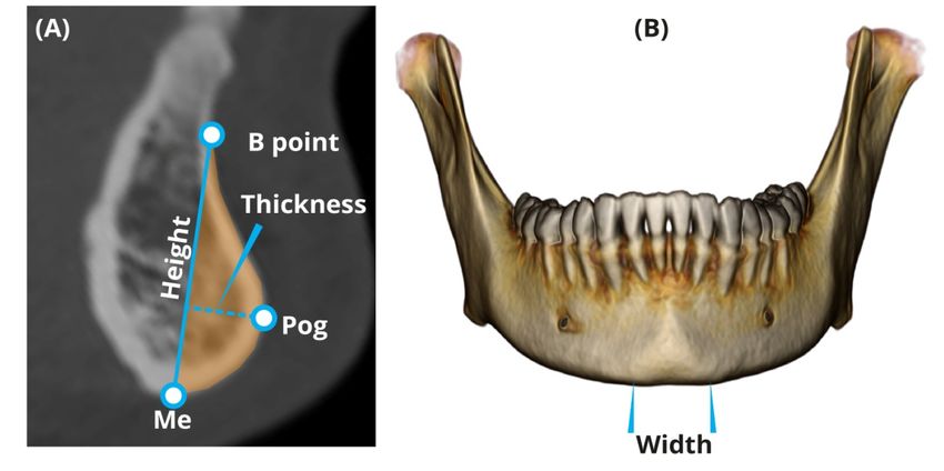
Int. J. Environ. Res. Public Health 2020, 17, 4249 2 of 15
Int. J. Environ. Res. Public Health 2020, 17, x 2 of 15
Figure
Figure 1. (A)
1. (A) ChinChin (mentum
(mentum osseum).
osseum). Frontal
Frontal viewview of the
of the mandible
mandible (using
(using the the volume
volume rendering
rendering
technique); chin borders (mental protuberance and bilateral mental tubercles) are denoted abydotted
technique); chin borders (mental protuberance and bilateral mental tubercles) are denoted by a
line. line.
dotted (B) Mandibular symphysis.
(B) Mandibular Midsagittal
symphysis. section;section;
Midsagittal symphysis borders borders
symphysis are denoted
are by dotted by
denoted lines.
dotted lines.
The mandibular symphysis isthe region where the two halves of the human fetal mandibular
corpus
Several arehypotheses
fused [4] (Figure
have been1B). As opposed
raised overtothe theyears
chin, totheelucidate
mandibular thesymphysis
reason foristhe found in all
chin's
hominoid mandibles and its morphology was functionally linked
appearance in humankind. The “sexual selection” hypothesis is among the most common; it states [5].
that theSeveral
appearance hypotheses
of the have
chin in been raisedand
humans overitsthe sizeyears
and to elucidate
shape variationthe reason
are duefor to the chin’s
sexual
appearance
selection, relatingin humankind. The “sexual
to facial attractiveness selection” hypothesis is among the most common; it states
[6–9].
that the appearance of the chin in
In modern dentistry, the chin and symphysis humans and itssize size and
and shape
shape variation are due torole
play an important sexual fromselection,
the
relating to facial attractiveness [6–9].
esthetic perspective [10]. Chin height and width are considered important factors in perceiving facial
In modern
attractiveness. dentistry,inthe
Deformities chinchin and symphysis
height (when greater sizethan
and shape
50% and play 58%an of important
the lower role from the
anterior
esthetic perspective [10]. Chin height and width are considered
facial height in males and females, respectively) were found to be the least attractive facial important factors in perceiving facial
attractiveness.
characteristic, andDeformities
consequently, in chin height was
surgery (when greater than
required 50% and
to correct the58%facialof the lower anterior
appearance [11]. facial
A
height in males and females, respectively) were found to be the least
squared contour of the chin in females is considered unattractive since it gives the face a masculine attractive facial characteristic,
and consequently,
appearance surgery was
and thus diminishes the required
appearance to correct the facial[8].
of femininity appearance
Narrowing[11]. A squared
genioplasty to contour
achieve of
the chin in
a feminine andfemales is considered
slim lower face is aunattractive
well-documented since it gives the face
procedure thata masculine
is practiced appearance
to improve andthe thus
diminishes the appearance of femininity [8]. Narrowing genioplasty
facial appearance and the lower facial contour [12,13]. Valenzano et al. [14] applied morphometric to achieve a feminine and slim
analysis(quantitative analysis of the chin and symphysis size and shape) regarding facialthe
lower face is a well-documented procedure that is practiced to improve the facial appearance and
lower facial contour
attractiveness. However, [12,13].
theyValenzano
failed to etfind al. [14]
an applied
association morphometric analysis(quantitative
between attractiveness and sexualanalysis
of the chin and symphysis size and
dimorphism of the lower face (including the chin). shape) regarding facial attractiveness. However, they failed to find
anQualitative
associationdifferences
between attractiveness
in the chin shape and sexual
between dimorphism
the sexesofare thewell
lower face (including
known the chin).
in anthropology:
Qualitative differences in the chin shape between the
males possess broad and prominent chins with a more square appearance and females possess sexes are well known in anthropology:
maleschins
pointed possess broad and
[15–19]. prominent
Thayer and Dobson chins with a more square
[9] compared appearance
the chin and females
shape between thepossess
sexes usingpointed
chins [15–19].
elliptical Thayer and
Fourier function Dobson
analysis [9] compared
(EFFA) and found thethat
chinsexual
shapedimorphism
between theexists sexes inusing elliptical
its shape.
However, there was a high degree of overlap in the chin shape between males and females due tothere
Fourier function analysis (EFFA) and found that sexual dimorphism exists in its shape. However, a
wasdegree
large a highof degree of overlap
variation in each in sex.
the chin shape between
Nevertheless, malesmales and females
possessed a tallerdue to a largeadegree
symphysis, more of
variationmentum
protrusive in each sex. Nevertheless,
osseum, and largermales possessed
tubercula a taller
laterale thansymphysis,
females did. a more
Garvin protrusive
and Ruffmentum [17]
used 3D surface laser scans to show that the absolute surface area and the volume of3D
osseum, and larger tubercula laterale than females did. Garvin and Ruff [17] used thesurface
chin are laser
scans to
sexually show that however,
dimorphic; the absolute thesurface area and the
sex differences volumenon-significant
became of the chin arewhen sexually they dimorphic;
were
however, the sex differences became non-significant when they
standardized for size. Garvin and Ruff's morphometric shape analysis revealed a relatively high were standardized for size. Garvin
and Ruff’s morphometric shape analysis revealed a relatively
overlap in the chin shape between the sexes. Males displayed relatively more prominent mental high overlap in the chin shape between
the sexes. Males
protruberances, lateraldisplayed
tubercles, relatively
and taller more
chinsprominent
than did mental
females.protruberances,
Coquerelleet al.lateral [20] and tubercles,
Franklinand
et al. [21,22] used the geometric morphometric method to find that the sexual dimorphismgeometric
taller chins than did females. Coquerelleet al. [20] and Franklin et al. [21,22] used the of the
chinmorphometric
and mandibular method to find that
symphysis the sexual dimorphism
is age-dependent. According of thetochin and mandibular
Coquerelleet al. [20],symphysis
there is is
age-dependent.
amarked According to
sexual dimorphism inCoquerelleet
symphysealal. [20], at
shape there is amarked
birth. sexual dimorphism
It dramatically decreases between in symphyseal
the
shape at birth. It dramatically decreases between the ages of 4–14
ages of 4–14 years, and increases again afterward. Adult dimorphism was similar to that of the years, and increases again afterward.
early
Adult dimorphism
postnatal stages;namely, wasmales
similarhad to that of the early postnatal
a square-shaped stages;namely,
chin compared with amales had a square-shaped
round-shaped chin in
females. However, Daegling [23] and Pampush [24] did not find any differences in Pampush
chin compared with a round-shaped chin in females. However, Daegling [23] and [24] did
chin thickness,
size, and shape between the sexes. Daegling showed that a substantial overlap of the chin shape
exists between the sexes in the midsagittal section.
Int. J. Environ. Res. Public Health 2020, 17, 4249                                                 3 of 15

not find any differences in chin thickness, size, and shape between the sexes. Daegling showed that a
substantial overlap of the chin shape exists between the sexes in the midsagittal section.
     Since the chin and symphysis are major components of the lower third of the face, it is essential to
determine whether their size and shape are sex-dependent for clinical and anthropological purposes.
Whereas some studies clearly showed the existence of sexual dimorphism in the chin and symphysis [9,17],
others did not conclude that it exists [23,24]. The inconsistent findings reported in the dental and
anthropological literature may be related to the different methodologies used to quantify the chin and
symphysis, and to the lack of age and size control; hence, the appropriateness of the “sexual selection”
hypothesis is questionable.
     This study aimed to compare the magnitude of sexual dimorphism in the chin and symphysis
size and shape using sets of measurements that are independent of jaw size, position, inclination, and
age. Assuming that the “sexual selection” theory is defensible, we would expect the following: (1)
sexual dimorphism will be more pronounced in the chin than in the symphysis, and (2) chin size and
shape will be more variable than symphysis size and shape.

2. Materials and Methods

2.1. Study Sample
      The studied population included 419 adults of Caucasian origin. The minimum number of
individuals was calculated using WinPepi software (v.11.65, J.H. Abramson, USA). It required at least
175 individuals per group (males and females) (total n = 350) to provide a significant difference with a
0.3 effect size for p < 0.05 and power = 80%. The included individuals had undergone a head and neck
CT scan (Brilliance 64, Philips Medical System, Cleveland, OH, USA) for medical diagnostic purposes
(unrelated to the present study) at Carmel Medical Center, Haifa, Israel. The following parameters
were used: slice thickness 0.9–3.0 mm, pixel spacing 0.3–0.5 mm, 120 kV, 250–500 mAs, the number of
slices 150–950, and matrix 512 × 512.
      The inclusion criteria were as follows: age ≥ 18 years, intact lower incisors, teeth at the centric
occlusion (maximum intercuspation), and the presence of at least two teeth of the posterior unit
(premolars and/or molars) on each side. The exclusion criteria were as follows: (1) the absence of
lower incisors; (2) dental implants and metal restorations that impede measurements; (3) evidence
of orthodontic treatment (e.g., brackets, appliances, and lingual fixed retainers); (4) previous surgery
in the head and neck region (based on medical files or signs on the skull); (5) prominent facial and
mandibular asymmetry, cranio-facial, temporo-mandibular joint, and muscle disorders; (6) trauma;
and (7) technically aberrant CT scans. The research was approved by the ethics committee of the
Carmel Medical Center (CMC 11-0066).

2.2. Measuring the Chin and Symphysis
     Before measuring the chin and symphysis, a standardized alignment of the skull parallel to
the Frankfort horizontal (FH) plane was performed. Landmarks of the chin and symphysis were
identified according to Caufield [25] and Swennenet al. [26]. All measurements were taken directly
from the CT scans using a multi-planner reformatting technique (“Extended Brilliance Workspace”
portal (v2.6.0.27) (Philips Medical Systems, Cleveland, OH, USA). A set of four parameters (linear and
cross-sectional area (CSA)) was developed to evaluate the chin size and shape, and five parameters
were developed for evaluating the symphysis, including angulations (Table 1). Most parameters of
the chin and symphysis were measured at the midsagittal section (Figures 2A and 3). Chin width
was measured using the volume-rendering model from a frontal view (Figure 2B). All of the CSAs
were extracted from the CT software using manual tracing of the anatomical borders of the structure.
The reliability of the measurements was assessed using 15 randomly selected CT scans of the head and
neck. To evaluate the intra-observer variation, a single researcher (T.S.T.) carried out the measurements
twice with a two-week interval between each attempt. The mean of the two measurement sets carried
Int. J. Environ. Res. Public Health 2020, 17, 4249 4 of 15
out for reliability assessment was utilized for further statistical analysis.To evaluate the inter-observer
variation, the measurements were carried out by an additional independent researcher (H.M.).
Table 1. Definitions of the chin and the symphysis measurements.
Measurement Definition
The distance between the most posterior midline point in the concavity of the
Height (mm)
mandibular symphysis (B point) and the menton
The maximum thickness of the chin, measured as the length of the perpendicular
Chin Thickness(mm)
line from pogonion to the chin height line
CSA (mm2 ) The portion of the symphysis CSA that is located anterior to the chin height line
Width (mm) The distance between the right and left mental tubercles
Height (mm) The distance between the most superior point on the alveolar bone and menton
Thickness (mm) The distance between the pogonion and the most posterior point on the symphysis
CSA (mm2 ) The total CSA of the symphysis in the midsagittal plane
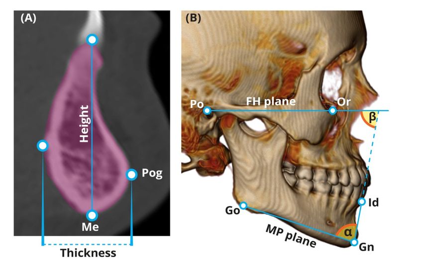
Symphysis Inclination of the symphysis relative to the MP: the angle (α angle) created
between the line passing from the infradentale (the midline point at the superior
Inclination (◦ )
tip of the septum between the mandibular central incisors) to the gnathion (Id-Gn
line), and the line passing from gonion to gnathion [27]
Inclination of the symphysis relative to the FH plane: the angle (β angle) measured
Orientation (◦ )
at the cross-point between the Id-Gn line and the FH plane (the porion-orbitale line)
Note: CSA- cross-sectional area; MP-mandibular plane; Id-infradentale; Gn-gnathion; FH- Frankfort horizontal.
Int. J. Environ. Res. Public Health 2020, 17, x 4 of 15
Figure 2.
Figure Measurementsof
2. Measurements of the
the chin.
chin. (A)
(A)Height,
Height,thickness,
thickness,and
andcross-sectional
cross-sectionalarea
area(CSA)
(CSA)(in
(in light
light
orange); Me—menton, Pog—pogonion. (B) Chin
orange); Me—menton, Pog—pogonion. (B) Chin width. width.Figure 2. Measurements of the chin. (A) Height, thickness, and cross-sectional area (CSA) (in light
orange); Me—menton, Pog—pogonion. (B) Chin width.
Int. J. Environ. Res. Public Health 2020, 17, 4249 5 of 15
Figure 3. Measurements of symphysis. (A) Height, thickness, and CSA (in light pink). (B) Symphysis
orientation (β angle) and inclination (α angle). Me—menton, Pog—pogonion, Po—porion, Or—orbitale,
Figure 3. Measurements of symphysis. (A) Height, thickness, and CSA (in light pink). (B) Symphysis
Go—gonion, Id—infradentale, Gn—gnathion, MP—mandibular plane, and FH—Frankfort horizontal.
orientation (β angle) and inclination (α angle). Me—menton, Pog—pogonion, Po—porion,
Or—orbitale, three
Additionally, Go—gonion,
indices wereId—infradentale, Gn—gnathion,
calculated: chin MP—mandibular
size index (%)—the plane, the
ratio between and
chin
FH—Frankfort horizontal.
CSA and the symphysis CSA, multiplied by 100; chin shape index (%)—the ratio between the chin
thickness and the chin height, multiplied by 100; and symphysis shape index (%)—the ratio between
Table 1. Definitions of the chin and the symphysis measurements.
the symphysis thickness and the symphysis height, multiplied by 100.
Measurement Definition
2.3. Size Factor
The distance between the most posterior midline point in the concavity of the
Height (mm)
All linear and CSA measurements were controlled
mandibular forpoint)
symphysis (B general
andmandibular
the menton size, as expressed by its
geometric mean (MGM—mandibular geometricthickness
The maximum mean). of
The
thelatter
chin, was calculated
measured as the based
length on a set of six
of the
Chin Thickness(mm)
perpendicular line from pogonion to the chin height line
linear measurements: ramus length and width, mandibular body length, gonial width, coronoid length,
andbigonial breadth, following the
Themethods
portion ofdescribed by Sella
the symphysis Tunis
CSA that et al. [28].
is located All to
anterior measurements
the chin height
CSA (mm2)
line using the Brilliance Workspace Portal (Philips v. 2.6.1.5, Philips,
were taken directly from the CT scans
Amsterdam, Netherlands). The square roots of the chin and symphysis CSAs were divided by the
MGM, following the principles presented in Jungers et al. [29].
2.4. Age Factor
Since the size and shape of the mandible are very dynamic [30] and may change considerably with
age, there was a need to locate measures that are age-dependent. Possible associations between the
individual’s chronological age and all chin and symphysis parameters were assessed using different
types of correlation analysis. Moreover, since different age distributions for males and females can
easily bias the results, we ensured that the samples of males and females were age-matched.
2.5. Orientation Factor
Previous studies suggested a possible association between chin morphometrics and the mandibular
plane angle (MPa) [31,32]. Therefore, in searching for possible associations between chin morphometrics
and other mandibular parameters, one needs to control for the MPa to avoid a potential confounder
effect. This is especially important when looking for an association between the chin and the symphysis
metrical characteristics and age since the MPa is age-dependent [33,34]. Initially, our measuring
methods of the chin and symphysis were constructed to be independent of MPa, i.e., the head was
positioned parallel to the FH plane and all of the landmarks and lines were drawn independent ofInt. J. Environ. Res. Public Health 2020, 17, 4249 6 of 15
the MP. Additionally, to control for the MPa, we used partial correlation analysis when searching for
associations between the chin and symphysis parameters and age. The MPa was measured following
Downs’ [35] method, relative to the FH plane.
2.6. Statistical Analysis
The data were recorded and analyzed using IBM SPSS statistics, version 20 software (IBM Corp.,
Armonk, NY, USA). The level of statistical significance was set at p < 0.05. The intraclass correlation
coefficient (ICC) was calculated to examine the reproducibility of the measurements; It was interpreted
according to the categorization method of Cicchetti [36]. One-sample Kolmogorov–Smirnov tests and
histograms were carried out to determine the normality of the distributions of the measurements.
Descriptive statistics (mean, SD, and range) were carried out for all parameters. The coefficient
of variation (CV) was defined as the standard deviation expressed as a percentage of the mean; it
was calculated for each sex separately to estimate the extent of the chin and symphysis variation.
The F-statistic and p-values were calculated to compare the chin and symphysis CVs between the sexes
according to Forkman [37] using MedCalc statistical software (version 19.3.1, MedCalc Software bv,
Ostend, Belgium; https://www.medcalc.org; 2016)).
The CV was classified according to the system devisedby Vazet al. [38]. Independent-sample
t-tests (two-tailed) were carried out to detect significant differences in the observed values of the chin
and symphysis between males and females, and to examine differences in the mean age between the
sexes. The Mann–Whitney test was carried out to detect significant differences in the controlled values
of the chin and symphysis and their indexes between the sexes. Pearson correlations were calculated to
test for associations between the chin and symphysis size and age. A partial correlation was calculated
between the chin and symphysis parameters and age, while controlling for the MPa, to eliminate its
potential role as a confounder. Linear regression analysis was carried out to evaluate the percentage of
the chin and symphysis variance that was accounted for by sex and size.
3. Results
3.1. Reliability Analysis
The intra-observer variation of all measurements showed excellent results (0.838 ≤ ICC≤ 0.986)
and the inter-observer variation showed good to excellent results (0.704 ≤ ICC ≤ 0.980) (p < 0.001).
3.2. Age Factor
The population studied included 419 adults: 203 males (48.4%) and 216 females (51.6%) between
18–96 years of age (Table 2). Males and females were age-matched, i.e., no significant difference was
found in the mean age between males and females (p = 0.052). Age was normally distributed in both
males and females (Kolmogorov–Smirnov test: p = 0.076 and p = 0.143, respectively).
Table 2. Descriptive statistics for chronological age (in years) by sex.
Statistics Males Females Total
Mean 51.46 55.39 53.48
SD 20.357 20.823 20.668
Minimum 18 18 18
Maximum 96 96 96
Range 78 78 78
n 203 216 216
Note: SD—standard deviation.
In both sexes, no significant associations were found between the chin parameters (observed
values) and age, except for a very weak association in males for chin width (r = 0.146) (Table 3).Int. J. Environ. Res. Public Health 2020, 17, 4249 7 of 15
Symphysis height was significantly negatively associated with age in both males (r = −0.371) and
females (r = −0.268), and its shape index was significantly positively associated with age (males
r = 0.366, females r = 0.275). Symphysis thickness and CSA were independent of age (p > 0.093).
Symphysis inclination (relative to the MP) did not vary with age, regardless of sex (p > 0.315), although
its orientation relative to the FH had a significant yet weak positive correlation with age (r = 0.247 and
r = 0.173 in males and females, respectively). We found a significant correlation between chin width and
the MPa (males r= −0.243, p < 0.001; females r = −0.416, p < 0.001). Regarding its potential confounder
effect, a partial correlation was calculated (controlling for the MPa). No significant associations between
the chin parameters and age were found after controlling for the MPa.
Table 3. Correlation coefficients between the chin and symphysis parameters and age by sex
(males n = 203, females n = 216).
Observed Controlled for the MPa b
Measurement Sex
r p-Value a r p-Value a
Male −0.077 0.275 −0.062 0.408
Height
Female 0.008 0.912 −0.010 0.894
Male 0.061 0.387 0.035 0.637
Thickness
Female −0.025 0.717 −0.083 0.246
Male 0.062 0.380 0.027 0.722
CSA
Female 0.022 0.748 −0.018 0.796
Chin
Male 0.146 0.038 0.110 0.140
Width
Female 0.094 0.170 0.048 0.497
Male 0.101 0.151 0.072 0.334
Shape index
Female −0.022 0.748 −0.103 0.147
Male 0.120 0.089 0.037 0.619
Size index
Female 0.048 0.486 0.009 0.901
Male −0.371Int. J. Environ. Res. Public Health 2020, 17, 4249 8 of 15
the chin height. The CV was independent of sex in four of the six traits studied, whereas the CV was
significantly greater in females in the two other traits (chin width and shape index).
Table 4. Morphometric characteristics of the male and female chin (males n = 203, females n = 216).
p-Values *
Chin Measurement Sex Mean SD Minimum Maximum Observed Controlled
Measures Measures
Male 21.58 3.102 13.30 28.900
Height (mm) 0.046 M)
Female 21.02 2.576 14.60 26.400
Male 4.00 0.991 1.40 7.100
Thickness (mm) 0.176 0.079
Female 3.86 1.054 1.00 6.800
Male 53.04 18.534 13.80 113.400
CSA (mm2 ) 0.120 0.001(F > M)
Female 50.32 17.181 11.70 110.500
Male 28.18 5.622 16.30 42.700
Width (mm) 0.085). No significant difference in the symphysis shape index was found between the sexes:
the symphysis-thickness-to-symphysis-height ratio was approximately 1:2 (Table 6). In contrast, the
symphysis orientation was significantly greater in males (80.24◦ ) than in females (78.30◦ ) (p = 0.011),
and the symphysis inclination was sex-independent (p = 0.905). This implied that males’ symphyses
were more lingually oriented than females’ symphyses. The overall results suggested that symphysis
size (the observed values) was significantly greater in males than in females. Nevertheless, when
controlling for the MGM, no significant differences existed in the symphysis morphometrics between
the sexes. Additionally, the symphysis shape was similar between males and females. The CV for all
symphysis parameters was found to be low in both sexes, ranging between 7.2% (symphysis inclination)
and 17.4% (symphysis CSA) (Table 7). Nevertheless, males exhibited a statistically significantly greater
variation in symphysis height and thickness compared with females (p < 0.044). No significantInt. J. Environ. Res. Public Health 2020, 17, 4249 9 of 15
differences between the sexes were observed for the CV of the symphysis CSA, shape index, orientation,
and inclination.
Table 6. Morphometric characteristics of the male and female symphysis (males n = 203,
females n = 216).
p-Values *
Symphysis
Sex Mean SD Minimum Maximum
Measurement Observed Controlled
Measures Measures
Male 33.28 3.303 24.60 42.600
Height (mm)Int. J. Environ. Res. Public Health 2020, 17, 4249 10 of 15
Table 8. Variation comparison of the chin and symphysis parameters in males and females (males
n = 203, females n = 216).
Chin and Symphysis Measurements a Sex F-Statistic p-Values *
Male 0.482Int. J. Environ. Res. Public Health 2020, 17, 4249 11 of 15
4.2. Sex Differences in Symphysis Size and Shape
Our findings clearly show the existence of sexual dimorphism in the observed symphysis metric
characteristics, i.e., males exhibit higher, thicker, and larger symphyses that are more lingually oriented
compared with females. However, we found that males and females possess similar symphyseal
shapes and similar sizes after controlling for mandibular size. Swastyet al. [39] reported higher
and thicker symphyses in males; however, they did not control for mandibular size. Similarly,
Aki et al. [40] reported higher and deeper symphyses in males than in females; the ratio between the
two (height/depth) was slightly greater in females. Since no difference between the sexes was found in
the symphysis inclination relative to the MP, we suggest that the male symphysis was more lingually
oriented relative to the FH plane due to a smaller mandibular angle, a finding already reported by
us [41].
4.3. Age Influence on Chin and Symphysis
A previous study suggested that the magnitude of sexual dimorphism in human’s mandibular
size and shape is age-dependent [20]. Although our study was retrospective, from the obtained data
correlated with age, it became clear that chin features are age-independent.
Similar findings were reported by Haskell [42]. In contrast to chin height, which remains
stable from the age of 18 years onward, symphysis height decreases with age. The decrease in the
symphysis height can explain the increase of its shape index, which leads to the development of a
more square-shaped symphysis with age in both sexes. An age-related reduction in alveolar bone crest
height was reported in the literature; this is usually attributed to deterioration in the periodontal status
or to tooth loss with age [43,44]. However, a reduction in alveolar bone crest height with age was also
found in individuals with healthy dental and periodontal status; mandibular incisors displayed the
greatest alveolar bone loss [45].
According to our study, symphysis orientation (measured relative to FH) changed significantly
with age. This implies that with age, the symphysis became more lingually oriented (retro lined) in
both males and females. Since no significant change in symphysis inclination (measured relative to the
MP) with age was evident, we hypothesized that the change in the symphysis orientation with age
was mainly due to a continuous forward rotation of the mandible over time (Figure 4). A reduction in
crown height (due to attrition/abrasion) and/or posterior tooth loss with age might lead to anterior
rotation of the mandible, and subsequently, may lead to a reduction in the MPa and to a lower anterior
facial height [46]. Age changes in the adult facial profile were studied by Formby et al. [33], who found
a decrease in the MPa relative to the cranial base with age in males. They attributed this phenomenon to
the mandibular forward rotation with age. In a longitudinal study, Pecora et al. [34] found differences
between males and females regarding the degree of mandibular rotation with age: the mandibles of
females undergo posterior rotation, whereas those of males undergo anterior rotation. This rotation of
the mandible is followed by a relative protrusion of the chin.Int. J. Environ. Res. Public Health 2020, 17, 4249 12 of 15
Int. J. Environ. Res. Public Health 2020, 17, x 12 of 15
Figure 4.
Figure Changes in
4. Changes in the
the symphysis
symphysis orientation
orientation and
and inclination
inclination with
with age.
age. The
Theillustration
illustrationon onthethe left
left
represents younger individuals, whereas the one on the right represents older individuals.
represents younger individuals, whereas the one on the right represents older individuals. No No change
in the symphysis
change inclination
in the symphysis relative relative
inclination to the MPto (α
theangle)
MP (αwas evident
angle) was (α 1 is similar
evident (α1 istosimilar
α2 ), whereas
to α2),
the symphysis orientation relative to the FH plane (β angle) increased with
whereas the symphysis orientation relative to the FH plane (β angle) increased with age βage (β1 < (β21);< this was
β2); this
probably due to a forward rotation of the mandible (arrow).
was probably due to a forward rotation of the mandible (arrow).
4.4. Evolutionary Implication: “Sexual Selection”
4.4. Evolutionary Implication: “Sexual Selection”
Based on the notion of its sex-specific characteristics, it has been suggested that the human
Based on the notion of its sex-specific characteristics, it has been suggested that the human chin
chin results from long-term sexual selection [8,9,16,47]. Facial appearance is extensively influenced
results from long-term sexual selection [8,9,16,47]. Facial appearance is extensively influenced by the
by the size and shape of the chin [10,48]. An interesting finding of our study relates to the sexual
size and shape of the chin [10,48]. An interesting finding of our study relates to the sexual
dimorphism in the size and shape of the chin. Contrary to the common notion that chin thickness
dimorphism in the size and shape of the chin. Contrary to the common notion that chin thickness is
is more pronounced in males, our findings suggest that no significant differences in chin thickness
more pronounced in males, our findings suggest that no significant differences in chin thickness
(observed and controlled) and chin shape existed between the sexes. Although the observed chin
(observed and controlled) and chin shape existed between the sexes. Although the observed chin
height dimension was greater in males, when controlling for mandibular size, females possessed
height dimension was greater in males, when controlling for mandibular size, females possessed
relatively higher chins. Moreover, when controlling for mandibular size, the chin CSA showed a
relatively higher chins. Moreover, when controlling for mandibular size, the chin CSA showed a
greater dimension in females than in males, and its size index (chin CSA/symphysis CSA) was smaller
greater dimension in females than in males, and its size index (chin CSA/symphysis CSA) was
in males compared with females, indicating that the chin was, in fact, smaller in males than in females.
smaller in males compared with females, indicating that the chin was, in fact, smaller in males than
Only chin width (the frontal aspect) was considerably greater in males than in females in both the
in females. Only chin width (the frontal aspect) was considerably greater in males than in females
observed and controlled measures.
in both the observed and controlled measures.
In the chin, except for chin width, mandibular size accounted for 6 to14% of the variance (it varied
In the chin, except for chin width, mandibular size accounted for 6 to14% of the variance (it
between the measures) and sex accounted for 1–2%. However, this phenomenon (variance explained
varied between the measures) and sex accounted for 1–2%. However, this phenomenon (variance
by size > variance explained by sex) was reversed regarding chin width, where sex accounted for 16%
explained by size > variance explained by sex) was reversed regarding chin width, where sex
of the variance and size for only2%. Notably, the chin width CV rate significantly differed between
accounted for 16% of the variance and size for only2%. Notably, the chin width CV rate significantly
males and females, i.e., males showed significantly lower variation rates (CV) compared with females,
differed between males and females, i.e., males showed significantly lower variation rates (CV)
suggesting a stronger selection. In the symphysis, mandibular size accounted for 24–33% of the
compared with females, suggesting a stronger selection. In the symphysis, mandibular size
variances of various traits, sex contributed only 4% to the height dimension, and null to thickness
accounted for 24–33% of the variances of various traits, sex contributed only 4% to the height
and CSA.
dimension, and null to thickness and CSA.
The symphysis only showed a pronounced sexual dimorphism in its observed values. Males
The symphysis only showed a pronounced sexual dimorphism in its observed values. Males
exhibited a greater symphysis size than did females (height, thickness, and larger CSA). However, no
exhibited a greater symphysis size than did females (height, thickness, and larger CSA). However,
sexual dimorphism existed after controlling for mandibular size. This suggests that the differences
no sexual dimorphism existed after controlling for mandibular size. This suggests that the
between the sexes could only be attributed to the greater and more robust mandibles of males.
differences between the sexes could only be attributed to the greater and more robust mandibles of
Additionally, the symphysis shape did not differ between males and females. As mentioned before,
males. Additionally, the symphysis shape did not differ between males and females. As mentioned
between 24–33% of the symphysis variance was explained by mandibular size, whereas only up to 4%
before, between 24–33% of the symphysis variance was explained by mandibular size, whereas only
was explained by sex.
up to 4% was explained by sex.
The above findings lend only partial support to the “sexual selection” theory, suggesting that
other factors may also be involved in chin formation. Therefore, after studying the chin size andInt. J. Environ. Res. Public Health 2020, 17, 4249 13 of 15
The above findings lend only partial support to the “sexual selection” theory, suggesting that other
factors may also be involved in chin formation. Therefore, after studying the chin size and shape, we
concluded that the possible input of other factors besides sexual selection should be considered. Future
studies are needed to reveal the magnitude and direction of chin and symphysis sexual dimorphism,
as well as the role of facial attractiveness in their expression.
5. Study Limitations
This is a retrospective study. The findings of the current study were based on a population of
Caucasian origin. A generalization of the results requires further study of the different populations
of different geographical regions. Information on previous orthodontic treatment was unavailable.
However, to avoid a possible confounding effect of the orthodontic treatment on the chin and symphysis
size and shape, individuals who showed indirect evidence of such treatment were excluded from
the study.
6. Conclusions
Chin width (the frontal view) was found to be a sexually selected trait; it can be considered as
a parameter for sex determination (males possess significantly wider chins than females do). Chin
thickness (the lateral view) is similar in both sexes. The symphysis was size-dependent and its size and
shape were sex-independent. The chin was found to be a more heterogeneous anatomical structure
than symphysis and it was sexually more dimorphic.
Author Contributions: Conceptualization, T.S.T. and I.H.; methodology, N.S.; software, A.D.V.; validation, H.M.
and T.S.T.; formal analysis, R.S.; investigation, T.S.T.; resources, N.S.; data curation, T.S.T.; writing—original draft
preparation, T.S.T.; writing—review and editing, T.S.T., I.H., and N.S.; visualization, T.S.T.; supervision, I.H.,
A.D.V., and N.S.; project administration, I.H.; funding acquisition, H.M. All authors have read and agreed to the
published version of the manuscript.
Funding: This research was funded by the Dan David Foundation, the Tassia and Dr. Joseph Meychan Chair for
the History and Philosophy of Medicine, and the Israeli Science Foundation (grant no.1116/16).
Acknowledgments: The authors wish to thank Ariel Pokhojaev for preparingthe illustrations inthis manuscript.
Additionally, we wish to thank Steve Manch for editing the English text.
Conflicts of Interest: The authors declare no conflict of interest. The funders had no role in the design of the
study; in the collection, analyses, or interpretation of data; in the writing of the manuscript; or in the decision to
publish the results.
References
1. Schwartz, J.H.; Tattersall, I. The human chin revisited: What is it and who has it? J. Hum. Evol. 2000, 38,
367–409. [CrossRef] [PubMed]
2. Cook, T.W. The human chin and human tooth change. Int. J. Orthod. Dent. Child. 1933, 19, 730–734.
[CrossRef]
3. Coquerelle, M.; Bookstein, F.L.; Braga, J.; Halazonetis, D.J.; Weber, G.W. Fetal and infant growth patterns
of the mandibular symphysis in modern humans and chimpanzees (pan troglodytes). J. Anat. 2010, 217,
507–520. [CrossRef] [PubMed]
4. Moore, K.L.; Argur, A.R. Essential Clinical Anatomy; Williams & Wilkins: Baltimore, MD, USA, 1996.
5. Daegling, D.J. Biomechanical scaling of the hominoid mandibular symphysis. J. Morphol. 2001, 250, 12–23.
[CrossRef]
6. Hershkovitz, P. The decorative chin. Bull. Field Mus. Nat. Hist. 1970, 41, 6–10.
7. Grammer, K.; Thornhill, R. Human (Homo sapiens) facial attractiveness and sexual selection: The role of
symmetry and averageness. J. Comp. Psychol. 1994, 108, 233–242. [CrossRef]
8. Barber, N. The evolutionary psychology of physical attractiveness: Sexual selection and human morphology.
Ethol. Sociobiol. 1995, 16, 395–424. [CrossRef]
9. Thayer, Z.M.; Dobson, S.D. Sexual dimorphism in chin shape: Implications for adaptive hypotheses. Am. J.
Phys. Anthropol. 2010, 143, 417–425. [CrossRef]Int. J. Environ. Res. Public Health 2020, 17, 4249 14 of 15
10. Czarnecki, S.T.; Nanda, R.S.; Currier, G.F. Perceptions of a balanced facial profile. Am. J. Orthod. Dentofac.
Orthop. 1993, 104, 180–187. [CrossRef]
11. Naini, F.B.; Donaldson, A.N.; McDonald, F.; Cobourne, M.T. Influence of chin height on perceived
attractiveness in the orthognathic patient, layperson, and clinician. Angle Orthod. 2012, 82, 88–95. [CrossRef]
12. Park, S.; Noh, J.H. Importance of the chin in lower facial contour: Narrowing genioplasty to achieve a
feminine and slim lower face. Plast. Reconstr. Surg. 2008, 122, 261–268. [CrossRef]
13. Capitan, L.; Simon, D.; Capitan-Canadas, F. Facial feminization surgery and facial gender confirmation
surgery. In Comprehensive Care of the Transgender Patient; Ferrando, C.A., Ed.; Elsevier: Philadelphia, PA,
USA, 2020.
14. Valenzano, D.R.; Mennucci, A.; Tartarelli, G.; Cellerino, A. Shape analysis of female facial attractiveness.
Vis. Res. 2006, 46, 1282–1291. [CrossRef] [PubMed]
15. Byers, S.N. Introduction to Forensic Anthropology; Press Pearson Education: New York, NY, USA, 2002.
16. Bass, W.M. Human Osteology: A Laboratory and Field Manual, 5th ed.; Missouri Archaeological Society:
Columbia, MO, USA, 2005.
17. Garvin, H.M.; Ruff, C.B. Sexual dimorphism in skeletal brow ridge and chin morphologies determined using
a new quantitative method. Am. J. Phys. Anthropol. 2012, 147, 661–670. [CrossRef] [PubMed]
18. Schutkowski, H. Sex determination of infant and juvenile skeletons: I. Morpho gnostic features. Am. J. Phys.
Anthropol. 1993, 90, 199–205. [CrossRef] [PubMed]
19. Loth, S.R.; Henneberg, M. Sexually dimorphic mandibular morphology in the first few years of life. Am. J.
Phys. Anthropol. 2001, 115, 179–186. [CrossRef] [PubMed]
20. Coquerelle, M.; Bookstein, F.L.; Braga, J.; Halazonetis, D.J.; Weber, G.W.; Mitteroecker, P. Sexual dimorphism
of the human mandible and its association with dental development. Am. J. Phys. Anthropol. 2011, 145,
192–202. [CrossRef]
21. Franklin, D.; Oxnard, C.E.; O’Higgins, P.; Dadour, I. Sexual dimorphism in the subadult mandible:
Quantification using geometric morphometrics. J. Forensic Sci. 2007, 52, 6–10. [CrossRef]
22. Franklin, D.; O’Higgins, P.; Oxnard, C.E.; Dadour, I. Sexual dimorphism and population variation in the
adult mandible: Forensic applications of geometric morphometrics. Forensic Sci. Med. Pathol. 2007, 3, 15–22.
23. Daegling, D.J. The human mandible and the origins of speech. J. Anthropol. 2012, 2012, 1–14. [CrossRef]
24. Pampush, J.D. Selection played a role in the evolution of the human chin. J. Hum. Evol. 2015, 82, 127–136.
[CrossRef]
25. Caufield, P.W. Tracing technique and identification of landmarks. In Radiographic Cephalometry: From Basics
to 3-D Imaging, 2nd ed.; Jacobson, A., Jacobson, R.L., Eds.; Quintessence Publishing: Chicago, IL, USA, 2006;
pp. 45–51.
26. Swennen, G.R.J.; Schutyser, F.; Hausamen, J.E. Three-Dimensional Cephalometry: A Color Atlas and Manual;
Springer: Berlin/Heidelberg, Germany, 2005.
27. Steiner, C.C. Cephalometrics for you and me. Am. J. Orthod. 1953, 39, 729–755. [CrossRef]
28. Sella-Tunis, T.; Pokhojaev, A.; Sarig, R.; O’Higgins, P.; May, H. Human mandibular shape is associated with
masticatory muscle force. Sci. Rep. 2018, 16, 6042. [CrossRef] [PubMed]
29. Jungers, W.L.; Falsetti, A.B.; Wall, C.E. Shape, relative size, and size-adjustments in morphometrics. Am. J.
Phys. Anthropol. 1995, 38, 137–161. [CrossRef]
30. Enlow, D.H.; Hans, M.G. Essentials of Facial Growth; Saunders: Philadelphia, PA, USA, 1996.
31. Sassouni, V. A classification of skeletal facial types. Am. J. Orthod. 1969, 55, 109–123. [CrossRef]
32. Björk, A. Prediction of mandibular growth rotation. Am. J. Orthod. 1969, 55, 585–599. [CrossRef]
33. Formby, W.A.; Nanda, R.S.; Currier, G.F. Longitudinal changes in the adult facial profile. Am. J. Orthod.
Dentofac. Orthop. 1994, 105, 464–476. [CrossRef]
34. Pecora, N.G.; Baccetti, T.; McNamara, J.A., Jr. The aging craniofacial complex: A longitudinal cephalometric
study from late adolescence to late adulthood. Am. J. Orthod. Dentofac. Orthop. 2008, 134, 496–505. [CrossRef]
35. Downs, W.B. Variations in facial relationships; their significance in treatment and prognosis. Am. J. Orthod.
1948, 34, 812–840. [CrossRef]
36. Cicchetti, D.V. Guidelines, criteria, and rules of thumb for evaluating normed and standardized assessment
instruments in psychology. Psychol. Assess. 1994, 6, 284–290. [CrossRef]
37. Forkman, J. Estimator and tests for common coefficients of variation in normal distributions. Commun. Stat.
Theory Methods 2009, 38, 233–251. [CrossRef]Int. J. Environ. Res. Public Health 2020, 17, 4249 15 of 15
38. Vaz, M.A.B.; Pacheco, P.S.; Seidel, E.J.; Ansuj, A.P. Classification of the coefficient of variation to variables in
beef cattle experiments. Ciênc. Rural 2017, 47, 1–4. [CrossRef]
39. Swasty, D.; Lee, J.; Huang, J.C.; Maki, K.; Gansky, S.A.; Hatcher, D.; Miller, A.J. Cross-sectional human
mandibular morphology as assessed in vivo by cone-beam computed tomography in patients with different
vertical facial dimensions. Am. J. Orthod. Dentofac. Orthop. 2011, 139, e377–e389. [CrossRef] [PubMed]
40. Aki, T.; Nanda, R.S.; Currier, G.F.; Nanda, S.K. Assessment of symphysis morphology as a predictor of the
direction of mandibular growth. Am. J. Orthod. Dentofac. Orthop. 1994, 106, 60–69. [CrossRef]
41. Tunis, T.S.; Sarig, R.; Cohen, H.; Medlej, B.; Peled, N.; May, H. Sex estimation using computed tomography of
the mandible. Int. J. Legal Med. 2017, 131, 1691–1700. [CrossRef]
42. Haskell, B.S. The human chin and its relationship to mandibular morphology. Angle Orthod. 1979, 49,
153–166.
43. Atwood, D.A. Bone loss of edentulous alveolar ridges. J. Periodontol. 1979, 50, 11–21. [CrossRef]
44. Van der Velden, U. Effect of age on the periodontium. J. Clin. Periodontol. 1984, 11, 281–294. [CrossRef]
45. Boyle, W.D.; Via, W.F.; McFall, W.T. Radiographic analysis of alveolar crest height and age. J. Periodontol.
1973, 44, 236–243. [CrossRef]
46. Zarb, G.A. Biomechanics of the edentulous state. In Prosthodontic Treatment for Edentulous Patients: Complete
Dentures and Implant Supported Prostheses, 12th ed.; Zarb, G.A., Bolender, C.L., Eckert, S.E., Jacob, R.F.,
Fenton, A.H., Mericske-Stern, R., Eds.; Mosby: St. Louis, MO, USA, 2004; pp. 6–23.
47. White, T.D.; Black, M.T.; Folkens, P.A. Human Osteology, 3rd ed.; Academic Press: San Diego, CA, USA, 2001.
48. Guyuron, B.; Michelow, B.J.; Willis, L. Practical classification of chin deformities. Aesthet. Plast. Surg. 1995,
19, 257–264. [CrossRef]
© 2020 by the authors. Licensee MDPI, Basel, Switzerland. This article is an open access
article distributed under the terms and conditions of the Creative Commons Attribution
(CC BY) license (http://creativecommons.org/licenses/by/4.0/).You can also read