What is angiogenesis? 09/05/2013
←
→
Page content transcription
If your browser does not render page correctly, please read the page content below
09/05/2013

Angiogenesi tumorale

                                                      What is
                                                      angiogenesis?

                       Angiogenesis: recruitment of endothelial cells
                       from existing vessels

                       Vasculogenesis: activation of endothelial cell
                       precursors

                                                                                1
09/05/2013
Int. Symp. "Angiogenesis. Key principles, Science, Technology, Medicine“
St. Gallen, Svizzera, 1991 Tumor Angiogenesis and Neovasculature
Prof. Pietro Gullino Dr. Steve Brem
Prof. Judah Folkman
A, Tumors less than 1 mm3 receive oxygen and nutrients by diffusion from host
Prof. Rakesh Jain vasculature. B, Larger tumors require new vessel network. Tumor secretes angiogenic
factors that stimulate migration, proliferation, and neovessel formation by endothelial
cells in adjacent established vessels. C, Newly vascularized tumor no longer relies
solely on diffusion from host vasculature, facilitating progressive growth.
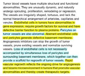
Abnormal Structural Features of Struttura dei vasi sanguigni normali
Tumor Microvasculature
AV = arteriovenous.
From Brown and Giaccia. Cancer Res. 1998;58:1408-1416, with permission.
Struttura vasi normali, segue Periciti
Cellule correlate alle cellule muscolari lisce vascolari.
pericita Queste cellule sono adiacenti e circondano l’endotelio, condividono
una lamina basale comune e hanno giunzioni di tipo “gap” con le
cellule endoteliali,
Non è ancora chiaro se queste cellule siano multipotenti, con la
capacità di differenziarsi sia in cellule muscolari lisce o persino
cellule endoteliali.
(Bergers G, Benjamin LE. Tumorigenesis and the angiogenic switch. Nat Rev Cancer. :401-410, 2003).
http://www.udel.edu/biology/Wags/histopage/vascularmodelingpage/circsystempage/capillaries/capillaries.html
2
09/05/2013

              http://www.medscape.org/viewarticle/461038_7                           http://www.medscape.org/viewarticle/461038_7

                                                                       Bergers & Benjamin. Nat Rev Cancer. 2003 Jun;3(6):401-10.

                                                                      The angiogenic balance (1)

                                                             Angiogenesis is orchestrated by a variety of activators and
                                                             inhibitors — only a few of which are listed in the picture above.
                                                             Activators of endothelial-cell proliferation and migration are
                                                             mainly receptor tyrosine kinase ligands, such as vascular
                                                             endothelial growth factor (VEGF), fibroblast growth factors
                                                             (FGFs), platelet-derived growth factor (PDGF) and epidermal
                                                             growth factor (EGF), but can also be of very different origin,
                                                             such as lysophosphatic acid (LPA).
                                                             EGF upregulates VEGF, FGF and interleukin-8, whereas LPA
                                                             upregulates VEGF levels.
                                                             The first described angiogenic inhibitor was
                                                             thrombospondin-1, which modulates endothelial-cell
                                                             proliferation and motility.

Bergers & Benjamin. Nat Rev Cancer. 2003 Jun;3(6):401-10.              Bergers & Benjamin. Nat Rev Cancer. 2003 Jun;3(6):401-10.

                                                                                                                                            3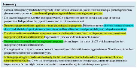
09/05/2013

          The angiogenic balance (2)

Remarkably, many inhibitory molecules, such as ‘statins’, are
derived from larger proteins that have no effect on
angiogenesis.
Among those that are listed are angiostatin (a fragment of
plasminogen that binds ATP synthase and annexin II), as well
as endostatin, tumstatin and canstatin (fragments of
collagens that bind to integrins).
In general, the levels of activators and inhibitors dictate
whether an endothelial cell will be in a quiescent or an
angiogenic state.
It is believed that changes in the angiogenic balance
mediate the angiogenic switch.

           Bergers & Benjamin. Nat Rev Cancer. 2003 Jun;3(6):401-10.              Bergers & Benjamin. Nat Rev Cancer. 2003 Jun;3(6):401-10.

   The classical angiogenic switch [interrutore].

The angiogenic switch is a discrete step in tumour development that
can occur at different stages in the tumour‐progression pathway,
depending on the nature of the tumour and its microenvironment.
Most tumours start growing as avascular nodules (dormant) (a) until
they reach a steady‐state level of proliferating and apoptosing cells.
The initiation of angiogenesis, or the ‘angiogenic switch’, has to occur
to ensure exponential tumour growth.
The switch begins with perivascular detachment and vessel dilation
(b), followed by angiogenic sprouting (c), new vessel formation and
maturation, and the recruitment of perivascular cells (d).
Blood‐vessel formation will continue as long as the tumour grows, and
the blood vessels specifically feed hypoxic and necrotic areas of the
tumour to provide it with essential nutrients and oxygen (e).
           Bergers & Benjamin. Nat Rev Cancer. 2003 Jun;3(6):401-10.              Bergers & Benjamin. Nat Rev Cancer. 2003 Jun;3(6):401-10.

                                                                             Normal vasculature (on right) is very
                                                                              orderly, unbranched, nearly parallel
                                                                           vessels compared with tumor vasculature
                                                                                           (on left)

                                                                                                                                                      4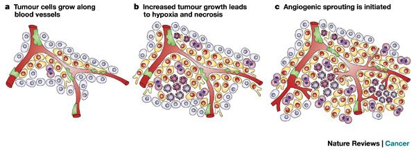
09/05/2013

  Typical structure of tumor vasculature

Note coiling,
irregularity,
size
heterogeneity
                                                              New blood-vessel formation. Blood vessels arise from pre-existing capillaries or post-
                                                              capillary venules in tumours (a). b | First, pericytes (green) detach and blood vessels dilate
                                                              before the basement membrane and extracellular matrix is degraded. c | This allows
                                                              endothelial cells (red) to migrate into the perivascular space towards angiogenic stimuli
                                                              produced by the tumour cells or host cells. d | Endothelial cells proliferate, loosely following
                                                              each other, and are presumably guided by pericytes. e | Behind the migration columns,
                                                              endothelial cells adhere to each other and create a lumen, which is accompanied by
                                                              basement-membrane formation and pericyte attachment. Finally, blood-vessel sprouts will
                                                              fuse with other sprouts to build new circulatory systems. Little is known about this fusion
                                                              mechanism.

Small tumors are not always avascular masses stimulating
vessel growth…some “co-opt” existing vessels, then
stimulate angiogenesis through hypoxia

                                                             Blood vessel co-option precedes angiogenesis in astrocytoma progression.
                                                             Astrocytomas first acquire their blood supply by co-opting existing normal brain blood vessels
                                                             without the necessity to initiate angiogenesis. They instead grow along blood vessels, without
                                                             a tumour capsule, eliciting an invasive character (a) . When grade III astrocytomas progress
                                                             into glioblastomas (GBM or grade IV astrocytoma), they become hypoxic and necrotic —
                                                             partly due to vessel regression and increased tumour-cell proliferation (b). These conditions,
                                                             in turn, induce formation of new blood vessels (angiogenic sprouting) (c) that supply the
                                                             tumour with the necessary metabolites. In fact, glioblastomas are partly defined by the
                                                             appearance of proliferating endothelial cells and a high blood-vessel density that distinguishes
                                                             grade IV tumours from the lower-grade astrocytomas

  Endothelial cells are aligned into three zones during
  angiogenesis.
 Basement   Parent      ZONE III    ZONE II         ZONE I
 membrane
            vessel

                                                                                             VEGF and FGF-2
                              Direction of growth

       Migrating cells       –                 –         +   Cathepsins,
       Dividing cells        –                 +         –   MMPs
       Maturing cells        +                 –
                             –

                                                                                                                                                                 5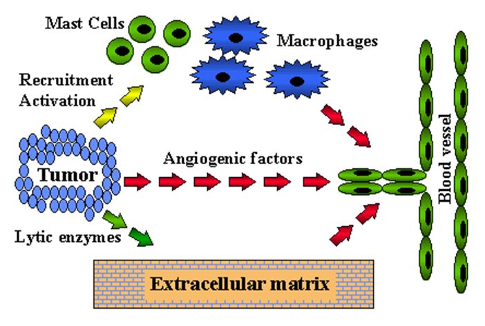
09/05/2013
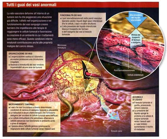
I VASI SANGUIGNI ANORMALI (sopra) complicano il caos all’interno del tumore ed
impediscono ai trattamenti di raggiungere le cellula tumorali. La possibilità di
“normalizzare” tali vasi (sotto) li rende una sorta di arma funzionale che può essere
utilizzata contro il tumore.
6
09/05/2013

                                                                                   (b) Untreated tumor showing a disorganized, anastomotic network
(a) Normal vasculature showing hierarchy of arterioles, capillaries and            of vessels that lacks the conventional hierarchy. Arterioles,
venules, which all have a characteristic size, shape and wall structure.           capillaries and venules are not discernable per se. Gaps are
Arterioles are enveloped by smooth muscle cells, and capillaries                   present between endothelial cells. Pericytes are irregularly
and venules are accompanied by pericytes. Thin basement                            shaped and loosely associated with endothelial cells. Basement
membrane surrounds all mural cells and endothelial cells.                          membrane has multiple layers in some places.

                                                              Baluk et al., 2005                                                                 Baluk et al., 2005

                                                                                                                         Scanning electron micrograph
                                                                                                                         showing
                                                                                                                         (a) the external (abluminal)
                                                                                                                             surface of an endothelial
                                                                                                                             sprout in a pancreatic islet
                                                                                                                             tumor from a RIP-Tag2
                                                                                                                             transgenic mouse. Multiple
                                                                                                                             filopodia (short arrows) extend
                                                                                                                             from the endothelial cell surface
                                                                                                                             near the tip of the sprout. A gap
                                                                                                                             is visible at an open endothelial
                                                                                                                             cell junction (long arrow).
                                                                                                                             Intravascular erythrocytes are
                                                                                                                             visible through the thin, nearly
                                                                                                                             transparent endothelium.
                                                                                                                         Scanning electron micrographs
(c) Vasculature of tumor after treatment with inhibitor of VEGF                                                          comparing
signaling. Many vessels have regressed, leaving empty sleeves of                                                         b) the smooth, tight endothelial cell
basement membrane (blue). Surviving vessels have a more normal                                                              monolayer covering the luminal
cylindrical shape. Loss of endothelial cells is not accompanied by                                                          surface of normal blood vessel
                                                                                                                            with
corresponding loss of pericytes. Some free pericytes are surrounded
                                                                                                                         c)    the disorganized endothelium of
by basement membrane but not accompanied by endothelial cells.
                                                                                                                              vessel in a RIP-Tag2 tumor.
                                                                                                                              Blood was removed by vascular
                                                                                                                              perfusion of fixative.
                                                              Baluk et al., 2005                                                                 Baluk et al., 2005

                                                                                                                                                                      7
09/05/2013

                                                                                     (d,e) Scanning electron micrographs showing pericytes on the surface of
                                                                                     irregularly shaped tumor vessels in RIP-Tag2 mice. As a reflection of their
                                                                                     loose association with tumor vessels, pericyte cell bodies are not located on the
                                                                Baluk et al., 2005   vessel wall, but some pericyte processes (arrows) contact endothelial cells.

                                                                                         I VASI SANGUIGNI in un letto vascolare del muscolo di topo
                                                                                       (sinistra) e all’interno di un tumore (destra) si differenziano
                                                                                       distintamente.
                                                                                         I vasi tumorali si ramificano in modo erratico, variano in diametro
                                                                                       lungo la loro lunghezza e sono di solito sovradimensionati – tutte
                                                                                       caratteristiche che contribuiscono ad un flusso ematico irregolare.
Changes produced in tumor blood vessels by inhibition of VEGF signaling. (a)
Scanning electron micrograph showing blood vessel in a RIP-Tag2 tumor treated
with inhibitor of VEGF-signaling for 7 days. The normalized vessel has a
cylindrical shape and is tightly enveloped by multiple pericyte processes                Jain R.K., Scientific American, Jan 2008

(arrows).

                                                                                                                                                                         8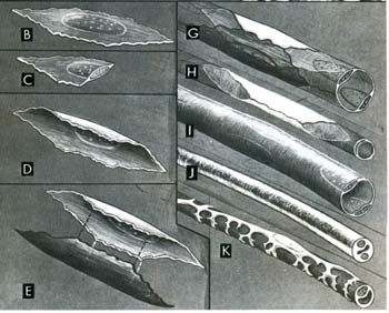
You can also read