Work in Progress: Tumor-Immune Interactions in Triple Negative Breast Cancer Brain Metastases
←
→
Page content transcription
If your browser does not render page correctly, please read the page content below
Work in Progress:

Tumor-Immune Interactions in Triple Negative
      Breast Cancer Brain Metastases

                   - Triple Negative Breast Cancer
                   - Breast-to-Brain Metastases
                       - TNBC
                       - Leptomeningeal Disease (LMD)
                   - Proposed Project Aims
                       - Rationale
                       - Preliminary Data
                       - Approach
                   - Current Work and Future Timeline

                                Maxine Umeh-Garcia, PhD, MSc.
                                              SCIT T32 Seminar
                               Hayden Gephart and Plevritis Labs
                                                April 22nd 2020
Breast Cancer
•   1 in 8 women in the U.S. will develop invasive breast cancer

•   In 2018, an estimated 268,600 new cases (invasive) and 62,930 (non-
    invasive) breast cancer are expected to be diagnosed in women in
    the U.S., of which about 41,760 women are expected to die

                  •   In women under 45, breast cancer is most common
                      in African-American women, and they are more
                      likely to die of breast cancer

                  •   Currently more than 3.1 million women with a history
                      of breast cancer in the U.S.

                  •   85% of breast cancers occur in women who have no
                      family history of breast cancer

                                                             SEER.CANCER.GOV
Triple Negative Breast Cancer (TNBC)
• TNBC is a heterogeneous group of tumors simply defined by the absence of
  estrogen (ER) and progesterone (PR) hormone receptors, and lack of
  overexpression of epidermal growth factor receptor 2 (ErbB2/Her2) gene
                                                             ErbB2/Her2
                                                                      ligand
• TNBC account for 10-20% of all
                                       ER      PR
  invasive breast cancers

• TNBC is associated with African-
  American race, younger age, higher
  tumor grade, and more advanced
                                     ER+/PR+
  tumor stage at diagnosis

• Chemotherapy is the only recommended systemic                Her2+
  treatment, however only 30% of TNBC patients
  achieve pCR. Patients who do not have 6-fold
  higher risk of relapse, and 12-fold higher risk of death

• Survival at 3 yrs is lower (68%) for metastatic TNBC patients
  compared to other metastatic breast cancer types (88%)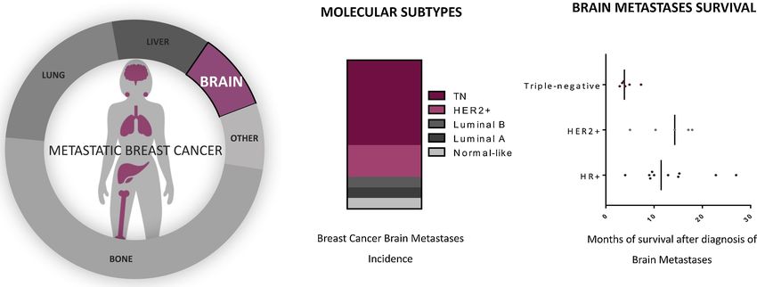
Triple Negative Breast Cancer (TNBC)

                           Liedtke C, Mazouni C, Hess KR et al.
TNBC in African-American Women
• Women of African ancestry have a disproportionately higher frequency
  (up to 79%) of TNBC, compared to women of European ancestry
    • TNBC frequency is consistently higher in women of African ancestry
      than any other racial/ethnic group

• In African-American women premenopausal status, increased parity
  (pregnancies), and shorter duration of breastfeeding are positively
  associated with increased risk of TNBC

• 5-year distant relapse-free survival is 62.8% for young black women,
  compared with 77% for young white women with equal access to health
  care (UK study)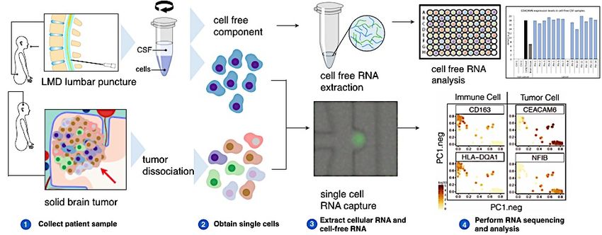
Dissertation Research –
What molecular mechanisms and/or
signaling pathways regulate TNBC
cells in vitro and TNBC tumors in vivo?        Primary TNBC

                              overexpression
                                 miR-127
                                                        reactivation of
TNBC Metastasis                                             LRIG1

                                      Postdoctoral Research –
                                      What molecular mechanisms drive
                                      shedding/dissemination, seeding,
                                      and outgrowth of TNBC metastases?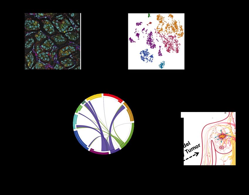
Breast Cancer Brain Metastasis
- Breast cancer brain metastasis (BCBM) occurs in 10-30% of metastatic breast
  cancer patients
     - Second leading cause of brain metastases following lung cancer

- Incidence of BCBM continues to increase
     - Prolonged patient survival
     - Improved imaging techniques

- Median survival ranges from 2 – 25.3 months
    - Few patients survive past 1 year
    - Associated with serve neurological decline

                        Before and After Surgical Resection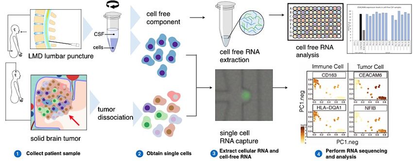
Breast Cancer Brain Metastasis

- BCBM Incidence and Survival is breast cancer subtype dependent

- Current treatment strategies:
    - Surgical resection
    - Whole brain radiation therapy (WBRT)
    - Stereotactic Radiosurgery
    - Chemotherapy
    - Targeted therapies (HR+: Tamoxifen, HER2+: Trastuzamab)

- Major challenge in treating BCBMs is the Blood-Brain-Barrier

Although there are ongoing clinical trails, no FDA-approved systemic treatments for BCBM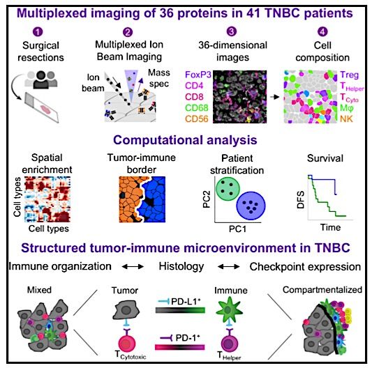
Leptomeningeal Disease (LMD)
- LMD is defined as tumor spread within the leptomeninges and subarachnoid space

- 10% of patients with solid cancers present with LMD

- Breast (TNBC), lung, and melanoma are the most common primary tumor sites in LMD
  patients

- LMD is diagnosed using MRI and CSF analysis
- LMD survival is extremely poor
    - Lung: 3 - 6 months
    - Breast: 3.5 – 4.4 months
    - Melanoma: 1.7 – 2.5 months

- Therapeutic strategies include intrathecal
  therapy (spinal canal and subarachnoid
  space to reach CSF), systemic therapy,
  and radiotherapy (WBRT)

- To date, there have been only 6
  randomized clinical trails specifically on
  treatment of LMD

- Understanding the molecular mechanisms that drive TNBC brain/LMD metastasis (seed
  – primary TNBC and soil – normal brain microenvironment) pose an unmet clinical need
“The Birth” of the Project

MY FIELD   SCIT T32

       NEW
     TRAINING

                                   Angelo et al, 2014, Nature Medicine
Project Hypothesis The spatial architecture of the tumor microenvironment reflects distinct tumor-immune interactions; these interactions prime systemic immune tolerance of disseminated tumor cells, enabling brain-specific metastases.
AIM 1: DETERMINE THE EXTENT TO WHICH THE STRUCTURED MICROENVIRONMENT
CORRELATES WITH PATIENT OUTCOMES BY GENERATING A TUMOR-IMMUNE
SPATIAL MAP OF TNBC BRAIN METASTASES.
RATIONALE:
1. Immune infiltration is associated with patient survival in specifically in TNBC
subtype
2. Angelo Lab – Immune landscape of 41 primary TNBCs using MIBI
Keren et al, 2018, Cell
3. The brain was previously thought to be an “immune-privileged” space so there
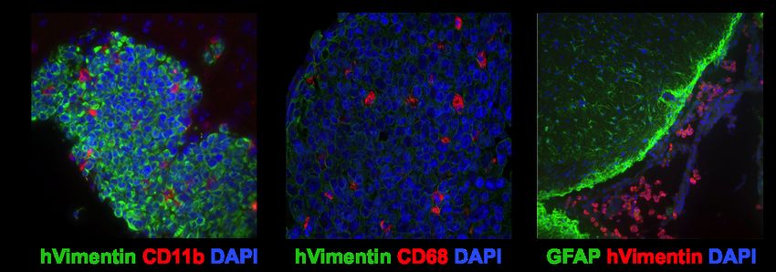
has been little interrogation of the immune landscape of TNBC brain metastasesAIM 1: DETERMINE THE EXTENT TO WHICH THE STRUCTURED MICROENVIRONMENT CORRELATES WITH PATIENT OUTCOMES BY GENERATING A TUMOR-IMMUNE SPATIAL MAP OF TNBC BRAIN METASTASES. PRELIMINARY DATA: 1. Presence of infiltrating immune cells in a mouse model of human TNBC brain metastases 2. Astrocytes increase the production of glial fibrillary acidic protein (GFAP) in the presence of TNBC leptomeningeal disease
AIM 1: DETERMINE THE EXTENT TO WHICH THE STRUCTURED MICROENVIRONMENT
 CORRELATES WITH PATIENT OUTCOMES BY GENERATING A TUMOR-IMMUNE
 SPATIAL MAP OF TNBC BRAIN METASTASES.

APPROACH

A. Construct an in-situ subcellular
   protein spatial map of the TNBC brain
   metastases microenvironment using
                                                                      MIBI
   MIBI on archival FFPE tissue samples.

B. Quantitate the composition and
   spatial architecture of the tumor-
   immune microenvironment using a
                                                       OR
   validated image analysis pipeline.
                                           MIXED        COMPARTMENTALIZED
C. Assess the extent to which the
   composition and spatial architecture
   correlates with CNS disease

                                            SURVIVAL
   progression, the likelihood of LMD                                    LMD
   development, and patient survival.                          and
                                                                     development?

                                                       TIME
A. Protein spatial map of the TNBC brain metastases microenvironment
Tumor-Immune Panel Keren et al, 2018, Cell
Tumor Immune Cell Types Immune Stroma
CD31 Vimentin
β-Catenin Lymphocytes Monocytes Antigen Regulation SMA
EGFR CD3 CD45RO CD11b Presentation Lag3
Keratin 6 CD4 CD20 CD11c HLA1 Cell Status
CD8 CD56 CD63 PD1
Keratin 17 HLA-DR dsDNA H3K27me3
CD56 CD16 CD68 CD209 PD-L1
Pan-keratin FoxP3 CD138 Neutrophils Ki-67 H3K9ac
IDO
p53 MPO pS6
Tumor-Brain, Immune-Brain, Metastasis, TNBC-Specific Panel
Brain Resident Cell Types
Neurons Astrocytes Microglia
NeuN GFAP CD45 CD45
TMEM119 OR
Iba1
P2RY12 Metastasis
Stemness
Interactions Nestin
TNBC- Tumor/Neurons Microglia/Macrophages CD133
Specific
CD47 ------------- SIRPa CD44
BRCA1 C3 ------------- CD11b MMP9
BRCA2 CD200 ------------- CD200R MMP1
SOX11 CD15
CX3CL1 ------------- CX3CR1AIM 2: IDENTIFY TUMOR-IMMUNE RECEPTOR-LIGAND PAIRS BY GENERATING A TRANSCRIPTOMIC PROFILE OF TNBC BRAIN METASTASES, AND DETERMINE IF THESE INTERACTIONS CORRELATE WITH TUMOR-IMMUNE SPATIAL ARCHITECTURE. RATIONALE: 1. MIBI panel is highly focused – unbiased approach to identify tumor-immune interactions (receptor-ligand pairs), which can be then be assessed by MIBI or traditional IHC 2. Identify novel targetable tumor-immune interactions for future therapies, beyond PD-1/PD-L1. PDCD1 (PD-1) PRELIMINARY DATA: 1. Assessed a few validated tumor- immune receptor-ligand pairs in GBMseq.org
AIM 2: IDENTIFY TUMOR-IMMUNE RECEPTOR-LIGAND PAIRS BY GENERATING A
 TRANSCRIPTOMIC PROFILE OF TNBC BRAIN METASTASES, AND DETERMINE IF THESE
 INTERACTIONS CORRELATE WITH TUMOR-IMMUNE SPATIAL ARCHITECTURE.

APPROACH

A. Build RNA expression profiles of TNBC
                                                               DISSOCIATE
   brain metastases (and healthy brain)                            and
   using single-cell RNA-sequencing        MET or NORMAL       SORT CELLS

B. Identify co-expression of genes that
   encode receptor-ligand pairs in
   tumor and immune cell populations
   using biocomputational approaches.
                                                                    SEQUENCING
                                                                        and
C. Assess the extent to which receptor-                               ANALYSIS
   ligand pairs correlate with tumor-
   immune spatial architecture.

                                                                            MIBI
                                                  RECEPTOR-LIGAND
                                                       PAIRS
AIM 3: DETERMINE IF TUMOR-IMMUNE INTERACTIONS IN PRIMARY TNBC PRIME TOLERANCE OF DISSEMINATED CELLS ENABLING METASTASES, AND DEFINE IF INTERACTIONS CORRELATE WITH RACE. RATIONALE: 1. Enk et al. – Altered function of dendritic cells in progressing versus regressing melanoma metastases. Hypothesized that this tolerance was a result of dendritic cells co-opted by the tumor, which possessed the ability to migrate from the primary tumor to the regional lymphatic organs. Suggests that the immune landscape of the primary tumor could contribute to systemic immune tolerance, enabling metastatic outgrowth PRELIMINARY DATA: 1. Does spatial organization of the tumor-immune microenvironment effect the odds of metastatic p = 0.13 potential in TNBC? Keren et al. study - Recurrence status on 38 of the 41 primary TNBC tumor samples. Tumors with compartmentalized spatial architecture were less likely to be associated with recurrence than tumors with mixed architecture
AIM 3: DETERMINE IF TUMOR-IMMUNE INTERACTIONS IN PRIMARY TNBC PRIME
 TOLERANCE OF DISSEMINATED CELLS ENABLING METASTASES, AND DEFINE IF
 INTERACTIONS CORRELATE WITH RACE.

APPROACH

A. Visualize the tumor-immune landscape in
   primary TNBC tumors using MIBI, and
   assess the extent to which it correlates
   with brain metastases and/or LMD
   development.
                                                                             ** Relevant
B. Identify tumor-immune interactions that                                  Targets from
   are differentially expressed between                                     Aims 1 & 2 **
   patients of differing racial backgrounds
   **Racial disparity in primary TNBC
   **Studies (limited) have identified differences in
     immune response based on patient race                      MIBI

C. Measure expression of relevant targets in
   human cerebrospinal fluid (CSF).
   **CSF can detect changes in brain tumors
   **Patient CSF can easily be collected/stored
   **HG lab is currently testing the stability and
                                                      IMMUNE             SPATIAL
     reproducibility of assay using CSF-derived RNA LANDSCAPE          ARCHITECTURE
Acknowledgments
² Melanie Hayden Gephart, MD, MAS
² Sylvia K. Plevritis, PhD
² Drs. M. Angelo, S. Napel, S. Quake, C. Curtis
² Gephart Lab Members
² Plevritis Lab Members
² Funding: NIH SCIT T32
Thank you for your attention!
Questions?You can also read