Prognostic factors in endometrial adenocarcinoma: FIGO staging and the CAP template - Richard J. Zaino, MD Hershey Medical Center Penn State ...
←
→
Page content transcription
If your browser does not render page correctly, please read the page content below
Prognostic factors in endometrial
         adenocarcinoma:
FIGO staging and the CAP template

         Richard J. Zaino, MD
        Hershey Medical Center
         Penn State University
             Hershey, PA
           rzaino@psu.edu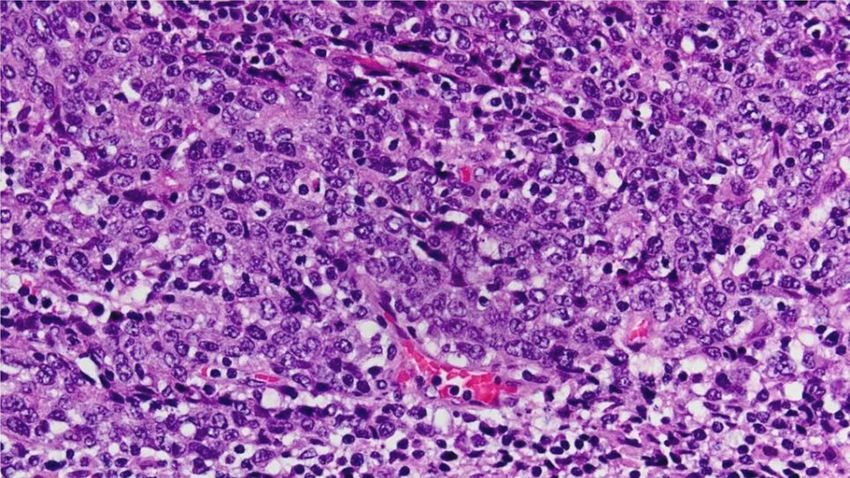
Disclosure
Consultant for Becker (NSF International)
 for cervical cancer screening
Objectives
1) Examine the changes and utility of the
   2008 FIGO staging scheme for endometrial
   cancer
2) Examine the application of and
   significance of the CAP template for
   endometrial cancer
3) Review the biology of the major types of
   endometrial adenocarcinoma
4) Examine prognostic factors in endometrial
   carcinoma
Surgical Staging of Corpus Cancer
            (FIGO,1988)
Stage           Characteristics
IA G123   tumor limited to endometrium
IB G123   invasion to inner half of myometrium
IC G123   invasion to outer half of myometrium
IIA       endocervical gland involvement
IIB       cervical stromal invasion
IIIA      tumor invades serosa, adnexa, or + peritoneal cyto
IIIB      vaginal metastases
IIIC      pelvic or para-aortic lymph node metastases
IVA       tumor invades bladder or bowel mucosa
IVB       distant, intraabdominal or inguinal node
          metastases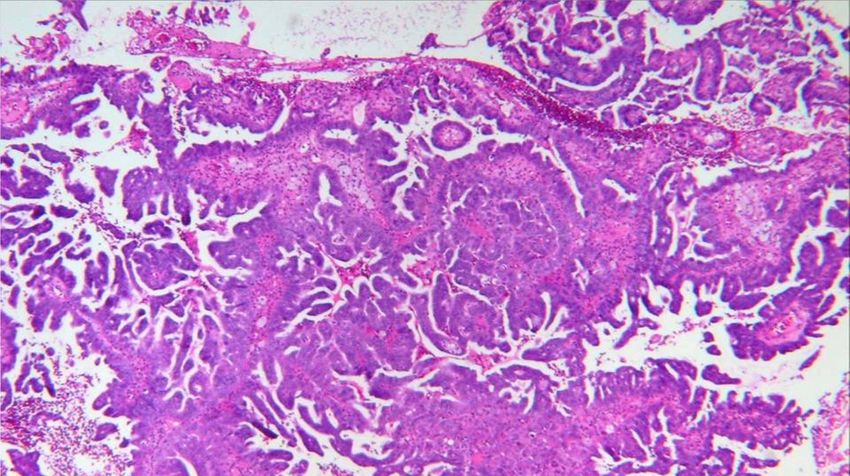
Surgical Staging of Corpus
             Cancer (FIGO, 2008)
Stage             Characteristics
IA G123     tumor to endometrium/inner half of myometrium
IB G123     invasion to outer half of myometrium
II          endocervical cervical stromal invasion
IIIA        tumor invades serosa, adnexa
IIIB        vaginal metastases or parametrial extension
IIIC1       pelvic lymph node metastases
IIIC2       para-aortic lymph node metastases
IVA         tumor invades bladder or bowel mucosa
IVB         distant, intraabdominal or inguinal node
            metastases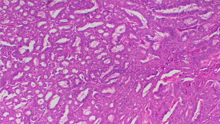
CAP approved
                    (so it must be good for us*)

Protocol for the Examination of Specimens from
  Patients with Carcinoma of the Endometrium

Based on AJCC/UICC TNM, 7th edition and FIGO
  2008 Annual Report

  Protocol web posting date: June, 2012
  Surgical Pathology Cancer Case Summary (Checklist)

*as a current member of the CAP committee on gyn tumor synoptic
  reports, these comments do not reflect the opinions of the CAP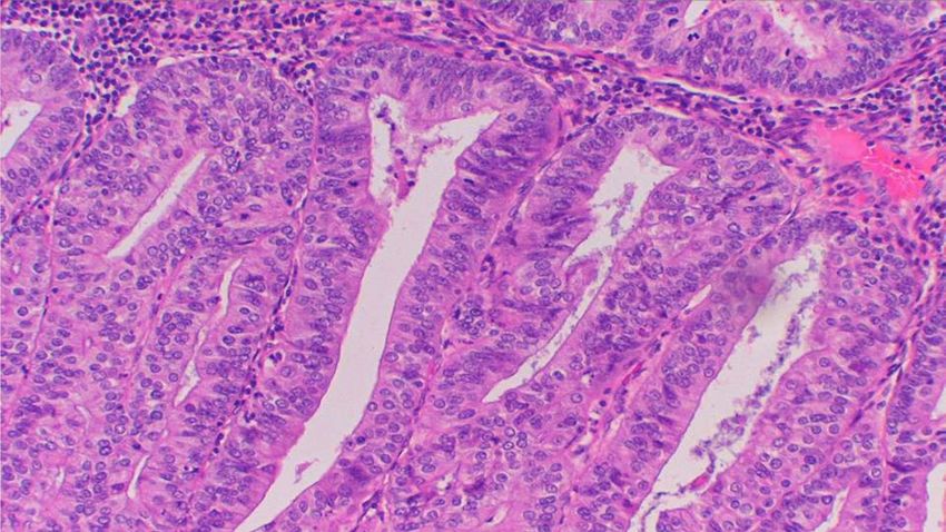
Tumor Size
Greatest dimension: ___ cm
*Additional dimensions: ___ x ___ cm
___ Cannot be determined (see Comment)

Histologic Type
___ Endometrioid adenocarcinoma, not otherwise characterized
___ Endometrioid adenocarcinoma, variant (specify):
___ Mucinous adenocarcinoma
___ Serous adenocarcinoma
___ Clear cell adenocarcinoma
___ Mixed carcinoma (specify types and percentages):
___ Squamous cell carcinoma
___ Transitional cell carcinoma
___ Small cell carcinoma
___ Undifferentiated carcinoma
___ Carcinosarcoma (malignant müllerian mixed tumor)
___ Other (specify):
Significance of maximum size of
  endometrial adenocarcinoma
Relative few studies addressing size, but
 prognostically significant

Mariani et al, 2001 and 2002
 size > 2cm is a predictor of lymphatic
 failure and distant failure by univariate
 analysis but not by multivariate analysis
Tumor Size
Greatest dimension: ___ cm
*Additional dimensions: ___ x ___ cm
___ Cannot be determined (see Comment)

Histologic Type
___ Endometrioid adenocarcinoma, not otherwise characterized
___ Endometrioid adenocarcinoma, variant (specify):
___ Mucinous adenocarcinoma
___ Serous adenocarcinoma
___ Clear cell adenocarcinoma
___ Mixed carcinoma (specify types and percentages):
___ Squamous cell carcinoma
___ Transitional cell carcinoma
___ Small cell carcinoma
___ Undifferentiated carcinoma
___ Carcinosarcoma (malignant müllerian mixed tumor)
___ Other (specify):
Pathologic classification of
endometrial adenocarcinomas
     1980              2012
adenocarcinoma   endometrioid
adenoacanthoma   endometrioid w squamous diff
adenosquamous    villoglandular
clear cell       secretory
                 mucinous
                 serous (UPSC)
                 clear cell
                 undifferentiated
                 dedifferentiated
                 mixed
uterine papillary serous carcinoma
papillaeSurvival in endometrial adenocarcinoma (all stages) Tumor type 5 yr survival Endometrioid 80 – 90% UPSC 10 – 30%
UPSC – patterns of spread
Author             sites of disease
Carcangiu    intrabdominal/small bowel
Mallipeddi   nodes, bowel, omentum, cyto
Lee          ovaries, nodes, peritoneum
Gitsch       cyto, nodes, omentum, liver, dia
Carcangiu    adnexa, peritoneum, omentum, nodes
Cirisano     nodes, ovaries, peritoneum, omentum
Wheeler      ovary, omentum, bowel
Goff         ovary, nodes, omentum, peritoneum
Sherman      nodes, cyto, ovary, omentum
Geisler      omentum, cyto, peritoneum, nodes
Immunohistochemistry
   Cell type                     ER/PR        p53
  Endometrioid                   +++^         -/+++*
  Villoglandular                 +++          -
  Serous                         -/+**        +++*
^ - in high grade
* +++ reflects with >80% positive or completely neg, if
  mutation not recognized by Ab
** often focal and weak
p53
Undifferentiated and
dedifferentiated carcinoma
Silva et al, Soslow et al
No gland formation (or bi-phasic with glands
in differentiated areas only)
Often appear discohesive
Usually keratin negative
Usually ER/PR negative
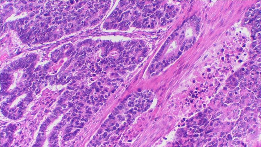
Very aggressive behaviorHistologic Grade (if applicable) (FIGO grading system applies to endometrioid and mucinous carcinomas only) ___ G1: 5% or less nonsquamous solid growth ___ G2: 6% to 50% nonsquamous solid growth ___ G3: More than 50% nonsquamous solid growth
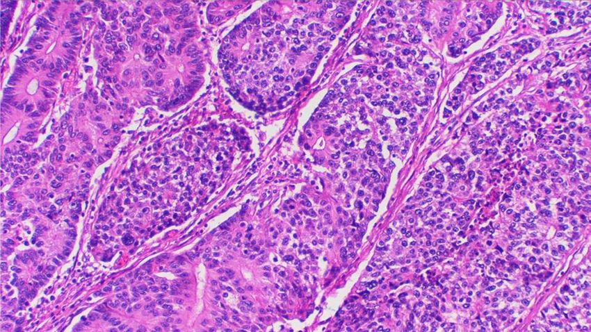
Grade 1
Grade 2
Grade 3
Grade 1 (architecture)
Grade 3 nuclei
Overall grade 2
Stage I adenocarcinoma of the
         endometrium
          (FIGO results, 2003)

   FIGO Grade              5 year survival
      1                    92%
      2                    88%
      3                    75%
Grading endometrial adenocarcinoma
Two grades versus three
FIGO – 3 grades, architecture +/- nuclear
GOG – 3 grades, architecture
Hachisuga -3 grades, nuclear (quantitative)
Taylor et al – 2 grades, architecture (10% solid)
Scholten – 2 grades, architecture (50% solid)
Lax – 2 grades, architecture (solid, pattern, necrosis)
Alkushi – 2 grades, architecture and nuclear
Each prognosticates wellReproducibility of grading
Inter-observer kappa
2 grade 3 grade
Alkushi (arch+nuclear) 0.76 0.61
Nielsen (arch) 0.70
Nielsen (nuclear) 0.55
Zaino (arch) 0.49
Zaino (nuclear) 0.57
Taylor (arch) 0.97 0.52
Lax (arch) 0.65 0.55
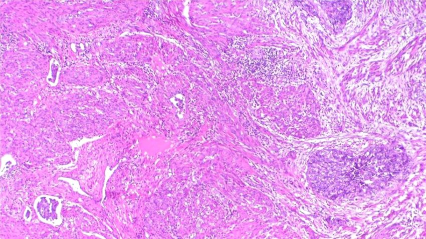
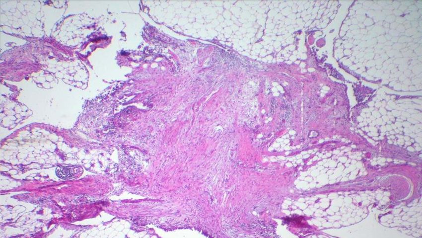
Scholten (arch, using Lax) 0.39 0.41Lymphatic invasion Lymph-vascular invasion ___not identified ___present ___indeterminate Prognostically important in almost every study 1) Use of immuno 2) Where to look
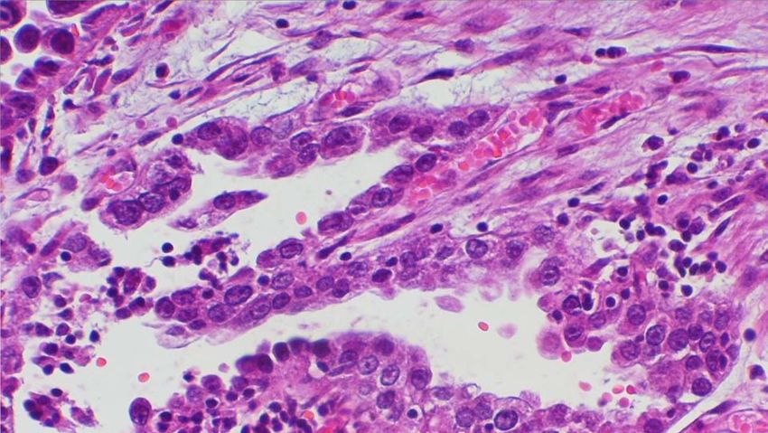
lymphatic invasion
vascular pseudo-invasion (VPI)
The Female Patient | VOL 35 SEPTEMBER 2010

 Gamal H. Eltabbakh, MD
Histologic artifacts in hysterectomy
     specimens. (Krizova, AJSP, 35:115-26, 2011)
              160 Malignant cases
Artifact RH NRLH         NRLH+UM          NLH
VPI        49%      7%        13%         0
Cleft      51      11         25          3
Intratub 40         9         25          3
 (DeLair*) 12       2         -           0
Crush      31       9         25          1
*DeLair et al, IJGP 32:188, 2013
FIGO 1988 Stage I Corpus Cancer 1) Is the distinction of non-invasive from inner half invasion reliable? 2) Should invasion be assessed in thirds or halves of myometrial thickness?
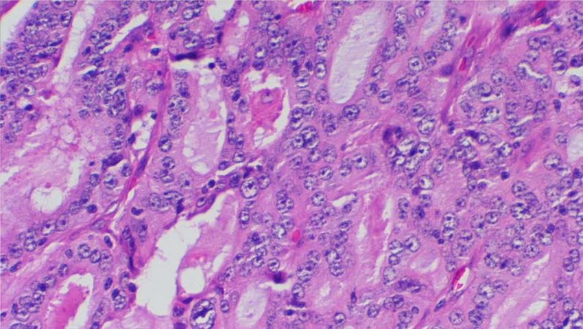
Superficial myometrial invasion
endometrium
myometrium
Superficial myometrial invasion
Stage I Corpus Cancer
     significance of invasion
Stage 5 year survival rates (FIGO, 2003)
IA 92% inability to distinguish inter-
IB 91% digitations from myo invasion
IC 81% outer half invasion highly
           significant
Primary Tumor (pT)
FIGO 2008
___ pTX [--]: Primary tumor cannot be assessed
___ pT0 [--]: No evidence of primary tumor
___ pT1a [IA]:Tumor limited to endometrium or invades less
than one-half of the myometrium
___ pT1b [IB]:Tumor invades greater than or equal to one-half
of the myometrium
___pT2 [II]: Tumor invades stromal connective tissue of the
cervix, but does not extend beyond uterus
___ pT3a [IIIA]:Tumor involves serosa and/or adnexa (direct
extension or metastasis)
___ pT3b [IIIB]:Vaginal involvement (direct extension or
metastasis) or parametrial involvement
___ pT4 [IVA]:Tumor invades bladder mucosa and/or bowel
mucosa (bullous edema is not sufficient to classify a tumor
as T4)Myometrial invasion or tumor
in adenomyosis
1) Multiple studies (Hall, Jacques, Mittal)
have demonstrated excellent prognosis
when carcinoma is confined to foci of
adenomyosis (superficial or deep)
2) Tumor confined to adenomyosis does not
increase the FIGO stage
3) What are the criteria for identification
of tumor in adenomyosis?Recognition of tumor in
adenomyosis
(Jacques and Lawrence, 1990)
Presence of endometrial stroma adjacent to
neoplastic glands in the myometrium
Presence of adjacent benign glands
Bulging expansion of endometrial-
myometrial junction or smoothly
rounded contour of entirely
intramyometrial foci
Absence of peritumoral desmoplasiaRecognition of tumor
in adenomyosis – CD10
(Sroden et al, 2003, Nascimento et al, 2003)
The presence of CD10 staining of small cells
adjacent to tumor is a sensitive but not
specific indicator of endometrial stromal
differentiation
More than 50% of myoinvasive endometrial
adenocarcinomas have CD10 positive
staining at least focally around the
tumorDifficulties in assessing the depth
of myometrial invasion in
endometrial carcinoma
Ali, Black and Soslow, (IJGP, 2007)
Depth of invasion – disagreement in 29% of cases
sources of disagreement
irregular endo-myometrial interface
exophytic tumor
smooth muscle metaplasia
tumor in adenomyosis
Most frequently - pathologists overestimate invasionFIGO 1988
Stage Characteristics
IA G123 tumor limited to endometrium
IB G123 invasion to inner half of myometrium
IC G123 invasion to outer half of myometrium
IIA endocervical gland involvement
IIB cervical stromal invasion
IIIA tumor invades serosa, adnexa, or + peritoneal cyto
IIIB vaginal metastases
IIIC pelvic or para-aortic lymph node metastases
IVA tumor invades bladder or bowel mucosa
IVB distant, intraabdominal or inguinal node
metastasesPrimary Tumor (pT)
FIGO 2008
___ pTX [--]: Primary tumor cannot be assessed
___ pT0 [--]: No evidence of primary tumor
___ pT1a [IA]:Tumor limited to endometrium or invades less
than one-half of the myometrium
___ pT1b [IB]:Tumor invades greater than or equal to one-half
of the myometrium
___pT2 [II]: Tumor invades stromal connective tissue of the
cervix, but does not extend beyond uterus
___ pT3a [IIIA]:Tumor involves serosa and/or adnexa (direct
extension or metastasis)
___ pT3b [IIIB]:Vaginal involvement (direct extension or
metastasis) or parametrial involvement
___ pT4 [IVA]:Tumor invades bladder mucosa and/or bowel
mucosa (bullous edema is not sufficient to classify a tumor
as T4)Stage II Corpus Cancer 5 year survival - 75%, lower than Stage I (FIGO results, 2003) More often associated with higher grade, deep myometrial invasion, and lymphatic invasion than Stage I Stage II is not a significant prognosticator by multivariate analysis
1988 Stage II Corpus Cancer
IIA   endocervical gland involvement
IIB   cervical stromal invasion

   Definitions applied in various publications:
IIA - surface epithelium only (Jordan)
IIA – gland involvement only (Fanning, Eltabbakh,
        Prat)
IIA – confined to endocervical epithelium (mucosa)
        (Clement and Young)
              but endocervix lacks a mucosa
              diagnostic reproducibility is low
              how does it involve glands only?
Cervical Involvement in Corpus
Cancer
(Zaino et al, Gyn Oncol, 2012)
Reproducibility and Prognostic Significance
for reproducibility
46 cases
6 pathologists (5 institutions) assessed
patterns of stromal vs gland involvement
for outcome (recurrence free survival)
200 cases and 200 matched stage I controlsReproducibility study Patterns of cervical involvement Kappa gland involvement 0.15 stromal invasion 0.28 vascular invasion only 0.09 contiguous spread 0.29 discontinuous spread 0.49 Kappa of 0-0.2 (slight), 0.2-0.4 (fair), 0.4-0.6 (moderate), 0.6-0.8 (substantial), 0.8-1.0 (almost perfect)
Results
Patterns of cervical involvement
            range of pathologists ID of feature

gland involvement                     63-91%
stromal invasion*                     30-78%
vascular invasion only                  0-9%
contiguous spread                     56-80%
discontinuous spread                  13-37%

*Definition of stage II (FIGO 2009)
Stage II Corpus Cancer
(Zaino et al, 2012)
Reproducibility
Reproducibility of distinction of gland involvement
from stromal invasion is poor
Kappa = 0.28
Prognostic Significance
Cervical involvement is not a significant
prognosticator by univariate or multivariate
analysisFIGO 2008 Primary Tumor (pT) ___ pT1a [IA]:Tumor limited to endometrium or invades less than one-half of the myometrium ___ pT1b [IB]:Tumor invades greater than or equal to one-half of the myometrium ___pT2 [II]: Tumor invades stromal connective tissue of the cervix, but does not extend beyond uterus ___ pT3a [IIIA]:Tumor involves serosa and/or adnexa (direct extension or metastasis) ___ pT3b [IIIB]:Vaginal involvement (direct extension or metastasis) or parametrial ___ pT4 [IVA]:Tumor invades bladder mucosa and/or bowel mucosa (bullous edema is not sufficient to classify a tumor as T4)
Frequency of positive
      Peritoneal cytology
      (no longer part of FIGO staging)

Uterine manipulator         13%
Without uterine manipulator 3%
Stage III Corpus Cancer
Stage IIIB – parametrial involvement or
vaginal metastases
Very rare, (less than 1% of corpus cancer
and about 2% of stage III pts)
Vaginal mets often associated with nodal
or distant metastases
Prognosis poor – 5 year survival about 25%FIGO 2008 Stage IIIC1 and IIIC2 Regional Lymph Nodes (pN) ___ pN1 [IIIC1]: Regional lymph node metastasis to pelvic lymph nodes ___ pN2 [IIIC2]: Regional lymph node metastasis to para- aortic lymph nodes, with or without positive pelvic lymph nodes Pelvic lymph nodes: Number examined: ___ Number involved: ___ Para-aortic lymph nodes: Number examined: ___ Number involved: ___
Stage III Corpus Cancer
Stage III C – pelvic/paraaortic nodal mets

(Mariani et al, 2002)
Stage IIIC often are also Stage IIIA/IIIB
5 year DFS – 33% Stage IIIC with IIIA/B
  mostly extranodal failures
5 year DFS – 68% Stage IIIC without IIIA/B
  mostly nodal failures
Stage III Corpus Cancer
Stage III C – pelvic/paraaortic nodal mets
5 year DFS – about 65-80% + pelvic node
5 year DFS – about 30% + paraaortic node

Significant survival differences between
  microscopic and grossly positive nodes,
  resected vs non-resected disease,
  radiated vs non-irradiated nodal beds,
  and capsular invasion and desmoplasia
Nodes with isolated tumor cells

Very few studies
Use of sentinel node examination
  currently undefined
Immunohistochemistry discloses isolated
  histiocyte-like cells (esp. with MELF)
Significance uncertain
Staging rule is undefined
Tentative staging conclusions
Stage IA can reliably be distinguished from Stage
  IB pathologically
Stage II is poorly defined pathologically and may
  not be prognostically significant
Stage III disease is heterogeneous
Stage IIIA alone is heterogeneous
  + adnexal spread diminishes survival (70%)
  + uterine serosa carries a worse prognosis (30%)
Tentative staging conclusions
Stage IIIB (parametrium/vaginal mets) is rare,
  with a poor prognosis (25%)
Stage IIIC (good to have split)
  IIIC1 + pelvic nodes significant (70%)
  IIIC2 + paraaortic nodes significantly worse
  (30%)
  (Stage IIIC limited to nodes usually fails in
  nodal area)
Surgical Staging of Corpus
             Cancer (FIGO, 2008)
Stage             Characteristics
IA G123     tumor to endometrium/inner half of myometrium
IB G123     invasion to outer half of myometrium
II          endocervical cervical stromal invasion
IIIA        tumor invades serosa, adnexa
IIIB        vaginal metastases or parametrial extension
IIIC1       pelvic lymph node metastases
IIIC2       para-aortic lymph node metastases
IVA         tumor invades bladder or bowel mucosa
IVB         distant, intraabdominal or inguinal node
            metastases
The future? 1) Pathologists need to be outspoken in the identification of characteristics that relate to prognosis and response to therapy. 2) Future refinements are needed in identification of cell types. 3) Pathologists need to identify features that can help guide individualized treatment for endometrial carcinoma
Objectives
1) Examine the changes and utility of the
   2008 FIGO staging scheme for endometrial
   cancer
2) Examine the application of and
   significance of the CAP template for
   endometrial cancer
3) Review the biology of the major types of
   endometrial adenocarcinoma
4) Examine prognostic factors in endometrial
   carcinoma
You can also read