World Journal of Clinical Cases - World J Clin Cases 2021 April 26; 9(12): 2696-2950 - NET
←
→
Page content transcription
If your browser does not render page correctly, please read the page content below
ISSN 2307-8960 (online)

World Journal of
Clinical Cases
World J Clin Cases 2021 April 26; 9(12): 2696-2950

                                                     Published by Baishideng Publishing Group Inc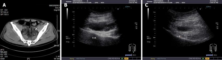
World Journal of
WJ C C                                                      Clinical Cases
Contents                                                    Thrice Monthly Volume 9 Number 12 April 26, 2021

          MINIREVIEWS
 2696     Standardization of critical care management of non-critically ill patients with COVID-19
          Wang CS, Gao Y, Kang K, Fei DS, Meng XL, Liu HT, Luo YP, Yang W, Dai QQ, Gao Y, Zhao MY, Yu KJ

 2703     Mediastinal lymphadenopathy in COVID-19: A review of literature
          Taweesedt PT, Surani S

 2711     Polycystic ovary syndrome: Pathways and mechanisms for possible increased susceptibility to COVID-19
          Ilias I, Goulas S, Zabuliene L

          ORIGINAL ARTICLE
          Clinical and Translational Research
 2721     Circulating tumor cells with epithelial-mesenchymal transition markers as potential biomarkers for the
          diagnosis of lung cancer
          Jiang SS, Mao CG, Feng YG, Jiang B, Tao SL, Tan QY, Deng B

          Retrospective Study
 2731     Management and implementation strategies of pre-screening triage in children during coronavirus disease
          2019 pandemic in Guangzhou, China
          Shi X, Cai YT, Cai X, Wen XL, Wang JY, Ma WC, Shen J, Wu JX, Liu HY, Sun J, He PQ, Lin Y, Zhao DY, Li PQ

 2739     Clinicopathological features of superficial CD34-positive fibroblastic tumor
          Ding L, Xu WJ, Tao XY, Zhang L, Cai ZG

 2751     Application of a rapid exchange extension catheter technique in type B2/C nonocclusive coronary
          intervention via a transradial approach
          Wang HC, Lu W, Gao ZH, Xie YN, Hao J, Liu JM

          SYSTEMATIC REVIEWS
 2763     Paradoxical relationship between proton pump inhibitors and COVID-19: A systematic review and meta-
          analysis
          Zippi M, Fiorino S, Budriesi R, Micucci M, Corazza I, Pica R, de Biase D, Gallo CG, Hong W

          META-ANALYSIS
 2778     Predictive risk factors for recollapse of cemented vertebrae after percutaneous vertebroplasty: A meta-
          analysis
          Ma YH, Tian ZS, Liu HC, Zhang BY, Zhu YH, Meng CY, Liu XJ, Zhu QS

        WJCC    https://www.wjgnet.com                         I                       April 26, 2021   Volume 9     Issue 12
World Journal of Clinical Cases
Contents
                                                            Thrice Monthly Volume 9 Number 12 April 26, 2021

          CASE REPORT
 2791     Malignant pheochromocytoma with cerebral and skull metastasis: A case report and literature review
          Chen JC, Zhuang DZ, Luo C, Chen WQ

 2801     Unresectable esophageal cancer treated with multiple chemotherapies in combination with
          chemoradiotherapy: A case report
          Yura M, Koyanagi K, Hara A, Hayashi K, Tajima Y, Kaneko Y, Fujisaki H, Hirata A, Takano K, Hongo K, Yo K, Yoneyama
          K, Tamai Y, Dehari R, Nakagawa M

 2811     Role of positron emission tomography in primary carcinoma ex pleomorphic adenoma of the bronchus: A
          case report
          Yang CH, Liu NT, Huang TW

 2816     Positive reverse transcription-polymerase chain reaction assay results in patients recovered from COVID-
          19: Report of two cases
          Huang KX, He C, Yang YL, Huang D, Jiang ZX, Li BG, Liu H

 2823     Laryngeal myxoma: A case report
          Yu TT, Yu H, Cui Y, Liu W, Cui XY, Wang X

 2830     Prostate stromal tumor with prostatic cysts after transurethral resection of the prostate: A case report
          Zhao LW, Sun J, Wang YY, Hua RM, Tai SC, Wang K, Fan Y

 2838     Intramuscular hematoma in rhabdomyolysis patients treated with low-molecular-weight heparin: Report
          of two cases
          Yuan SY, Xie KF, Yang J

 2845     Partial response to Chinese patent medicine Kangliu pill for adult glioblastoma: A case report and review
          of the literature
          Sun G, Zhuang W, Lin QT, Wang LM, Zhen YH, Xi SY, Lin XL

 2854     Behcet’s disease manifesting as esophageal variceal bleeding: A case report
          Xie WX, Jiang HT, Shi GQ, Yang LN, Wang H

 2862     Successful endoscopic surgery for emphysematous pyelonephritis in a non-diabetic patient with
          autosomal dominant polycystic kidney disease: A case report
          Jiang Y, Lo R, Lu ZQ, Cheng XB, Xiong L, Luo BF

 2868     Robotically assisted removal of pelvic splenosis fifty-six years after splenectomy: A case report
          Tognarelli A, Faggioni L, Erba AP, Faviana P, Durante J, Manassero F, Selli C

 2874     Pulmonary alveolar proteinosis complicated with nocardiosis: A case report and review of the literature
          Wu XK, Lin Q

 2884     Detection of EGFR-SEPT14 fusion in cell-free DNA of a patient with advanced gastric cancer: A case report
          Kim B, Kim Y, Park I, Cho JY, Lee KA

        WJCC    https://www.wjgnet.com                        II                          April 26, 2021   Volume 9   Issue 12
World Journal of Clinical Cases
Contents
Thrice Monthly Volume 9 Number 12 April 26, 2021
2890 Timing of convalescent plasma therapy-tips from curing a 100-year-old COVID-19 patient using
convalescent plasma treatment: A case report
Liu B, Ren KK, Wang N, Xu XP, Wu J
2899 Torsades de pointes episode in a woman with high-grade fever and inflammatory activation: A case report
Qiu H, Li HW, Zhang SH, Zhou XG, Li WP
2908 Salivary duct carcinoma of the submandibular gland presenting a diagnostic challenge: A case report
Uchihashi T, Kodama S, Sugauchi A, Hiraoka S, Hirose K, Usami Y, Tanaka S, Kogo M
2916 Allogeneic hematopoietic stem cell transplantation in a 3-year-old boy with congenital pyruvate kinase
deficiency: A case report
Ma ZY, Yang X
2923 Congenital bilateral cryptorchidism in an infant conceived after maternal breast cancer treatment: A case
report
Hu WK, Liu J, Liu RX, Liu XW, Yin CH
2930 Sclerosing polycystic adenosis of the submandibular gland: Two case reports
Wu L, Wang Y, Hu CY, Huang CM
2937 Budd-Chiari syndrome associated with liver cirrhosis: A case report
Ye QB, Huang QF, Luo YC, Wen YL, Chen ZK, Wei AL
2944 Separated root tip formation associated with a fractured tubercle of dens evaginatus: A case report
Wu ZF, Lu LJ, Zheng HY, Tu Y, Shi Y, Zhou ZH, Fang LX, Fu BP
WJCC https://www.wjgnet.com III April 26, 2021 Volume 9 Issue 12World Journal of Clinical Cases
Contents
Thrice Monthly Volume 9 Number 12 April 26, 2021
ABOUT COVER
Editorial Board Member of World Journal of Clinical Cases, Jing Liu, MD, PhD, Chief Doctor, Professor, Department
of Neonatology and NICU, Beijing Chaoyang District Maternal and Child Healthcare Hospital, Beijing 100021,
China. liujingbj@live.cn
AIMS AND SCOPE
The primary aim of World Journal of Clinical Cases (WJCC, World J Clin Cases) is to provide scholars and readers from
various fields of clinical medicine with a platform to publish high-quality clinical research articles and
communicate their research findings online.
WJCC mainly publishes articles reporting research results and findings obtained in the field of clinical medicine
and covering a wide range of topics, including case control studies, retrospective cohort studies, retrospective
studies, clinical trials studies, observational studies, prospective studies, randomized controlled trials, randomized
clinical trials, systematic reviews, meta-analysis, and case reports.
INDEXING/ABSTRACTING
The WJCC is now indexed in Science Citation Index Expanded (also known as SciSearch ®), Journal Citation
Reports/Science Edition, Scopus, PubMed, and PubMed Central. The 2020 Edition of Journal Citation Reports®
cites the 2019 impact factor (IF) for WJCC as 1.013; IF without journal self cites: 0.991; Ranking: 120 among 165
journals in medicine, general and internal; and Quartile category: Q3. The WJCC's CiteScore for 2019 is 0.3 and
Scopus CiteScore rank 2019: General Medicine is 394/529.
RESPONSIBLE EDITORS FOR THIS ISSUE
Production Editor: Ji-Hong Liu; Production Department Director: Xiang Li; Editorial Office Director: Jin-Lei Wang.
NAME OF JOURNAL INSTRUCTIONS TO AUTHORS
World Journal of Clinical Cases https://www.wjgnet.com/bpg/gerinfo/204
ISSN GUIDELINES FOR ETHICS DOCUMENTS
ISSN 2307-8960 (online) https://www.wjgnet.com/bpg/GerInfo/287
LAUNCH DATE GUIDELINES FOR NON-NATIVE SPEAKERS OF ENGLISH
April 16, 2013 https://www.wjgnet.com/bpg/gerinfo/240
FREQUENCY PUBLICATION ETHICS
Thrice Monthly https://www.wjgnet.com/bpg/GerInfo/288
EDITORS-IN-CHIEF PUBLICATION MISCONDUCT
Dennis A Bloomfield, Sandro Vento, Bao-Gan Peng https://www.wjgnet.com/bpg/gerinfo/208
EDITORIAL BOARD MEMBERS ARTICLE PROCESSING CHARGE
https://www.wjgnet.com/2307-8960/editorialboard.htm https://www.wjgnet.com/bpg/gerinfo/242
PUBLICATION DATE STEPS FOR SUBMITTING MANUSCRIPTS
April 26, 2021 https://www.wjgnet.com/bpg/GerInfo/239
COPYRIGHT ONLINE SUBMISSION
© 2021 Baishideng Publishing Group Inc https://www.f6publishing.com
© 2021 Baishideng Publishing Group Inc. All rights reserved. 7041 Koll Center Parkway, Suite 160, Pleasanton, CA 94566, USA
E-mail: bpgoffice@wjgnet.com https://www.wjgnet.com
WJCC https://www.wjgnet.com IX April 26, 2021 Volume 9 Issue 12World Journal of
WJ C C Clinical Cases
Submit a Manuscript: https://www.f6publishing.com World J Clin Cases 2021 April 26; 9(12): 2838-2844
DOI: 10.12998/wjcc.v9.i12.2838 ISSN 2307-8960 (online)
CASE REPORT
Intramuscular hematoma in rhabdomyolysis patients treated with
low-molecular-weight heparin: Report of two cases
Shi-Yang Yuan, Kai-Fan Xie, Jian Yang
ORCID number: Shi-Yang Yuan Shi-Yang Yuan, Jian Yang, Department of Critical Care Medicine, Nanping First Hospital
0000-0002-3390-4255; Kai-Fan Xie Affiliated to Fujian Medical University, Nanping 353000, Fujian Province, China
0000-0003-3292-1317; Jian Yang
0000-0001-5265-1698. Kai-Fan Xie, Department of Respiratory Medicine, 907 Hospital of the Joint Logistics Team,
Nanping 353000, Fujian Province, China
Author contributions: Yuan SY was
the patients’ neurosurgeon, Corresponding author: Jian Yang, MSc, Chief Doctor, Department of Critical Care Medicine,
reviewed the literature, and Nanping First Hospital Affiliated to Fujian Medical University, No. 317 Zhongshan Road,
contributed to manuscript drafting; Yanping District, Nanping 353000, Fujian Province, China. doctoryj@163.com
Xie KF reviewed the literature and
contributed to manuscript drafting;
Yang J was responsible for the Abstract
revision of the manuscript for BACKGROUND
important intellectual content; all Rhabdomyolysis is a serious complication of heat stroke. Unlike that in acute
authors provided final approval of kidney injury, the risk of muscle bleeding in rhabdomyolysis is often ignored and
the version to be submitted. can substantially increase via the widespread use of anticoagulants, leading to the
formation of intramuscular hematoma.
Supported by Natural Science
Foundation of Fujian Province of CASE SUMMARY
China, No. 2020J011312. During the summer, a middle-aged man and an elderly man were diagnosed with
heat stroke, rhabdomyolysis, and acute renal impairment. Low-dose enoxaparin
Informed consent statement: sodium was initiated for prophylaxis of deep vein thrombosis after the disease
Written informed consent was was stabilized with continuous renal replacement therapy. After that, the patients'
obtained from the patients for hemoglobin decreased progressively, and no obvious intracranial, thoracic,
publication of this report and any digestive, or skin bleeding tendency was found. However, one of the patients had
accompanying images. hip muscle pain, and computed tomography and color ultrasound confirmed that
the patients separately had lumbar back and hip intermuscular hematoma. After
Conflict-of-interest statement: The
discontinuation of anticoagulant drugs and monitoring of the steady increase in
authors have nothing to disclose.
hemoglobin, the intermuscular hematomas were gradually absorbed. Following
CARE Checklist (2016) statement: the use of prophylactic anticoagulation therapy, the patients' hemoglobin showed
The authors have read the CARE a progressive downward trend. Hematoma formation in the lumbosacral and
Checklist (2016), and the buttock muscles was confirmed after excluding bleeding in typical regions (such
manuscript was prepared and as the digestive tract, thoracic cavity, and abdominal cavity). Anticoagulant drugs
revised according to the CARE were discontinued immediately, and nutritional support was increased.
Checklist (2016). Subsequently, the hemoglobin levels gradually increased, and the hematoma
volumes gradually decreased.
Open-Access: This article is an
open-access article that was
CONCLUSION
selected by an in-house editor and
Patients with rhabdomyolysis have a risk of muscle bleeding, and inappropriate
use of anticoagulants may lead to an increased risk or even to the formation of an
WJCC https://www.wjgnet.com 2838 April 26, 2021 Volume 9 Issue 12Yuan SY et al. Intramuscular hematoma in rhabdomyolysis patients
fully peer-reviewed by external intermuscular hematoma. When continuous blood loss is found in the body, the
reviewers. It is distributed in possibility of bleeding in the muscles and more typical sites should be considered.
accordance with the Creative
Commons Attribution Key Words: Rhabdomyolysis; Heat stroke; Anticoagulant drugs; Intramuscular hematoma;
NonCommercial (CC BY-NC 4.0) Continuous renal replacement therapy; Case report
license, which permits others to
distribute, remix, adapt, build
©The Author(s) 2021. Published by Baishideng Publishing Group Inc. All rights reserved.
upon this work non-commercially,
and license their derivative works
on different terms, provided the Core Tip: We report two patients with rhabdomyolysis caused by heat stroke who
original work is properly cited and developed an intermuscular hematoma during prophylactic anticoagulation therapy.
the use is non-commercial. See: htt We used heparinized continuous renal replacement therapy, and the patients’
p://creativecommons.org/License conditions gradually stabilized after a few days. However, in the use of anticoagulants
s/by-nc/4.0/ to prevent deep vein thrombosis, the two patients had a progressive decline in
hemoglobin. After comprehensive examination, common blood loss was excluded,
Manuscript source: Unsolicited such as gastrointestinal bleeding, retroperitoneal hematoma, and pleural and abdominal
manuscript hematoma, and the diagnosis of intramuscular hematoma of the lumbosacral region and
buttock was finally confirmed.
Specialty type: Medicine, research
and experimental
Country/Territory of origin: China Citation: Yuan SY, Xie KF, Yang J. Intramuscular hematoma in rhabdomyolysis patients
treated with low-molecular-weight heparin: Report of two cases. World J Clin Cases 2021;
Peer-review report’s scientific 9(12): 2838-2844
quality classification URL: https://www.wjgnet.com/2307-8960/full/v9/i12/2838.htm
Grade A (Excellent): 0 DOI: https://dx.doi.org/10.12998/wjcc.v9.i12.2838
Grade B (Very good): 0
Grade C (Good): C
Grade D (Fair): 0
Grade E (Poor): 0
INTRODUCTION
Received: November 23, 2020
Rhabdomyolysis, a serious complication of heat stroke, is correlated with
Peer-review started: November 23,
mitochondrial abnormalities, glucose and lipid metabolism, and inflammatory
2020
myopathy. Usually, it manifests as muscle soreness, stiffness, muscle weakness, brown
First decision: January 7, 2021 urine, soy sauce urine, etc. Muscle swelling, compartment syndrome, and even acute
Revised: January 16, 2021 kidney injury may occur in the later stage[1]. When rhabdomyolysis is encountered in
Accepted: February 22, 2021 clinical practice, our focus should be on clearing the accumulation of metabolites
Article in press: February 22, 2021 caused by the decomposition of myocytes after acute necrosis and on protecting organ
Published online: April 26, 2021 function as soon as possible. However, the risk of muscle bleeding caused by
rhabdomyolysis is often ignored. The risk of deep vein thrombosis in critically ill
P-Reviewer: Cheng Q patients is of increasing concern, as it is associated with increased use of
S-Editor: Zhang L anticoagulants[2]. In rhabdomyolysis patients, the improper use of anticoagulants may
lead to a significant increase in the risk of muscle bleeding, leading to the formation of
L-Editor: Wang TQ
intramuscular hematoma. At present, there are no reports of intermuscular hematoma
P-Editor: Liu JH caused by rhabdomyolysis due to heat stroke. We report two patients with
rhabdomyolysis caused by heat stroke who presented with an intermuscular
hematoma under anticoagulant therapy.
CASE PRESENTATION
Chief complaints
Case 1: A 49-year-old man visited our emergency room (ER) complaining of syncope
and persistent high fever.
Case 2: A 66-year-old man visited the ER complaining of fever, fatigue, and syncope.
History of present illness
Case 1: The patient fainted while working in the field and was taken to the ER by his
family. The monitored vital signs showed a temperature of 40 °C, and his body
temperature did not decline, even after conventional antipyretic drugs and physical
methods were used.
Case 2: Four days prior, the patient had symptoms of influenza, which led to fever and
WJCC https://www.wjgnet.com 2839 April 26, 2021 Volume 9 Issue 12Yuan SY et al. Intramuscular hematoma in rhabdomyolysis patients
fatigue. While taking a bath, he fainted in the bathroom behind closed doors. After
waking up, he could not climb out of the tub on his own. The next day, he was found
by his family and was brought to the ER. This period lasted approximately 27 h.
History of past illness
Case 1: Prior to this, the patient had no underlying disease.
Case 2: Four years previous, the patient had been diagnosed with hypertension and
received 5 mg amlodipine tablets once a day. His blood pressure was consistently
monitored and controlled.
Personal and family history
There were no special circumstances in the patients' personal and family history.
Physical examination
Case 1: In the ER, the monitored vital signs showed a temperature of 40 °C, heart rate
of 93 bpm, respiratory rate of 32 breaths per minute, blood pressure of 98/61 mmHg,
and oxygen saturation in room air of 98%. Physical examination revealed that all the
skin of the body was red with scattered red spots and a Glasgow Coma scale of 3/15,
without any other pathological signs.
Case 2: The patient’s vital signs were stable. On physical examination, it was found
that the skin was bruised at compression sites such as the chest and knee, and wet
rales were observed on auscultation at the bottom of both lungs.
Laboratory examinations
Case 1: Blood analysis revealed mild leukocytosis (13.65 × 10⁹/L) with predominant
neutrophils (80.8%) with a platelet count of 96 × 109/L and normal hematocrit.
Prothrombin and partial thromboplastin times were normal, and D-dimers were
slightly increased at 1511.00 µg/L. Serum C-reactive protein was increased at 9.37
mg/L (normal range < 12 mg/L). Blood biochemistry revealed that peripheral blood
glucose was 8.7 mmol/L, creatinine was 192 µmol/L, uric acid was 905 µmol/L,
alanine aminotransferase was 67 IU/L, aspartate aminotransferase was 123 IU/L,
creatine kinase (CK) was 5460 IU/L, CK myocardial isoenzyme was 162.2 IU/L, lactate
dehydrogenase was 450 IU/L, and myoglobin was 56800 ng/L. Electrocardiogram
revealed sinus tachycardia. Arterial blood was normal.
Case 2: Blood analysis revealed mild leukocytosis (16.12 × 10⁹/L) with predominant
neutrophils (90.1%) and normal hematocrit and platelet count. Prothrombin and
partial thromboplastin times were normal, and D-dimers were slightly increased at
1535.00 µg/L. Serum C-reactive protein was increased at 247 mg/L, and procalcitonin
was ≥ 100 ng/mL. Blood biochemistry revealed that peripheral blood glucose was 6.0
mmol/L, creatinine was 487.3 µmol/L, uric acid was 1446 µmol/L, alanine
aminotransferase was 428.7 IU/L, aspartate aminotransferase was 2590.6 IU/L, CK
was 179785 IU/L, lactate dehydrogenase was 4274.7 IU/L, and CK myocardial
isoenzyme was 3925 IU/L. Electrocardiogram findings and arterial blood were
normal.
Imaging examinations
Case 1: In the ER, computed tomography (CT) of the head and lung showed no
obvious abnormality.
Case 2: In the ER, CT of the head showed no obvious abnormality, but CT of the lung
showed diffuse exudative lesions in both lower lobes and the medial segment of the
right middle lobe.
Further diagnostic work-up
Case 1: In the intensive care unit (ICU), after several days of ICU treatment, the
patient's vital signs were basically stable, and myocardial enzymatic indexes were
significantly improved compared with those before treatment. To further effectively
prevent deep vein thrombosis (DVT), low-molecular-weight heparin was used.
However, subsequent monitoring of routine blood tests revealed a slow and
progressive decline in hemoglobin levels. At this time, the patient did not complain of
body pain and discomfort. We excluded digestive system diseases leading to blood
loss through stomach content and fecal occult blood examinations, and no obvious
WJCC https://www.wjgnet.com 2840 April 26, 2021 Volume 9 Issue 12Yuan SY et al. Intramuscular hematoma in rhabdomyolysis patients
pleural or peritoneal effusion was found by bedside ultrasound examination. In
addition, the coagulation function was generally normal. We continuously monitored
blood analysis and further refined CT examinations of the head, lungs, and whole
abdomen. The CT results showed that the left lumbosacral muscles were locally
swollen and less homogeneous in density, suggesting that the lumbosacral muscle
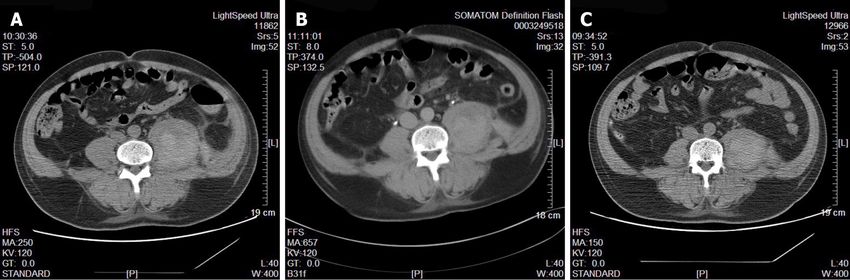
hemorrhage had formed an intermuscular hematoma (Figure 1A).
Case 2: Similar to case 1, we started prophylactic anticoagulant therapy when the
patient's condition became stable after treatment in the ICU. A similar progressive
decrease in hemoglobin occurred, and the patient complained of obvious buttock
muscle soreness. We immediately improved CT and bedside color Doppler
examination of the buttocks. CT revealed a new uneven density in the soft tissues of
both hips (Figure 2A). Bedside color Doppler ultrasound indicated the formation of
bilateral gluteal intermuscular hematoma.
FINAL DIAGNOSIS
Case 1
The final diagnoses were heat stroke, rhabdomyolysis, acute kidney disease injury,
and lumbosacral intermuscular hematoma.
Case 2
The final diagnoses were heat stroke, rhabdomyolysis, acute kidney disease injury,
pulmonary infection, hypertension, and buttock intermuscular hematoma.
TREATMENT
Both patients were diagnosed with rhabdomyolysis and acute kidney disease injury.
We immediately performed continuous renal replacement therapy (CRRT) to remove
accumulated metabolites, manage volume, etc. Heparinized anticoagulation was used,
and the activated partial thromboplastin time was monitored to adjust the heparin
dose. After admission to the ICU, we continued to monitor vital signs, provide organ
function and nutritional support, prevent DVT, and perform other measures. In
addition, we dynamically monitored the indicators of blood inflammation, liver and
kidney function, electrolytes, and coagulation function and adjusted the drug regimen
in a timely manner. Case 1's core body temperature remained high on admission to the
ICU, and we added a variety of physical cooling methods to rapidly reduce and
maintain the core body temperature to a normal range. At the time of admission to the
ICU, case 2 showed clear evidence of pulmonary infection, and his procalcitonin level
was significantly elevated, so he was given additional broad-spectrum antibiotic
therapy. The procalcitonin levels were continuously monitored to guide antibiotic
therapy. After treatment, the condition of the two patients gradually stabilized, and
CRRT was stopped on the 3rd and 8th d for cases 1 and 2, respectively. To continue
prophylactic anticoagulant therapy, enoxaparin sodium (0.3 mL) was added once a
day for case 1 and subcutaneously every other day for case 2. However, subsequent
monitoring of routine blood tests revealed a slow and progressive decline in
hemoglobin levels. Initially, we thought that the cause of the hemoglobin reduction
might have been related to inadequate nutrient intake and high body fat consumption
caused by infection. At this time, we excluded digestive system diseases leading to
blood loss through stomach content and fecal occult blood examinations, and no
obvious pleural or peritoneal effusion was found by bedside ultrasound examination.
No obvious damage was found in the dynamic monitoring of coagulation function.
We offered an enhanced enteral nutrition regimen, but continued monitoring of
routine blood tests still indicated a persistent decrease in hemoglobin. To this end, we
needed to consider additional possibilities. We then performed CT scans of the head,
chest, and whole abdomen. The cause of the persistent blood loss was finally found to
be the formation of a lumbosacral intermuscular hematoma and buttock intermuscular
hematoma. When the cause of hemorrhage was clear, we immediately stopped using
anticoagulant drugs and infused suspended red blood cells appropriately to correct
the anemia. CT and bedside color Doppler ultrasonography were used to check the
absorption of the intramuscular hematoma, and we continued to monitor routine
blood tests until the condition was stable. Follow-up monitoring of the patients
WJCC https://www.wjgnet.com 2841 April 26, 2021 Volume 9 Issue 12Yuan SY et al. Intramuscular hematoma in rhabdomyolysis patients
Figure 1 Outcome of lumbosacral intramuscular hematoma in case 1. A: For the first time, it was demonstrated that the left lumbosacral muscle was
swollen and less homogeneous in density; B and C: Subsequent computed tomography reexamination revealed that the volume of the left lumbosacral intramuscular
hematoma was significantly smaller than before.
Figure 2 Outcome of buttock intramuscular hematoma in case 2. A: For the first time, it was demonstrated that the density of soft tissue on both sides of
the buttocks was uneven; B and C: Subsequent bedside color Doppler reexamination revealed that the volume of the buttock intramuscular hematoma was
significantly smaller than before.
routinely found that the hemoglobin steadily increased, and CT and color Doppler
ultrasound suggested a gradual decrease in the volume of the intramuscular
hematoma (Figure 1B and C and Figure 2B and C).
OUTCOME AND FOLLOW-UP
The two patients were transferred to the general ward after hemoglobin stabilization.
A few weeks later, color Doppler ultrasonography showed that the lumbosacral and
gluteal intermuscular hematoma was basically absorbed.
DISCUSSION
Heat stroke is a life-threatening clinical syndrome characterized by an increase in core
temperature of > 40 °C and abnormalities in the central nervous system, such as
altered mental status, convulsions, or coma, and is associated with multiple organ
damage due to an imbalance in body heat production and dissipation caused by
exposure to a thermal environment and/or intense exercise[1]. According to the
different causes and susceptible populations, heat stroke can be divided into classical
heat stroke and exertional heat stroke. Due to the involvement of thermal injury
factors, rhabdomyolysis caused by heat stroke is significantly different from general
exercise rhabdomyolysis. The increase in CK within 24 h of onset of the former is often
not prominent and then gradually increases, often peaking 5-7 d after onset, but its
peak value is higher than that of exercise rhabdomyolysis, reaching up to 400000 U/L.
Myoglobin is often > 1000 ng/mL, and the peak can reach 70000-80000 ng/mL[3].
WJCC https://www.wjgnet.com 2842 April 26, 2021 Volume 9 Issue 12Yuan SY et al. Intramuscular hematoma in rhabdomyolysis patients
According to the classification of heat stroke, cases 1 and 2 were diagnosed with
exertional heat stroke and classical heat stroke, respectively. CK and myoglobin in case
1 peaked on the second day of admission, with the highest values reaching 61400 IU/L
and 77847 ng/mL, respectively. Case 2 showed peak CK and myoglobin values of
264600 IU/L and 326376 ng/mL, respectively, on the day of admission. We used
continuous renal replacement therapy in time after the patient entered the ICU;
otherwise, these values would have been higher.
Venous thromboembolism, including DVT and pulmonary embolism, is a disease
with high morbidity and mortality[4]. In recent years, increasing attention has been
given to the prevention of VTE, which means that the use of anticoagulants has
become increasingly frequent, especially for critically ill patients in the ICU. However,
we know that anticoagulants and antiplatelet drugs lead to a certain risk of bleeding.
Common bleeding-related risks include thrombocytopenia[5], active gastrointestinal
bleeding[6], retroperitoneal hematoma[7], etc., but muscle bleeding is easily ignored
because it is relatively rare. A retrospective study by Hiraga et al[8] showed that the
incidence of muscle hematoma was 0.4% among 694 patients with ischemic stroke who
received antithrombotic therapy. The initial symptoms of hematoma included pain (n
= 3) and syncope (n = 1), and the patients were not correctly diagnosed at the onset of
hematoma formation. Artzner et al[9] conducted a two-center retrospective study to
analyze the incidence of spontaneous iliopsoas muscle hematoma among 40 ICU
inpatients, of whom 50% received dialysis and 95% received prophylactic or
therapeutic doses of heparin. Watanabe et al[10] reported a patient treated with warfarin
for atrial fibrillation complicated with bilateral lumboiliac hematoma, rhabdomyolysis,
and acute kidney injury, which was attributed to improper use of warfarin. The two
cases reported here were both heat stroke patients who had rhabdomyolysis and acute
kidney injury and were treated with heparin-based CRRT. After CRRT treatment was
completed and no coagulation dysfunction was found, an appropriate dose of
enoxaparin sodium prophylactic anticoagulant therapy was used. Subsequently, the
levels of hemoglobin continued to decrease, and no obvious bleeding sites were found.
For one patient, the formation of a left psoas major intermuscular hematoma was
observed when examining the bleeding sites. The other patient complained of right
gluteal pain, which was confirmed by CT and color Doppler ultrasound. Immediately
after the discovery of the intramuscular hematoma, the patient was discontinued from
enoxaparin sodium anticoagulation therapy and transfused to correct the anemia.
Subsequently, his hemoglobin level stabilized, and the extent of the intramuscular
hematoma decreased.
CONCLUSION
Given the diagnosis of and treatment experience with case 1, we used prophylactic
anticoagulation with subcutaneous enoxaparin with a further dose reduction of 0.3 mL
every other day. However, muscle bleeding remained at the noninjected site. Given
the results with the above two cases, we have reason to believe that rhabdomyolysis
patients have a risk of muscle bleeding and that improper use of anticoagulants may
increase this risk or even lead to the formation of intramuscular hematomas. In
addition, when continuous blood loss is found in the body, the possibility of bleeding
in the muscles and more typical sites should be considered.
REFERENCES
1 Liu SY, Song JC, Mao HD, Zhao JB, Song Q; Expert Group of Heat Stroke Prevention and
Treatment of the People’s Liberation Army; and People’s Liberation Army Professional Committee
of Critical Care Medicine. Expert consensus on the diagnosis and treatment of heat stroke in China.
Mil Med Res 2020; 7: 1 [PMID: 31928528 DOI: 10.1186/s40779-019-0229-2]
2 Reynolds PM, Van Matre ET, Wright GC, McQueen RB, Burnham EL, Ho PJM, Moss M, Vandivier
RW, Kiser TH; Colorado Pulmonary Outcomes Research Group (CPOR). Evaluation of Prophylactic
Heparin Dosage Strategies and Risk Factors for Venous Thromboembolism in the Critically Ill
Patient. Pharmacotherapy 2019; 39: 232-241 [PMID: 30592541 DOI: 10.1002/phar.2212]
3 People’s Liberation Army Professional Committee of Critical Care Medicine. Expert consensus
on standardized diagnosis and treatment for heat stroke. Mil Med Res 2016; 3: 1 [PMID: 26744628
DOI: 10.1186/s40779-015-0056-z]
4 Tran HA, Gibbs H, Merriman E, Curnow JL, Young L, Bennett A, Tan CW, Chunilal SD, Ward CM,
Baker R, Nandurkar H. New guidelines from the Thrombosis and Haemostasis Society of Australia
WJCC https://www.wjgnet.com 2843 April 26, 2021 Volume 9 Issue 12Yuan SY et al. Intramuscular hematoma in rhabdomyolysis patients
and New Zealand for the diagnosis and management of venous thromboembolism. Med J Aust 2019;
210: 227-235 [PMID: 30739331 DOI: 10.5694/mja2.50004]
5 Greinacher A. CLINICAL PRACTICE. Heparin-Induced Thrombocytopenia. N Engl J Med 2015;
373: 252-261 [PMID: 26176382 DOI: 10.1056/NEJMcp1411910]
6 Li L, Geraghty OC, Mehta Z, Rothwell PM; Oxford Vascular Study. Age-specific risks, severity,
time course, and outcome of bleeding on long-term antiplatelet treatment after vascular events: a
population-based cohort study. Lancet 2017; 390: 490-499 [PMID: 28622955 DOI:
10.1016/S0140-6736(17)30770-5]
7 Sbrana F, Pasanisi EM. A massive retroperitoneal hematoma during low-molecular-weight-heparin
therapy. Intern Emerg Med 2016; 11: 153-154 [PMID: 26078200 DOI: 10.1007/s11739-015-1269-6]
8 Hiraga A, Nakagawa Y, Kamitsukasa I, Suzuki T, Kuwabara S. Muscle haematoma due to
antithrombotic treatment for ischaemic stroke. J Clin Neurosci 2015; 22: 1160-1163 [PMID:
25882254 DOI: 10.1016/j.jocn.2015.01.023]
9 Artzner T, Clere-Jehl R, Schenck M, Greget M, Merdji H, De Marini P, Tuzin N, Helms J, Meziani
F. Spontaneous ilio-psoas hematomas complicating intensive care unit hospitalizations. PLoS One
2019; 14: e0211680 [PMID: 30794573 DOI: 10.1371/journal.pone.0211680]
10 Watanabe Y, Koutoku H, Nagata H, Oda Y, Kikuchi H, Kojima M. Rhabdomyolysis with Acute
Kidney Injury Caused by Bilateral Iliopsoas Hematoma in a Patient with Atrial Fibrillation. Intern
Med 2019; 58: 2887-2890 [PMID: 31243202 DOI: 10.2169/internalmedicine.2749-19]
WJCC https://www.wjgnet.com 2844 April 26, 2021 Volume 9 Issue 12Published by Baishideng Publishing Group Inc
7041 Koll Center Parkway, Suite 160, Pleasanton, CA 94566, USA
Telephone: +1-925-3991568
E-mail: bpgoffice@wjgnet.com
Help Desk: https://www.f6publishing.com/helpdesk
https://www.wjgnet.com
© 2021 Baishideng Publishing Group Inc. All rights reserved.You can also read