11th Annual Scientific Symposium - Ultrahigh Field Magnetic ...
←
→
Page content transcription
If your browser does not render page correctly, please read the page content below
11th Annual Scientific Symposium

Ultrahigh Field Magnetic Resonance:
Clinical Needs, Research Promises and Technical Solutions

         Local organizers: Thoralf Niendorf (MDC & Charité, Berlin), Lucio Frydman (Weizmann Institute of Science,
         Rehovot, Israel), Jeanette Schulz-Menger (Charité, Berlin), Michal Neeman (Weizmann Institute of Science,
         Rehovot, Israel), Min Chi Ku (MDC, Berlin), Sebastian Schmitter (PTB, Berlin), Sonia Waiczies (MDC, Berlin)

        September 03rd - 04th 2020
        Virtual Meeting at the Max Delbrück Center,
        Berlin, Germany

                                                                          www.uhf-mr.de
                                                             Max Delbrück Center for Molecular Medicine
                                                                           in the Helmholtz Association
       www.mdc-berlin.de                                    Robert-Rössle-Straße 10, 13125 Berlin, Germany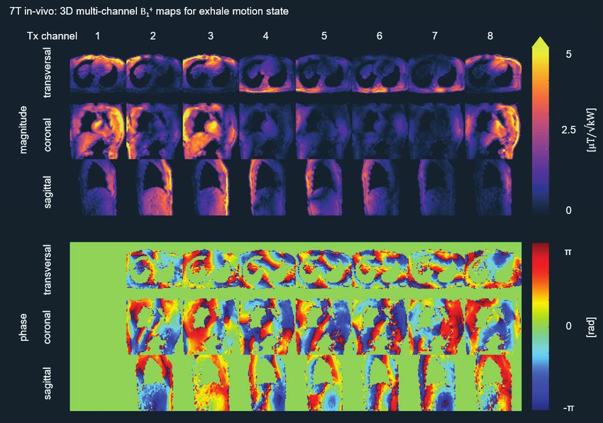
11th Annual Scientific Symposium

Dear colleagues and friends,

Very warm welcome to Berlin.

We are mostly excited by the larger audience that has joined us for the 11th
Scientific Symposium on Clinical Needs, Research Promises and Technical
Solutions in Ultrahigh Field Magnetic Resonance, due the “convenience” of
having a virtual meeting. The silver lining of the corona pandemic is that we are
quickly learning how to become more digitally connected. Limitations that have
kept scientists from linking with others at the other end of the world are con-
nected to travel (jetlag, time), environmental concerns (carbon footprint) and ex-
penses (flights, outrages conference fees) that have been prohibitive for many of
us; these have been narrowed down to the triviality of time-zone differences. For
scientists this is of little consequence, most of us are anyway renowned night owls.

Imaging bridges a crucial gap in space and time in life science and medicine: from
atomic to anatomic objects to whole body imaging, from picoseconds to years in
population studies. New molecular and cellular insights are obtained from imag-
ing. These findings should be integrated with data science into a coherent picture
of tissues, organs and organisms for early interception of disease. These funda-
mental developments call for hitherto unavailable research frameworks, interna-
tional partnership and collaborative culture to promote strong ties across multiple
research domains and imaging modalities; connecting nanoscopic views, length
scales, time scales and mesoscopic pictures with mechanistic insights and macro-
scopic function of biological and clinical importance.

To meet this goal the Max Delbrück Center for Molecular Medicine; the Weizmann
Institute of Science in Rehovot, Israel; the Humboldt University of Berlin and the
Charité-University Medicine, Berlin have joined forces to establish a Helmholtz
International Research School (HIRS) on imaging from the NAno to the MEso (iN-
AMES, https://www.mdc-berlin.de/inames). The this year’s symposium marks the of-
ficial start of iNAMES. These efforts are complemented by the Helmholtz Imaging
Platform (HIP), with the Max Delbrück Center being a HIP core in collaboration
with the DESY Hamburg and DKFZ Heidelberg. iNAMES and HIP are acting as
springboards to intensify scientific interactions in imaging, data sciences, informa-
tion technologies and digital engineering research fields.
11th Annual Scientific Symposium

The field of Magnetic Resonance (MR) has evolved rapidly over the past quarter
of a century, allowing for an ever growing number of applications across a broad
spectrum of basic, translational and clinical research. One important development
which is in the spotlight of MR research is Ultrahigh Field Magnetic Resonance
(UHF-MR). The pace of discovery is heartening and a powerful motivator to trans-
fer the lessons learned at ultrahigh fields from basic research into the clinical
scenario. These efforts are fueled by the unmet clinical needs and the quest for
advancing the capabilities of diagnostic MR imaging – today.

The development of UHF-MR is moving forward at an amazing speed that is break-
ing through technical barriers almost as fast as they appear. UHF-MR has become
an engine for innovation in experimental and clinical research. With more than
60.000,000 MR examinations already performed at 7.0 Tesla, the reasons for
moving UHF-MR into clinical applications are more compelling than ever. Images
from these instruments have revealed new aspects of the anatomy, functions and
physio-metabolic characteristics of the brain, heart, joints, kidneys, liver, eye, and
other organs/tissues, at an unparalleled quality. UHF-MR has a staggering number
of potential uses in neuroscience, neurology, radiology, neuroradiology, cardiol-
ogy, internal medicine, oncology, nephrology, ophthalmology and other related
clinical fields. As they are developed, we will push the boundaries of MR physics,
biomedical engineering and biomedical sciences in many other ways.

Realizing these opportunities, we are very much delighted to very warm welcome
you at the 11th Annual Scientific Symposium on Clinical Needs, Research Promises
and Technical Solutions in Ultrahigh Field MR. The symposium is a collaboration
between the Max Delbrück Center for Molecular Medicine, the Charité, the German
Metrology Institute (PTB) and the Weizmann Institute of Science, Rehovot, Israel.
The symposium is designed to provide an overview of state-of-the-art (pre)clinical
UHF-MR, to discuss the clinical relevance of UHF-MR, to explore future directions
of UHF-MR, to foster explorations into UHF-MR and to initiate local, regional, na-
tional and international collaboration and last but not least to provide plenty of
opportunities to engage into fruitful exchange with peers and colleagues.

For the scientific program we are very much honored to present extraordinary
speakers including MR technology leaders, distinguished clinical experts and
emerging scientists – all bridging disciplinary boundaries and stimulating the im-

                                          3
11th Annual Scientific Symposium
aging community to throw further weight behind the solution of unsolved problems
and unmet clinical needs. The scientific program is tailored to survey current trends
in UHF-MR technology, to provide an overview of state-of-the-art (pre)clinical UHF-
MR, to discuss MR safety topics, to demonstrate the clinical relevance of UHF-MR,
to explore future directions of UHF-MR and to brainstorm about physiometabolic
imaging technologies. We wish to acknowledge the passion and dedication of the
many imaging students who put their heads together to brainstorm on the topics
covered in the scientific program.

We would like to draw your attention to the electronic posters, all being made
readily available for viewing and download. We wish to thank those of you who
walked the extra milage and submitted poster contributions. We really appreciate
your efforts. Thanks to your valuable feedback we have also included several slots
of 2 min enlightening poster power presentations into the program. This will give
a large number of poster presenters the opportunity to be in the spotlight of the
audience. Please support the poster presenters and do not miss to vote for the best
poster.

We cordially invite to the advertisements included in the digital program booklet
ready for viewing and download. It also behooves us to emphasize that this is the
right moment to acknowledge our generous sponsors who provided marvelous sup-
port to the symposiums scientific and educational activities. Thank you very much to
our sponsors and supporters.

  Thoralf Niendorf             Lucio Frydman              Jeanette Schulz-Menger       Michal Neemann
 MDC & Charité, Berlin   Weizmann Institute of Science,       Charité, Berlin       Weizmann Institute of
                               Rehovot, Israel                                      Science, Rehovot, Israel

                    Min-Chi Ku              Sebastian Schmitter           Sonia Waiczies
                    MDC, Berlin                 PTB, Berlin                MDC, Berlin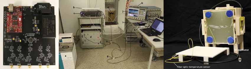
www.mdc-berlin.de

 11th Annual Scientific Symposium

Table of Contents

Organization............................................................................................     3

Program...................................................................................................   4

Poster Abstracts......................................................................................  	 13

Sponsors.................................................................................................. 90
MRI.TOOLS GmbH

RF Coils & Accesories for Animal and Human Imaging
We provide succesfully tested products as well as custom
solutions that exactly meet your needs and specifications.
Our portfolio encompasses clinical and preclinical RF coils for
field strength from 1.5 T to 11.5 T and for the nuclei of your
choice.
With our coils, we provide a certificate issued by a notified
body which confirms compliance with IEC 60601-1 and IEC
60601-2-33

Examples from our portfolio
32 channel modular transceiver array for cardiac/body imaging at 7.0 Tesla

8 channel transceiver array for carotid imaging at 3.0 Tesla

8 channel 1H knee RF coil for pTX application at 7.0 Tesla

6 channel transceiver array for eye imaging at 3.0 Tesla and 7.0 Tesla

4 channel 23Na and 4 channel 1H RF coil for cardiac/liver imaging at 7.0 Tesla
19
     F/1H transceiver array for lung imaging at 3.0 Tesla and 7.0 Tesla

small monkey volume RF coil for brain imaging at 11.7 T
19
     F/1H volume RF coil for mouse imaging at 9.4 T

... and more

Device Testing and Certification
           We offer dedicated consulting and testing
           services with the focus on in-house-built
           hardware including custom RF coils.

               We prepare all documents and procedures including the
               certificate issued by a notified body for your IRB or local
               safety board.
                                                                             Contact us:
                                                                             www.mritools.de
                                                                             info@mritools.de
                                                                             +49 30 9489 2582

                                                                 2
Organization

Organizers:
Thoralf Niendorf          Berlin, Germany (MDC & Charité)
Lucio Frydman             Rehovot, Israel (Weizmann Institute of Science)
Jeanette Schulz-Menger Berlin, Germany (Charité & Helios Klinikum)
Michel Neeman             Rehovot, Israel (Weizmann Institute of Science)
Min-Chi Ku		              Berlin, Germany (MDC)
Sebastian Schmitter       Berlin, Germany (PTB)
Sonia Waiczies            Berlin, Germany (MDC)

Conference Office:
Lien-Georgina Dettmann, Matthias Runow & Timkehet Teffera
E-Mail: MRSymposium@mdc-berlin.de
Max Delbrück Center for Molecular Medicine in the
Helmholtz Association
Robert-Rössle-Straße 10
13125 Berlin
Phone: +49 (0)30 9406/3720/4255/2719

Office Prof. Niendorf:
Carolin Heydrich
E-Mail: Carolin.Heydrich@mdc-berlin.de
Max Delbrück Center for Molecular Medicine in the Helmholtz Association
Robert-Rössle-Straße 10
13125 Berlin
Phone: +49 (0)30 9406 4505

                                         3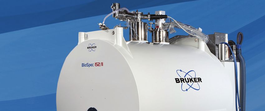
Program 3rd September 2020

11TH ANNUAL SCIENTIFIC SYMPOSIUM ON ULTRAHIGH
FIELD MAGNETIC RESONANCE

 Time     Time    Time
                            DAY 1
 (CEST)   (EDT)   (CST)
 12:00    06:00   18:00    Welcome
                           chair: Thoralf Niendorf, Berlin, Germany
                                  Sonia Waiczies, Berlin, Germany
 12:15    06:15   18:15    KEYNOTE: Why all the Fuss About Ultrahigh Field MRI: New
                           Directions in Optical Imaging from the Nanoscopic to the
                           Mesoscopic
                           Katrin Heinze, University of Würzburg

SCIENTIFIC SESSION I
GETTING TO THE MATTER OF THE HEART: CLINICAL NEEDS AND RESEARCH PROMISES OF
CARDIOVASCULAR UHF-MR
chair: Lucio Frydman, Rehovot, Israel
       Thoralf Niendorf, Berlin, Germany

 12:35    06:35   18:35    Ten Reasons for Doing Cardiac MRI at 7.0 T: .
                           Dreams.versus.Reality
                           El-Sayed Ibrahim, Medical College of
                           Wisconsin, Milwaukee, USA
 12:55    06:55   18:55    Physiometabolic Probing of the Heart with Multinuclear .
                           MR at 7.0T
                           Tanja Platt, DKFZ Heidelberg, Germany
 13:15    07:15   19:15    LUNCH BREAK / BREAKFAST BREAK
 14:00    08:00   20:00    Size Matters: High Density RF Array for Cardiac MR at 7.0 T
                           Thomas Eigentler, Max Delbrück Center for Molecular Medicine,
                           Berlin, Germany
 14:20    08:20   20:20    Rich Opportunities for Discovery: Vascular and Body MRI .
                           at 7.0 T Revisited
                           Natalie Schön, PTB-Physikalisch-Technische Bundesanstalt,
                           Berlin, Germany

                                              4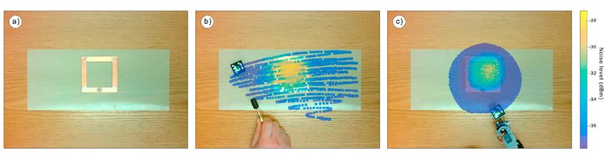
Program 3rd September 2020
14:40 08:40 20:40 POSTER POWER SESSION (5 x 2 min)
Identifying Radiation Therapy Effect on Cardiac Function Using
Ultrahigh Field 9.4 T MRI
El-Sayed Ibrahim, Medical College of Wisconsin, Milwaukee, USA
Towards Carotid Arteries Characterization: Time-Resolved 3D
Flow-MR Fingerprinting Implementation at 7.0 T
Lisa Leroi, PTB-Physikalisch-Technische Bundesanstalt, Berlin,
Germany
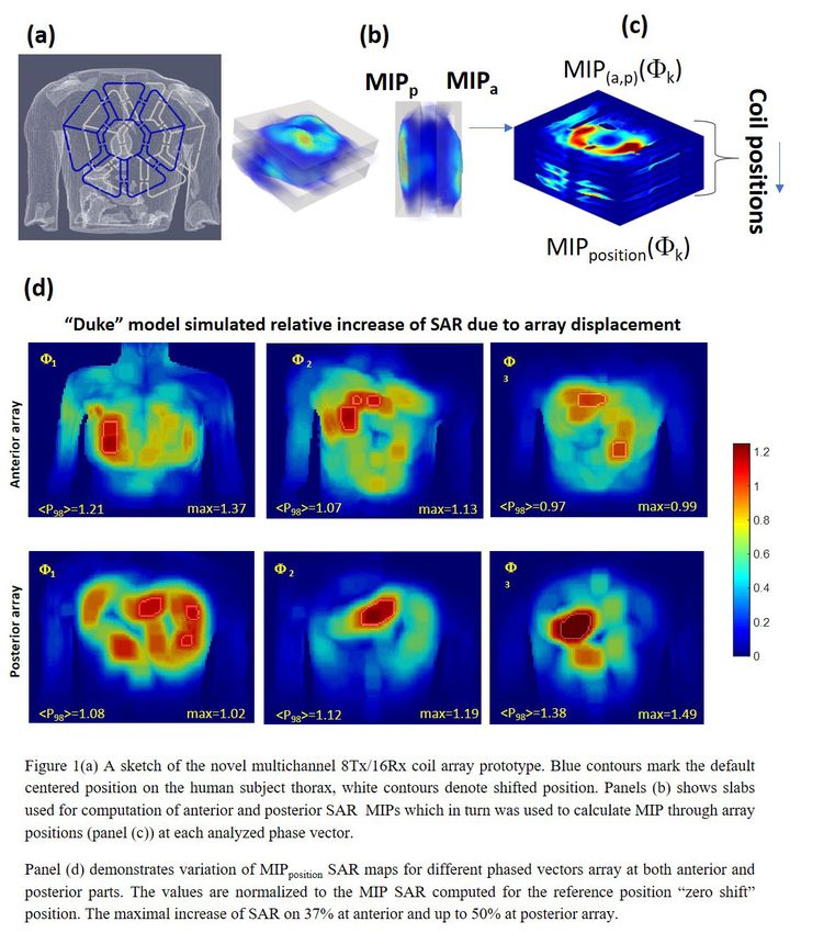
SAR distribution for the Transceiver Array for Human Cardiac
MRI at 7.0 T: Variations due to Displacement on Thorax.
Terekhov Maxim, University Hospital Würzburg, Würzburg,
Germany
Cardiac Functional Imaging Using Ultrahigh Field 7.0 T MRI
El-Sayed Ibrahim, Medical College of Wisconsin, Milwaukee, USA
Whole-Heart 4D Flow MRI at 7.0 T Using Self-Gated Respiratory
Motion Correction
Jean Pierre Bassenge, PTB-Physikalisch-Technische
Bundesanstalt, Berlin, Germany
14:50 08:50 20:50 PANEL DISCUSSION: CARDIAC MRI at UHF
chair: Min-Chi Ku, Berlin, Germany
Sebastian Schmitter, Berlin, Germany
15:05 09:05 21:05 COFFEE BREAK / RELAXATION WITH LIVE MUSIC
SCIENTIFIC SESSION II
GETTING TO THE MATTER OF THE BRAIN: CLINCIAL NEEDS AND RESEARCH PROMISES FOR
NEUROVASCULAR UHF-MR AND RELATED FIELDS
chair: Michal Neeman, Rehovot, Israel
Paula Ramos Delgado, Berlin, Germany
15:30 09:30 21:30 Diffusion and Functional Measurements at High and Ultrahigh
Fields. A Preclinical Perspective
Lucio Frydman, Weizmann Institute of Science, Rehovot, Israel
5
Program 3rd September 2020

15:50   09:50   21:50   Speed Saves: Simultaneous Parametric T2 and T2* Mapping in
                        Healthy Subjects and in Patients with Multiple Sclerosis
                        Carl Herrmann, Max Delbrück Center for Molecular Medicine,
                        Berlin, Germany

                        Laura Lewis, Boston University,
                        USA
16:30   10:30   22:30   Metabolic Profiling of the Human Brain: Clinical High Resolution
                        3D-MR Spectroscopic Imaging at 7.0 T
                        Eva Heckova, Medical University Vienna, Austria
16:50   10:50   22:50   POSTER POWER SESSION (5 x 2 min)
                        Kinetic Oscillatory Stimulation in the Nasal Cavity Elicits
                        Functional Brain Activation in the Limbic System of the Brain
                        Xia Li, China Jiliang University, Hangzhou, China
                        Postmortem Human Brain 9.4 T Ultrahigh Field MRI: the UTAP Study
                        Stijn Michielse, Maastricht University Medical Center, The
                        Netherlands
                        MRI Reveals Brain Ventricle Expansion in Pediatric Patients with
                        Acute Disseminated Encephalomyelitis
                        Jason Millward, Max Delbrück Center for Molecular Medicine in
                        the Helmholtz Association, Berlin, Germany
                        In-vivo Repeatability of SPECIAL Based Single-voxel Spectroscopy
                        Using Different Adiabatic Inversion Pulses
                        Layla Tabea Riemann, PTB-Physikalisch-Technische Bundesanstalt,
                        Berlin, Germany
                        1H NMR Based Serum and Urine Bio-Fluids Metabolic Profile
                        Correlates with the Neurological Recovery in Treated Acute Spinal
                        Cord Injury (ASCI) Subjects: A Prospective Case Control Study
                        Alka Singh, King George’s Medical University, Daliganj, Lucknow, India
17:00   11:00   23:00   PANEL DISCUSSION: BRAIN MRI at UHF
                        chair: Ariane Fillmer, Berlin, Germany
                               Henning Reimann, Berlin, Germany
17:15   11:15   23:15   ADJOURN – End of Day 1

                                          6
                                          6
Program 4th September 2020
Time Time Time DAY 2
(CEST) (EDT) (CST)
12:00 06:00 18:00 Welcome
chair: Thoralf Niendorf, Berlin, Germany
Sonia Waiczies, Berlin, Germany
12:15 06:15 18:15 KEYNOTE: 17.6 T MRI of the Perivascular Network: Imaging Brain
Waste Clearance Paths
Malisa Sarntinoranont, University of Florida, Gainesville, USA
SCIENTIFIC SESSION III
TRANSLATIONAL RESEARCH: FROM BLUE SKY EXPLORATIONS EN ROUTE TO .
CLINICAL APPLICATIONS
chair: Min-Chi Ku, Berlin, Germany
Philipp Selenko, Rehovot, Israel
Tissue MR Imaging
Paula Ramos Delgado, Max Delbrück Center for Molecular
Medicine, Berlin, Germany
12:55 06:55 18:55 Fluoride Nanocrystals: Tracers for In Vivo Hot Spot 19F MRI
Amnon Bar-Shir, Weizmann Institute of Science, Rehovot, Israel
13:15 07:15 19:15 LUNCH BREAK / BREAKFAST BREAK
14:00 08:00 20:00 Pushing.the.Boundaries.of.Chemical.Exchange.Saturation.
Transfer.(CEST) pH.Imaging.at.Ultrahigh.Magnetic.Fields
Dario Longo, Italian National Research Council (CNR), Torino, Italy
14:20 08:20 20:20 MR-Synchronized Optogenetic, Visual and Auditory Stimulation
to Probe Sensory Processing in Rats Using Functional MRI at 7.0 T
Ed Wu, The University of Hong Kong, China
14:40 08:40 20:40 Sea to Summit: Progress in Preclinical MR at Ultrahigh and
Extreme Magnetic Fields
Wulf-Ingo Jung, Bruker Biospin MRI GmbH, Ettlingen, Germany
7
7Program 4th September 2020
15:00 09:00 21:00 POSTER POWER SESSION (5 x 2 min)
Deep CEST MR Fingerprinting at 7.0 T Reveals Tumor Apoptotic
Response to Oncolytic Virotherapy In Vivo
Or Perlman, Massachusetts General Hospital and Harvard
Medical School, Charlestown, MA, USA
Characterization of Fluorinated Pesticides using Fluorine (19F)
MR Methods
Salina Skenderi, Max Delbrück Center for Molecular Medicine in
the Helmholtz Association, Berlin, Germany
Towards Non-Invasive Imaging of MS Disease-Modifying 19F Drugs
Fatima Sherazi, Max Delbrück Center for Molecular Medicine in
the Helmholtz Association, Berlin, Germany
Performance of Compressed Sensing for Detecting Low SNR
19F MRI in Experimental Autoimmune Encephalomyelitis using
Prospective Undersampling
Ludger Starke, Max Delbrück Center for Molecular Medicine in
the Helmholtz Association, Berlin, Germany
Point Spread Function Mapping Eliminates Image Distortion
From Renal Echo-Planar_Imaging: Preliminary Results from a
9.4T Animal MR System
Kaixuan Zhao, Max Delbrück Center for Molecular Medicine in
the Helmholtz Association, Berlin, Germany
15:10 09:10 21:10 PANEL DISCUSSION: TRANSLATIONAL MRI at UHF
chair: Sonia Waiczies, Berlin, Germany
Jason Millward, Berlin, Germany
15:25 09:25 21:25 COFFEE BREAK / RELAXATION WITH LIVE MUSIC
SCIENTIFIC SESSION IV
LOOKING AT THE HORIZON
chair: Amnon Bar-Shir, Rehovot, Israel
Sebastian Schmitter, Berlin, Germany
15:50 09:50 21:50 Body MR Imaging at 10.5 Tesla – Toys for Boys?
Kamil Ugurbil, University of Minnesota, Minneapolis, USA
8Program 4th September 2020
16:10 10:10 22:10 Following the Footsteps of Neurological Disease with UHF MRI
Priti Balchandani, BioMedical Engineering and Imaging Institute at
Mount Sinai, New York, USA
16:30 10:30 22:30 (UHF-)MR Image Reconstruction: How Deep Learning Will Shape .
the Future of MRI
Chen Qin, University of Edinburgh, Edinburgh, UK
16:50 10:50 22:50 Thermal MR: Generalization of the Time- and Frequency Multiplexed
Problem of Radiofrequency Induced Heating Intervention
Andre Kühne, MRI.TOOLS GmbH, Berlin, Germany
17:10 11:10 23:10 Sea to Summit: Progress in Clinical Ultrahigh Field MRI
Robin Heidemann, Siemens, Erlangen, Germany
17:30 11:30 23:30 POSTER POWER SESSION (5 x 2 min)
Rapid Mapping of Radiofrequency Coil Fields with Computer Vision
Egor Kretov, Max Delbrück Center for Molecular Medicine in the
Helmholtz Association, Berlin, Germany
Respiration-Resolved 3D Multi-Channel Absolute B1+ Mapping .
of the Body at 7.0 T
Sebastian Dietrich, PTB-Physikalisch-Technische Bundesanstalt,
Berlin, Germany
Evaluation of Static 2D and 3D Parallel Transmission of the Human
Heart at 7.0 T
Christoph Stefan Aigner, PTB-Physikalisch-Technische Bundesanstalt,
Berlin, Germany
B0 Robust Adiabatic Multi-Band Inversion Pulses
Christoph Stefan Aigner, PTB-Physikalisch-Technische Bundesanstalt,
Berlin, Germany
Multi-Channel RF Power and Phase Supervision Systems .
Technology for Thermal Magnetic Resonance: Development,
Evaluation and Application
Haopeng Han, Max Delbrück Center for Molecular Medicine in the
Helmholtz Association, Berlin, Germany
17:40 11:40 23:40 PANEL DISCUSSION: FUTURE of UHF MRI
chair: Christoph Aigner, Berlin, Germany
Ludger Starke, Berlin, Germany
17:55 11:55 23:55 END OF SYMPOSIUM
9Go further than ever before with
Bruker Ultra-high Field MRI
The Bruker BioSpec series offers multi-purpose high field MRI/
MRS research systems, designed for the emerging market of
preclinical veterinary imaging and molecular MRI.
Superconducting Magnet System
• Actively-shielded superconducting wide-bore magnet
• Nitrogen free (no cryogenic maintenance by customer)
• Cryo-refrigerator (Minimal helium consumption, long hold-times,
long maintenance intervals)
• Reduced stray field
BioSpec 152/11 USR/R
Field strength: 15.2 Tesla
Diameter of clear bore: 110 mm
BioSpec 117/16 USR
Field strength: 11.7 Tesla
Diameter of clear bore: 160 mm
BioSpec 117/11 USR Customer Insights
Field strength: 11.7 Tesla
Diameter of clear bore: 110 mm How Mass Spectrometry Techniques
are Propelling the Advancements of
Single Cell Biology
Mass Spectrometry
Ultra-High Field NMR
Innovation with IntegrityPoster Abstracts
(In alphabetical order)
12P1
Evaluation of static 2D and 3D parallel transmission of the
human heart at 7T
Christoph Stefan Aigner1, Sebastian Dietrich1, Sebastian Schmitter1
Physikalisch-Technische Bundesanstalt (PTB), Braunschweig and Berlin, Germany
1
Introduction
Three-dimensional human heart imaging at ultra-high fields is highly challenging
due to respiratory and cardiac motion-induced artefacts as well as spatially het-
erogeneous B1+ profiles1. Various methods have been proposed to address the
problem of spatial heterogeneities of the B1+ fields, including parallel transmission
(pTx)1. So far, however, the lack of 3D human abdominal B1+ maps at 7 T hindered
the development of 3D imaging methods in the body. In this study, we investigate
the feasibility of static pTx for 2D/3D heart flip angle homogenization at 7 T based
on relative 3D B1+ maps2. In addition, in vivo data of 6 subjects of different body
sizes were acquired with 3D GRE radial phase encoding (RPE)3,4 using subject spe-
cific static phase-only pTx.
Methods
MRI was performed on a 7T scanner (Siemens, Erlangen, Germany) with a com-
mercial 8Tx/32Rx thorax coil (MRI.TOOLS, Berlin, Germany) certified by a notified
body to comply with the local SAR limits in first level controlled mode of 20 W/kg
(IEC 606012-33). Six healthy volunteers (2f/4m, 21-35 years) with a wide range of
BMI (20-34 kg/m2) were scanned in the supine position according to an approved
IRB protocol.
Relative, 3D channel-wise B1+ estimates were computed from a 3D free breath-
ing GRE RPE3,4 scan following references2,6. The channel-wise superposition of the
resulting B1+ maps were then used to manually draw the region-of-interest (ROI)
of the heart on a slice-by-slice basis (9-16 slices). The ROI was used as a binary
mask for static shimming solving a cost function that optimized the homogeneity
measured by the coefficient of variation (CV). Two different shim solutions (phase-
only, magnitude and phase) were computed for 1 slice (ROI1), 3 slices (ROI3) and
the whole heart (ROIH) with a slice gap of 8 mm.
The subjects were scanned in the same MRI session additionally with a high-reso-
lution 3D RPE-GRE free-breathing sequence using the optimized ROI3 static phase-
only shim. The 3D dataset was reconstructed for an isotropic voxel size of 1.4 mm
with a respiratory motion surrogate retrieved from a 1D projection in the head-
feet direction in the k-space center. The underlying motion field was estimated via
13image registration based on the respiratory resolved reconstructions and used to
correct the respiratory motion5.
Results and Discussion
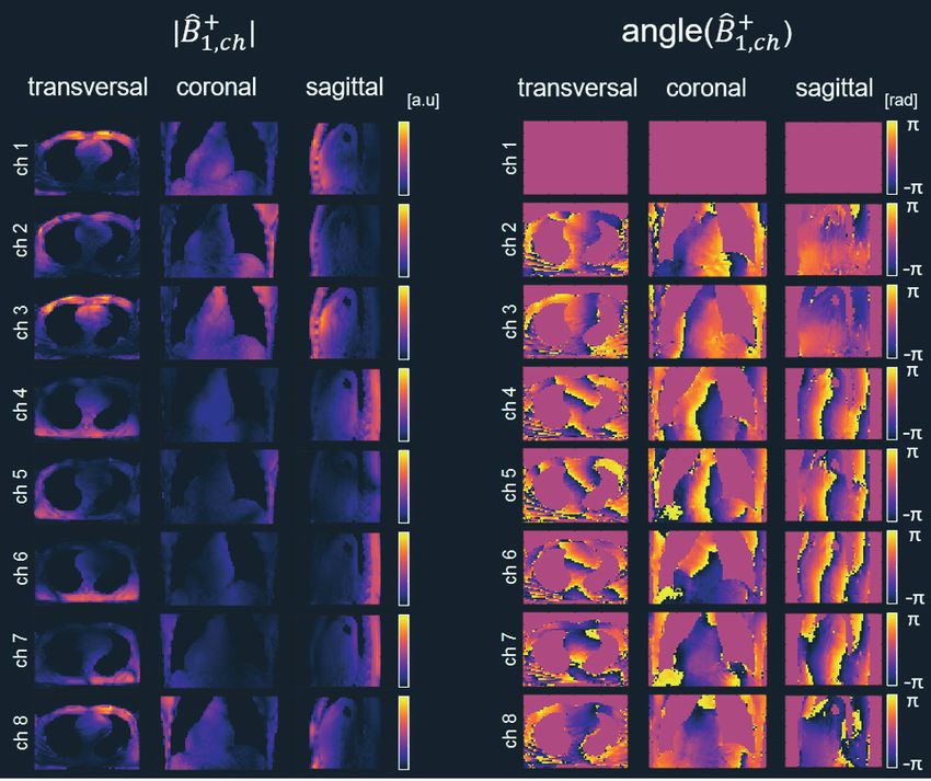
Figure 1 shows the 4 mm isotropic 3D relative B1+ maps of subject 1 for all 8 trans-
mit channels. Both, the magnitude and the phase are free of breathing artefacts,
despite acquiring the underlying 3D GRE data under free breathing. The same
observation has been made for all 6 subjects. Figure 2 shows the B1+ predictions of
the optimized static phase-only phase shims for ROI1 and, ROI3 and ROIH. Whereas
the central slice has comparable B1+ homogeneity, the shim of ROI3 and ROIH
shows more pronounced B1+ variations. Figure 3 summarizes and compares the
coefficient of variation (CV) of the nominal FA for each ROI and subject. Across all
subjects, the phase-only shims resulted in a CV of 21.9% (ROIH), 18.8% (ROI3) and
17.2% (ROI1) which could be reduced by approximately 2.5% for the magnitude and
phase shims. Figure 4 qualitatively validates the 3D B1+ prediction of the optimized
phase-only ROI3 shim setting using the reconstructed 3D GRE RPE measurements.
The experimental GRE measurements reproduce well the nominal flip-angle pat-
terns of the B1+ predictions. The remaining signal drop in AP-direction results from
receive-profile, which has not been corrected in the plots.
Conclusion
This study demonstrates in 6 subjects that 3D relative B1+ mapping can be used
to investigate and reduce FA heterogeneities via pTx across the entire 3D heart
volume at 7 T. However, it also shows that the degree of freedom of static shims
is not enough to compensate FA heterogeneities below an acceptable CV level of
about 15% with the used coil, which is likely achievable with dynamic pTX such as
2D slice selective spokes or 3D non-selective kT-points pulses.
Figure 1: 3D relative estimated
B1+ maps (left: magnitude, right:
phase-angle) of subject 1 at 7
T. Four channels are located on
the chest and four channels are
positioned under the subject’s
back. The phase angle of channels
2 to 8 are computed relative to
channel 1.
14Figure 2: 3D view of the complex
sum of estimated B1+ maps with
optimized ROI1, ROI3 and ROIH
static phase setting of subject 1.
Figure 3: Coefficient of variation
(CV) for optimized static shim
settings of all subjects (left: phase-
only, right: magnitude and phase).
Three different settings have been
optimized and evaluated for an
ROI of 1 slice (ROI1), 3 slices (ROI3)
and the entire heart volume
(ROIH).
Figure 4: B1+ predictions and
reconstructed respiration
corrected 3D GRE images for
subjects 1 with optimized
subject-specific ROI3 static
phase-only pTx. The 3D images
are free of breathing artifacts
and demonstrate, despite some
differences close to the coil
elements, the validity of the
B1+ maps and the resulting B1+
predictions with the optimized
phase-only shim. The remaining
signal changes in AP direction in
the measured views are a result
of receive (B1-) variations.
15P2
B0 robust adiabatic multi-band inversion pulses
Christoph Stefan Aigner1, Sebastian Schmitter1
Physikalisch-Technische Bundesanstalt (PTB), Braunschweig and Berlin, Germany
1
Introduction
Adiabatic RF pulses generate homogeneous flip angles (FA) despite inhomogene-
ities of the transmit magnetic radio frequency (RF) field (B1+) at the cost of high RF
peak amplitudes and RF power1. To tackle this problem, multiple methods exist to
reduce RF peak amplitudes2 or a combined reduction of RF peak amplitudes and
RF power3,4. In this work, we extend the design of adiabatic multi-band (MB) inver-
sion pulses5, where RF power becomes an even stronger problem, for variable
slice selective gradient shapes and combine it with low power gradient offset inde-
pendent adiabatic (GOIA4) pulses. The proposed GOIA MB pulses result in smooth
slice selective gradients with highly increased bandwidth and matched multi-band
RF waveforms. Phantom and in vivo experiments at 7T validate the simulations
and demonstrate the B1+ and B0 robust inversion of multiple slices.
Theory and Methods
Adiabatic MB pulses can be designed with a superposition of frequency offset
shifted adiabatic single-band (SB) pulses. The frequency offset is computed as a
function of the slice selective gradient, to correct for the time-variable slice se-
lective gradient shapes, and phase scrambling is used to reduce RF peak ampli-
tudes6,7,8. Two different adiabatic inversion pulses, hyperbolic secant (HS)1 with
a constant slice selective gradient and GOIA4,5 with a time varying slice selective
gradient were designed for a pulse duration of 12 ms and a spectral bandwidth
of 1.25 kHz (HS) and 20 kHz (GOIA). Gradients were designed for a slice thick-
ness of 20 mm. Three SB pulses, inverting at positions -50, 0, +50 mm measured
from isocenter were superposed to form an MB pulse with an optimized phase
scrambling scheme7 (MB3ps). The MB3ps inversion pulses were implemented in a
pulse sequence followed by an orthogonal slice selective excitation and cartesian
phase encoding and read-out. High-resolution scans (TR/TE = 2000/4.8 ms, FOV
= 256x128 mm, matrix = 384×192) have been acquired on a 7T system (Siemens,
Erlangen, Germany) with a 1Tx32Rx Nova Medical head coil. Each scan was per-
formed twice (without and with the adiabatic inversion pulse) and the inversion
profiles were calculated by the complex difference of the two images.
16Results and Discussion
Figure 1 compares HS-MB3ps and the proposed GOIA-MB3ps pulse which results
in a 16 times higher bandwidth with only a 2-fold RF power increase. As expected,
phase scrambling of adiabatic MB pulses resulted in a sub-linear scaling of the
peak B1 amplitude and as expected from theory a linear scaling of RF power de-
mand with the number of bands. While there are strong RF magnitude and phase
variations, the GOIA Gs waveform is smooth and does not require correction of
gradient imperfections8,9. Figure 2 shows simulated inversion profiles (scaled from
0 to 1) of the adiabatic multiband pulses for a B1 variation of 0-150% and a B0 offset
range of 0-500Hz shows a similar transition to the adiabatic threshold and a dif-
ferent impact of B0 variations. For instance, a 500Hz B0 offset shifts the inversion
profiles by 12 mm for HS and only by 1.2 mm for the GOIA pulse with a maximal
error of 2.5% at 500Hz. Figure 3 shows inversion slice profile measurements in a
phantom bottle and a healthy volunteer at a 7T system. The reconstructed inver-
sion slices were computed from two measurements without and with the inver-
sion pulse and the resulting difference of both complex images has been normal-
ized by a reference scan. The reconstructed data clearly shows a precise inversion
of the proposed GOIA MB3ps example despite B1+ and B0 variations.
Conclusion
The Bloch simulations and measurements on a 7T system showed the potential
of using phase scrambled adiabatic GOIA-MB pulses for simultaneous inversion of
multiple slices and will serve as a basis to reduce chemical shift artefacts and voxel
bleeding in multi-voxel spectroscopy.
Figure 1: Magnitude and slice selective gradient of phase scrambled adiabatic Hyperbolic
Secant (first row) and GIOA (second row) based multi-band pulses and simulated inversion
profiles for the central slice and the entire spatial domain.
17Figure 2: Simulated impact of
B1 and B0 off-resonance scaling
on the adiabatic multi-band
pulses. The simulations in
the first row were performed
assuming an ideal B0 field with
a fixed B0 off-resonance of
0 Hz. The simulations in the
second row are performed
with a fixed B1 scaling of 1.5.
The cross sections indicate the
excellent B1 and B0 robustness
of the proposed GOIA MB3ps
example.
Figure 3: Inversion slice profile
measurements in a phantom
bottle and a healthy volunteer
using the adiabatic HS- and
GOIA-MB3ps pulses from
Figure 1 at a 7T system. The
reconstructed inversion slices
were computed from two
measurements without and
with the inversion pulse and
have been normalized by a
reference scan.
1) Tannus A, Garwood M. Adiabatic pulses. NMR Biomed, 10: 423-434, 1997.
2) Balchandani P, Pauly J and Spielman D, MRM, 64: 843-851, 2010.
3) Warnking JM and Pike GB, MRM, 52: 1190-1199, 2004.
4) Andronesi OC, Ramadan S, Ratai EM, Jennings D, Mountford CE and Sorensen AG, JMR,
203(2): 283-293, 2010.
5) Goelman G and Leigh JS, JMR, 101(2): 136-146, 1993.
6) Barth M, Breuer F, Koopmans PJ, Norris DG and Poser BA. MRM, 75(1):63-81, 2016.
7) Wong EC, ISMRM 20, 2209, 2012
8) Abo Seada S, Price AN, Schneider T, Hajnal J.V., Malik SJ, MRM, 81: 362- 376. 2019
9) Aigner CS, Rund A, Abo Seada S, Malik SJ, Hajnal JV, Kunisch K and Stollberger R. MRM, 83(2):
561-574. 2020
18P3
Whole-heart 4D flow MRI at 7T using self-gated respiratory
motion correction
Jean Pierre Bassenge1,2, Sebastian Dietrich2, Christoph Aigner2, Christoph Kolbitsch2,
Sebastian Schmitter2
Working Group on Cardiovascular Magnetic Resonance, Experimental and Clinical
1
Research Center, a joint cooperation between the Charité Medical Faculty and the
Max‐Delbrück Center for Molecular Medicine, Berlin, Germany,
Physikalisch‐Technische Bundesanstalt (PTB), Braunschweig and Berlin, Germany
2
Introduction
4D flow MRI, the three-dimensional and ECG-gated acquisition of blood veloc-
ity vector fields, is of interest in a multitude of cardiovascular conditions. [1]
Acquisitions at ultra-high field strengths such as 7T offer large potential for 4D
flow due to increased signal-to-noise ratio and more localized coil sensitivity pro-
files, which improve parallel imaging. [2,3] Nevertheless, thoracic 4D flow MRI is
more challenging at 7T compared to lower field strengths due to highly inhomo-
geneous B1+ fields. In the worst cases, B1+ inhomogeneities yield signal dropouts
that can impair both acquisitions at the target region and of navigators for respira-
tory motion compensation. In previous publications , thoracic 4D flow MRI at 7T
was limited to the aorta, and prospective respiratory-gating was performed using
cross-hair navigator pairs at the intersection of liver and lung, which requires dy-
namic switching between separate B1+ shim sets for target region and navigator
[2,3]. Here, we investigate the feasibility of 4D flow MRI at 7T in a large FOV cover-
ing aorta, heart and pulmonary vessels, using a single B1+ shim set in combination
with self-gated retrospective motion correction.
Methods
After IRB approval and giving informed consent, a healthy volunteer (m35, BMI
27.8) was scanned at 7T (Magnetom 7T, Siemens, Erlangen, Germany) in pTx mode
using a commercial 32Rx/8Tx thorax coil (MRI.Tools, Berlin, Germany). Relative
B1+ estimates were acquired for each Tx channel following [5] (3D acq., 3.9mm
isotr. res., TA=3:25). A 3D region of interest was manually selected to calculate a
magnitude and phase B1+ shim set that optimizes flip angle homogeneity across
the heart and the aorta. 4D flow data was obtained with this shim set using a
GRE sequence with symmetric referenced velocity encoding (VENC=1.5m/s) in a
sagittal slab (width 200mm). The sequence employs radial phase encoding [6],
i.e. one cartesian read-out dimension (kx: head-feet, FOV=400mm) and two non-
19cartesian phase-encode dimensions (ky: ant.-post. and kz: right-left). For respira-
tory self-gating, the k-space center (ky=kz=0) was updated every 400ms. Further
parameters: 2.5mm isotr. res., TR=5.0ms, TE=2.9ms, nom. FA=24°, BW=640Hz/px,
TA=13:37.
A surrogate respiratory signal was calculated from coil PCA of the k-space center
projections, and was used to bin the acquired data into 8 respiratory phases. Non-
rigid motion-transformation-fields were calculated to co-register all respiratory
phases to the end-expiration phase. Subsequently, all data was binned into 24
cardiac phases using the recorded ECG-signal, and the motion-fields were used
in an iterative kt-SENSE reconstruction to obtain a motion-compensated 4D flow
data set. [4]
For reference, a 2D cartesian flow sequence was acquired in a breathhold
(6mm slice thickness, 2.5mm in-plane res., prosp. ECG-trig., temp. res. 34.8ms,
VENC=1.5m/s through-plane), using the same B1+ shim set as for the 4D flow se-
quence. In addition, a validation scan for the respiratory self-gating was conduct-
ed, in which the 4D flow sequence was acquired for 52s during which the volun-
teer followed a respiration paradigm (Fig. 2).
Results
Fig.1 shows the impact of the applied B1+ shim for the compensation of signal
dropouts in this subject. Fig.2 demonstrates that the self-gating signal reflects the
subject’s respiration. A pathline visualization of the obtained whole-heart 4D flow
vector field at peak systole is presented in Fig.3 along with derived wall-shear-
stress values. While the time-resolved blood flow curves match those of the 2D
reference (Fig.4), peak flow was slightly underestimated by 4.2% in the ascending
and by 5.5% in the descending aorta.
Discussion
Our preliminary results indicate that whole-heart 4D flow MRI with 100% respira-
tory navigator efficiency and predictable scan time at 7T is feasible. While a single
shim set was sufficient in this subject, further studies with more subjects and a
range of BMI are needed to investigate the robustness of the method.
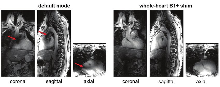
20Fig. 1: Acquired field-of-view (FOV) without and with subject specific B1+ shimming. In the non-
optimized default mode, dropouts can be observed across the heart and the left-ventricular
outflow tract (red arrows). The default mode was set by the manufacturer to provide sufficient
B1+ throughout the entire ascending and descending aorta. Using a subject specific whole-heart
B1+ shim, signal dropouts were minimized in the target region.
Fig. 2: Respiratory self-navigator (red curve) obtained from a 4D flow scan using the whole-
heart B1+ shim as shown above. In order to validate the self-gating procedure, the subject was
following a 52 seconds respiration paradigm, starting in exhale breathhold. After 15 seconds,
the subject received directions via headphones to breath in and breath out for 4 and 6 seconds
respectively.
Fig. 3: Left: Pathline visualisation of
whole-heart 4D flow vector field at peak
systole acquired with whole-heart B1+
shim and motion-compensation. Right:
Corresponding wall-shear-stress (WSS)
values. Post-processing was done using
the 4D flow prototype of Circle CVI42.
21Fig. 4: Flow curves of the motion-compensated 4D flow scan in comparison to the 2D flow
reference scan.
[1] Markl et al. “4D flow MRI”, Journal of Magnetic Resonance Imaging 36.5 (2012), 1015-1036.
[2] Hess et al. “Aortic 4D flow: Quantification of signal‐to‐noise ratio as a function of field
strength and contrast enhancement for 1.5 T, 3T, and 7T.” Magnetic Resonance in Medicine
73.5 (2015): 1864-1871.
[3] Schmitter et al. “Towards high‐resolution 4D flow MRI in the human aorta using kt‐GRAPPA
and B1+ shimming at 7T.” Journal of Magnetic Resonance Imaging 44.2 (2016): 486-499.
[4] Kolbitsch et al. ‘Respiratory motion corrected 4D flow using golden radial phase encoding.’,
Magnetic Resonance in Medicine 83.2 (2020): 635-644.
[5] Dietrich et al. ‘Respiration-Resolved 3D Multi-Channel B1 mapping of the body at 7T.’ Proc.
28th Annu. Meet. ISMRM, Paris, France: Abstract 2507; 2020.
[6] Prieto et al. “3D undersampled golden‐radial phase encoding for DCE‐MRA using inherently
regularized iterative SENSE.” Magnetic Resonance in Medicine 64.2 (2010): 514-526.
22P4
Respiration-Resolved 3D Multi-Channel Absolute B1+ Mapping
of the Body at 7T
Sebastian Dietrich1, Christoph Aigner1, Johannes Mayer1, Christoph Kolbitsch1, Tobias
Schäffter1, 2, Sebastian Schmitter1
Physikalisch‐Technische Bundesanstalt (PTB), Braunschweig and Berlin, Germany,
1
Division of Imaging Sciences and Biomedical Engineering, King’s College London,
2
London, United Kingdom
Introduction
A major challenge of ultra-high field body MRI is the spatially inhomogeneous
transmit (Tx) radio frequency field (B1+) that induces spatial contrast variations
and in the worst cases areas with no signal. This problem has been addressed by
combinations of multi-channel Tx coils, spatial mapping of B1+ or the flip angle (FA),
and by B1+ shimming or parallel transmission [1,2].
Since channel-wise mapping of the B1+ fields over a 3D body volume is highly chal-
lenging, most applications in the body so far are applied as (fast 2D) slice selective
method and performed under a breath-hold [3, 4]. Nevertheless, 3D applications
in the human body require the FA to be adjusted over a 3D volume and, thus, the
knowledge of the underlying 3D B1+ maps is essential.
To close this gap we propose a free-breathing method for 3D absolute, channel-
wise respiration-resolved B1+ mapping of the human body at 7T. The method is an
extension of a technique proposed initially for the human brain [5] to estimate the
B1+ maps of the individual Tx channels, which has also been applied in 2D as rela-
tive B1+ mapping method to the human body.
Methods
The method requires two different GRE-based free-breathing scans that are ob-
tained using a 3D radial phase-encoded (RPE) trajectory [6, 7] (Fig 1a) . The first
acquisition, termed RPE-GRE (cf. Fig 1b) acquires channel-wise 3D images for each
Tx channel where only a single Tx channel is active that is togged between scans
while all receive channels Rx acquiere the signal. The RPE-GRE data is used to
compute relative B1+ maps for each Tx channel and respiratory state following [5]
by assuming that the sum of |B1+| of all Tx channels equals the sum of |B1-| of all
Rx channels. Since these scans provide only relative estimates of the individual B1+
maps, a second, RPE-based actual flip angle (AFI) [8, 9] scan is obtained, termed
RPE-AFI that is acquired using an efficient B1+-shim setting ϕeff that maximizes
23the B1+ field in the anterior section of the heart (ROIheart). This map was used to
derive a calibration factor λ in ROIheart, that converts the channel-wise relative B1+
maps to absolute B1+ for each respiratory state. Data was reconstructed into 5
respiratory motion states using a NUFFT iterative SENSE reconstruction [10, 11].
The method was tested on 11 volunteers on a 7T (Magnetom 7T, Siemens,
Germany) and a 32Rx/8Tx channel body coil (MRI.Tools, Berlin, Germany) accord-
ing to an approved IRB protocol and after providing informed consent. All subjects
were asked to breath regularly except for two subjects that have been asked to
deliberately breath deeply to test the method. Acquisition parameters are listed
in Table 1.
Results
Respiration-resolved in-vivo 3D B1+ maps of the 8 Tx channels in one of the 11
subjects are illustrated in Fig.2 for 3 different orientations. As can be seen, clean
estimated B1+ maps free from motion artefacts are obtained for the magnitude as
well as for the phase for all transmit channels.
Fig.3, displays B1+ maps for 2 orientations and 3 respiratory motion states for Tx
channel 3 and 4 demonstrate B1+ variations of up to 1.0±0.5 μT/√kW (28±18 %)
between inhale and exhale across the heart for Tx channel 3.
A head-feet motion of right hemi-diaphragm of 2.8±0.6 cm was measured across
the two deeply breathing subjects.
Discussion & Conclusion
In this work we present a novel technique to retrieve channel-wise, motion-re-
solved absolute 3D B1+ maps of the human body at 7T. Although the maps in-
herently contain a proton density bias, similar non-respiration-resolved maps ob-
tained in 2D have been used successfully for cardiac pTx and other applications
at 7T. Ideally, a direct merging of RPE-AFI and RPE-GRE is preferable, which was
unfeasible because of insufficient B1+ amplitudes available in the body center. The
present method circumvents this limitation by using AFI data only in regions with
high B1+ amplitude for calibration purposes.
24fig.1: RPE trajectory with cartesian data acquisition along read out direction kx on a radial grid
in phase encoding plane ky - kz. Between successive radial lines the angle is increased by the
golden angle, k-space data is retrospectively binned into different respiratory motion states
with the help of a motion surrogate. Each bin is reconstructed and RPE-GRE data for different
motion states can be acquired (a). (b) Workflow of proposed B1+ mapping approach which uses
acquired AFI data with efficient shim setting φeff (1) and calculated magnitude of the sum of
estimated channel-wise B1+ for efficient shim setting (2,3) to calculate calibration factor λ in
ROIheart (4) resulting in channel-wise estimated absolute B1+ maps (5)
fig.2: B1+ magnitude and phase distribution of 8 Tx channel in 3 different orientations in exhale
motion state for one volunteer performing deep breathing.
25fig.3: B1+ magnitude distribution in 2 orientations, obtained from a breath deeply multi-channel
acquisition for 2 of the 8 Tx channels in 3 different respiratory motion states; Line plots show
B1+ values across the heart for motion state exhale (red), half-inhaled(gray) and inhale(black),
with maximum difference of 1.0 μT/√kW (28%) for Tx channel 3.
tab.1: Aqusition parameter for RPE-AFI and RPE-GRE with repetition time (TR), echo time (TE),
aqusition time (TA), oversampling as factor of phase encoding points additionally acquired
compared to a fully sampled cartesian scan with the same field of view (FOV) and voxel size,
nominal flip angle (FA), band width (BW) and sampling time (TS) for k-space center points
aquiered for self-navigation.
1. Kerr et al. Proc ISMRM: Abstract 16, 2008.
2. Padormo et al. NMR in Biomedicine 29: 1145-1161, 2016.
3. Metzger et al. Magnetic Resonance in Medicine 69: 114-126(2013)
4. Schmitter et al. Magnetic Resonance in Medicine 70: 1210-1219 (2013)
5. Van de Moortele et al. Proc. ISMRM: Abstract 367, 2009.
6. Prieto et al. Magnetic Resonance in Medicine 68: 514-526, 2010.
7. Buerger et al. Trans Med Imaging 31: 805-815, 2012.
8. Yarnykh. Magentic Resonance in Medicine 57: 192-200, 2007.
9. Dietrich et al. Proc. ISMRM: Abstract 4560, 2019.
10. Jackson et al. IEEE Transactions on Medical Imaging 10: 473-478, 1991.
11. Pruessmann et al. Magnetic Resonance in Medicine 46: 638-651, 2001.
26P5
Multi-Channel RF Power and Phase Supervision Systems
Technology for Thermal Magnetic Resonance: Development,
Evaluation and Application
Haopeng Han1, Thomas Wilhelm Eigentler1, Eckhard Grass2,3, Thoralf Niendorf1,4,5
Berlin Ultrahigh Field Facility (B.U.F.F.), Max Delbrück Center for Molecular Medicine
1
in the Helmholtz Association, Berlin, Germany,
2
IHP Leibniz-Institut für innovative Mikroelektronik, Frankfurt (Oder), Germany,
3
Institute of Computer Science, Humboldt-Universität zu Berlin, Berlin, Germany,
Experimental and Clinical Research Center (ECRC), a joint cooperation between the
4
Charité Medical Faculty and the Max Delbrück Center for Molecular Medicine, Berlin,
Germany, 5MRI.TOOLS GmbH, Berlin, Germany
Introduction
Thermal Magnetic Resonance integrates radio frequency induced thermal inter-
vention and in vivo temperature mapping using MR thermometry to permit su-
pervised targeted in vivo temperature modulation1. To achieve precise energy fo-
cal point formation, accurate thermal dose control and safety management, the
transmitted RF signals’ amplitude and phase need to be supervised and regulated
in real-time. Supervision modules implemented previously2, 3, 4 only monitor the
RF signal power level for SAR supervision. Here we propose a multi-channel pow-
er and phase supervision module which provides real-time RF power and phase
monitoring and regulation.
Methods
Our hardware implements four RF input channels and a reference signal input
channel and supports a frequency range of 10MHz to 2.7GHz. Figure 1 shows the
block diagram. The reference signal was split into five length- and impedance-
matched routes. Each RF input signal was routed to the input A of an AD8302
(Analog Devices, MA, USA) chip. All voltage outputs from the meter chips were
fed to a 16-channel, 16-bit analog-to-digital converter AD7616 (Analog Devices).
The whole system is managed by a field programmable gate array chip XC7Z020
(Xilinx, CA, USA) which is the core of a system-on-module unit AES-Z7MB-7Z020-
SOM-I-G (Avnet, AZ, USA).
A graphical user interface (Figure 2) was developed to monitor the measured RF
power and phase information. The phase meters were calibrated with a 4-chan-
nel arbitrary waveform generator (M8190A, Keysight, CA, USA). The power meters
27were calibrated using a power signal generator (SMGL, R&S, Munich, Germany).
The supervision module was integrated with a home-built 32-channel RF signal
generator5, a home-built power amplifier and a directional coupler to form a feed-
back control loop. Basic PID controllers were implemented to regulate the power
amplifier so that its output maintains a stable power level and phase. Ten minute
tests were conducted with the control loop open and closed to examine the ability
of the supervision module.
Heating experiments were conducted with the control loop either open or closed.
A self-grounded bow-tie antenna6 was applied to an agarose phantom placed into
the isocenter of a 7.0T MR scanner (Magnetom, Siemens Healthineers, Erlangen,
Germany). The heating paradigm (Fin=400MHz, Pin=42.5dBm at the port of the
antenna) was applied for 10 minutes for each experiment. MR thermometry using
the PRFS approach (TR=99ms, TE1=2.7ms, TE2=6.7ms, Voxel size=1x1x5mm3) was
conducted before and after heating.
Results
Figure 3 illustrates the supervision module hardware and its operation in RF heat-
ing experiments. After calibration, the supervision module demonstrated a power
detecting range of 100dBm and a phase detecting range of 180°. Figure 4 high-
lights the power and phase monitoring/regulating results from two 10-min tests.
When the loop was open, there were spikes (>1.5dBm) and substantial deviations
over time from the setting point, both having a major impact on the fidelity of the
focal energy point in Thermal MR. For the closed-loop experiment, the power level
was stabilized around the setting point, with a maximum deviation of 0.5dBm. The
signal phases stayed stable for both open- and closed-loop experiments.
Proof-of-principle was demonstrated in RF heating experiments using a phan-
tom setup. Figure 5 shows the MR thermometry results. The applied RF heating
paradigm induced maximum temperature increases of 7.0°C and 8.5°C with the
control loop open and closed, respectively, in an area close to the antenna. A tem-
perature rise difference of 1.5°C was observed.
Discussion and Conclusion
This work shows that the proposed multi-channel RF power and phase supervi-
sion module is suitable for Thermal MR. Early applications using a phantom setup
at 7.0T demonstrate the module’s feasibility and efficiency for real-time monitor-
ing and regulation of RF power and phase. This provides the technological basis
for a Thermal MR hardware system using an integrated RF applicator for anatomi-
cal imaging, temperature mapping and RF heating.
28Figure 1: System block diagram. There is no difference on the signal propagation delay
between the four RF input signals. Two serial peripheral interface (SPI) buses were routed
from the FPGA to the conditioning chips (ADL5330, AD5683, Analog Devices, MA, USA; F1956,
IDT, CA, USA) for configuration. Customized FPGA logic utilizing direct memory access (DMA)
was developed to interface the ADC chip. Digital low-pass filters were also implemented in
the FPGA. The data was transferred through the Ethernet port using user datagram protocol
(UDP).
Figure 2: Graphical user interface (GUI). The GUI was developed in Haskell and runs on the
host computer for monitoring the measurements, and communicates with the supervision
module through an Ethernet connection. There is a UDP sever running on the ARM processer
inside the FPGA on the supervision module. Users can configure the conditioning chips on the
left side of the GUI. The RF power and phase information is displayed on the right of the GUI.
Progress bars were used to show the relative relationship between channels.
29Figure 3: The supervision module (left). The heating experiment setup (middle): a) water
cooling system, b) 8-channel clock distributor (CDA-2990, National Instruments, TX, USA), c) RF
signal generator, d) RF power amplifier, e) panel filter, f) directional coupler (BDC0810-50/1500,
BONN Elektronik, Holzkirchen, Germany), g) power splitter (ZFSC-2-1W-S+, Mini-Circuits, NY,
USA), h) network router, i) supervision module, j) oscilloscope (DPO7254, Tektronix, OR, USA),
k) power sensor (NRP18T, R&S, Munich, Germany). The rectangular phantom (178x163x116
mm3) (right).
Figure 4: Power and phase monitoring without (left) and with (right) regulation. The blue curves
are power levels measured using the supervision module; the red curves are the power levels
measured with the power sensor; the green curves are the recorded signal phases using
the supervision module. The RF signal power level at the output of the amplifier was set to
45dBm. The RF signal phase with respect to the reference was set to zero at the beginning and
changed to 90° after 150 seconds. Both the supervision module and the power sensor were
set to averaging over 1024 samples.
30Figure 5: MR thermometry results with the feedback control loop open and closed. A
transversal slice in the middle of the phantom aligned with the center of the RF applicator
was selected for MR thermometry. The left figure shows the temperature mapping without
the supervision module regulating the power amplifier. A maximum temperature rise of 7.0°C
was observed. The right figure shows the result with the supervision in the loop. A maximum
temperature rise of 8.5°C was observed.
1. Winter, L., et al., Thermal magnetic resonance: physics considerations and electromagnetic
field simulations up to 23.5 Tesla (1GHz). Radiat Oncol. 2015 Sep 22;10:201. doi: 10.1186/
s13014-015-0510-9
2. Orzada S, et al. (2019) A 32-channel parallel transmit system add-on for 7T MRI. PLOS ONE
14(9): e0222452. https://doi.org/10.1371/journal.pone.0222452
3. Yan, X., et al., A Multi-Channel Real-Time Power Monitoring System for SAR Estimation Using
FPGA in High Field MRI. Proc Intl Soc Mag Reson Med, 2016, #3662
4. El-Sharkawy, et al., A multichannel, real‐time MRI RF power monitor for independent SAR
determination. Med. Phys., 39: 2334-2341. doi:10.1118/1.3700169
5. Han, H., et al., Design, Implementation, Evaluation and Application of a 32-Channel Radio
Frequency Signal Generator for Thermal Magnetic Resonance Based Anti-Cancer Treatment.
Cancers 2020, 12, 720
6. W.Eigentler, T., et al., Wideband Self-Grounded Bow-Tie Antenna for Thermal MR. NMR
Biomed. 2020, 41, e4274
31P6
Identifying radiation therapy effect on cardiac function using
ultrahigh field 9.4T MRI
El-Sayed Ibrahim1, Dhiraj Baruah1, Jason Rubenstein1, Rachel Schlaak1, Anne Frei1,
Carmen Bergom1
Medical College of Wisconsin
1
Introduction
Lung cancer is the leading cause of cancer-related deaths, accounting for about
25% of all cancer deaths. About one-third of lung cancer patients are treated with
radiation therapy (RT), where the incidence of cardiac complications in lung cancer
patients after RT is as high as 33%. The current paradigm for cardiotoxicity detec-
tion and management relies primarily upon the assessment of LV ejection fraction
(EF), which may not reflect the underlying advancement of subclinical cardiovascu-
lar disease. Therefore, there is a need for identifying new markers capable of early
detection of RT-induced cardiotoxicity in lung cancer. In this study, we investigate
the capabilities of ultrahigh field MRI for identifying early development of RT-
induced subclinical cardiac dysfunction in a small-animal model of lung cancer RT.
Methods
A total of 22 salt-sensitive adult rats were divided into two groups: control (n=7)
and RT (n=15). The RT group received image-guided localized whole-heart RT to 24
Gy using 3 equally-weighted fields, and then was divided in two groups that were
imaged at 8 weeks post-RT and at 10 weeks post-RT. All rats (control and RT) were
imaged when they are around the same age of 20 weeks on a Bruker 9.4T Biospec
MRI scanner with 30-cm bore diameter and equipped with 4-element surface coil.
The MRI scan included cine and tagging sequences. The cine images were ana-
lyzed to measure EF, end-diastolic volume (EDV), and myocardial mass. The tagged
images were analyzed to measure myocardial circumferential (Ecc) strain, radial
(Err) strain, and longitudinal (Ell) strains.
Results
Global cardiac function was normal in all rats (Figure 1), with increased EF and
myocardial mass in the RT rats compared to controls. EF and mass were 67±7%,
78±2, 79±3% and 0.38±0.04g, 0.49±0.04g, 0.56±0.04g in the control, 8-week post-
RT, and10-week post-RT rats, respectively. EDV values were slightly smaller in the
RT rats. EDV = 0.29±0.02ml, 0.26±0.03ml, and 0.26±0.02ml in the control, 8-week
post-RT, and10-week post-RT rats, respectively. Despite normal global function,
32strain analysis (Figure 2) showed reduced (absolute) values in the RT rats com-
pared to controls, where changes post-RT in Ecc were more than changes in Err
or Ell. Global Ecc = -14.1±2.2%, -10.2±0.7%, and -8.4±0.5%; global Err = 23.3±4.0%,
21.7.2±3.7%, and 20.9.2±6.0%; and global Ell = -15.6±1.8%, -12.3±1.0%, and
-12.7±1.9% in the control, 8-week post-RT, and10-week post-RT rats, respective-
ly. The strain measurements showed larger change between the control and the
8-week post-RT rats compared to the change between the 8-week and 10-week
post-RT rats. Err showed wider range of values, especially at the basal and apical
sites, compared to Ecc and Ell ranges of values.
Discussion
The most interesting finding in this study is the increased EF post-RT, where EF
increased from 67% in the control rats to 78% and 79% in the 8-week and 10-
week post-RT rats. This was accompanied by significant ventricular hypertrophy:
29% and 47% corresponding increases in myocardial mass, compared to control
values. This reflects the nature of cardiac remodeling to maintain global function
in the face of acute injuries from RT. Despite the normal global function, the MRI
tagging-generated regional cardiac function parameters revealed deteriorated
myocardial contractility. Specifically, myocardial strain showed to be a sensitive
marker for detecting subclinical cardiac dysfunction, where different strain com-
ponents helped characterize the nature of abnormal contractility patterns.
Conclusion
In conclusion, regional cardiac functional imaging and tissue characterization by
MRI provides detailed information about myocardial contractility pattern post-RT
and allows for early detection of induced cardiotoxicity before global cardiac func-
tion is affected.
Figure 1. Cine images showing end−
diastolic (ED) and end−systolic (ES)
images in both control and RT rats.
The images show preserved cardiac
function post−RT, along with cardiac
remodeling and hypertrophy (solid
arrow) compared to controls. Note
pleural and pericardial effusions in
RT (dotted arrows).
33You can also read