A Case of Cutaneous Myiasis Caused by Cordylobia anthropophaga Larvae in a Korean Traveler Returning from Central Africa
←
→
Page content transcription
If your browser does not render page correctly, please read the page content below
ISSN (Print) 0023-4001
                                                                                                                                          ISSN (Online) 1738-0006

                                                                                                         Korean J Parasitol Vol. 56, No. 2: 199-203, April 2018
▣ CASE REPORT                                                                                                        https://doi.org/10.3347/kjp.2018.56.2.199

        A Case of Cutaneous Myiasis Caused by Cordylobia
       anthropophaga Larvae in a Korean Traveler Returning
                       from Central Africa

                                Joo Yeon Ko1, In-Yong Lee2, Byeong Jin Park1, Jae Min Shin1, Jae-Sook Ryu3,*
       1
        Department of Dermatology, Hanyang University College of Medicine, Seoul 04763, Korea; 2Department of Environmental Medical Biology, and
        Institute of Tropical Medicine, Yonsei University College of Medicine, Seoul 03722, Korea; 3Derpartment of Environmental Biology and Medical
                                           Parasitology, Hanyang University College of Medicine, Seoul 04763, Korea

     Abstract: The cutaneous myiasis has been rarely reported in the Republic of Korea. We intended to describe here a case
     of furuncular cutaneous myiasis caused by Cordylobia anthropophaga larvae in a Korean traveler returned from Central
     Africa. A patient, 55-year-old man, had traveled to Equatorial Guinea, in Central Africa for a month and just returned to
     Korea. Physical examinations showed 2 tender erythematous nodules with small central ulceration on the left buttock and
     thigh. During skin biopsy, 2 larvae came out from the lesion. C. anthropophaga was identified by paired mouth hooks
     (toothed, spade-like, oral hooklets) and 2 posterior spiracles, which lack a distinct chitinous rim. Although rarely described
     in Korea until now, cutaneous myiasis may be encountered more frequently with increasing international travel and ex-
     change workers to tropical areas.

     Key words: Cordylobia anthropophaga, cutaneous, myiasis, Central Africa

                            INTRODUCTION                                                     creasing travel opportunity in endemic tropical areas. There-
                                                                                             fore, we intended to report here a case of furuncular myiasis
   Myiasis is characterized as the infestation of any living tissue                          caused by C. anthropophaga larvae in a Korean traveler return-
of vertebrate animal including human that can involve cutane-                                ing from Equatorial Guinea.
ous, enteric, ophthalmic, nasopharyngeal, auricular, and uro-
genital systems by larvae of the order of Diptera (true flies) [1].                                               CASE RECORD
Cutaneous myiasis is the most common form and it is clini-
cally divided into 3 types, i.e., wound, migratory (creeping)                                   A 55-year-old man presented to the dermatology outpatient
and furuncular, depending on the species of infesting larvae                                 clinic at Hanyang University Seoul Hospital, with 1-week his-
[2]. Among the 3 clinical types of cutaneous myiasis, the fu-                                tory of 2 painful erythematous nodules on the left buttock and
runcular type is mainly caused by the larvae of Cordylobia an-                               upper thigh (Fig. 1). He was usually very healthy with no un-
thropophaga, Dermatobia hominis, Cuterebra sp., and Wohlfahrtia                              derlying systemic diseases but had a traveling history to Equa-
sp. [1]. This dermatological disease by C. anthropophaga larvae                              torial Guinea, Central Africa for a month and just returned to
is commonly occurred in tropical Africa through skin contact                                 Korea. On physical examination, there were 2 tender, erythem-
with the ground contaminated with the fly larvae [3]. Until                                  atous indurated nodules with small central ulceration.
now, total 3 imported cases of cutaneous myiasis have been                                      Laboratory tests demonstrated normal white blood cell
reported in the Republic of Korea (Korea) [4-6]. However, this                               count (6,700/mm3) with neutrophils (54.7%), normal hemo-
exotic disease may be encountered more frequently with in-                                   globin (12.9 g/dl) and platelet count (288,000/mm3). The
                                                                                             tests also revealed normal aspartate aminotransferase, creati-
• Received 11 January 2018, revised 19 February 2018, accepted 3 March 2018.                 nine and erythrocyte sedimentation rate (ESR) with values of
* Corresponding author (jsryu@hanyang.ac.kr)                                                 20 U/L (normal: 7-38), 1.07 mg/dl (0.5-1.2), 15 mm/hr (0-
© 2018, Korean Society for Parasitology and Tropical Medicine
                                                                                             20), respectively. However, the C-reactive protein (CRP) level,
This is an Open Access article distributed under the terms of the Creative Commons
Attribution Non-Commercial License (http://creativecommons.org/licenses/by-nc/4.0)           1.2 mg/dl (< 0.5), was more or less increased.
which permits unrestricted non-commercial use, distribution, and reproduction in any
medium, provided the original work is properly cited.                                           Histopathologic examination of the left buttock showed

                                                                                       199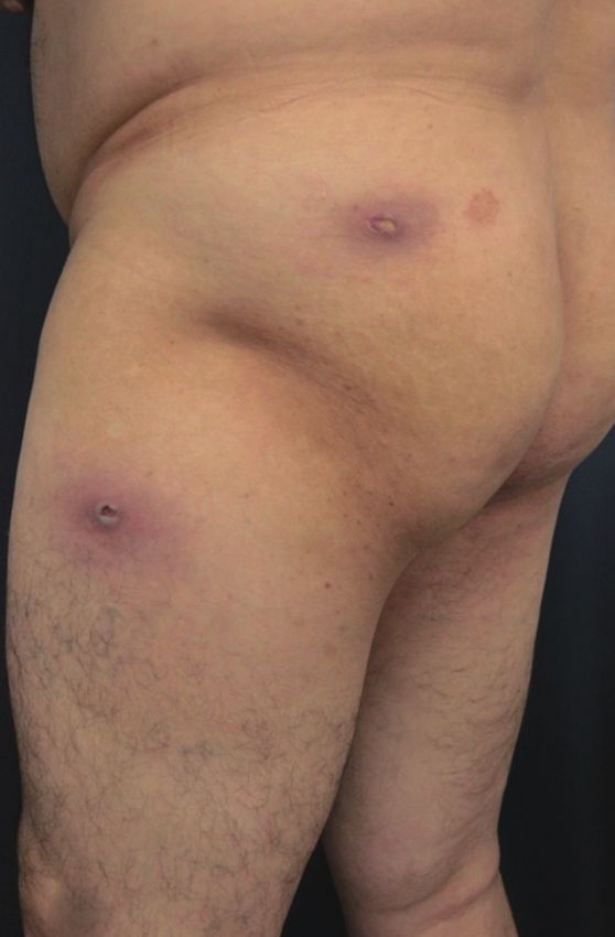
200 Korean J Parasitol Vol. 56, No. 2: 199-203, April 2018
B
A C
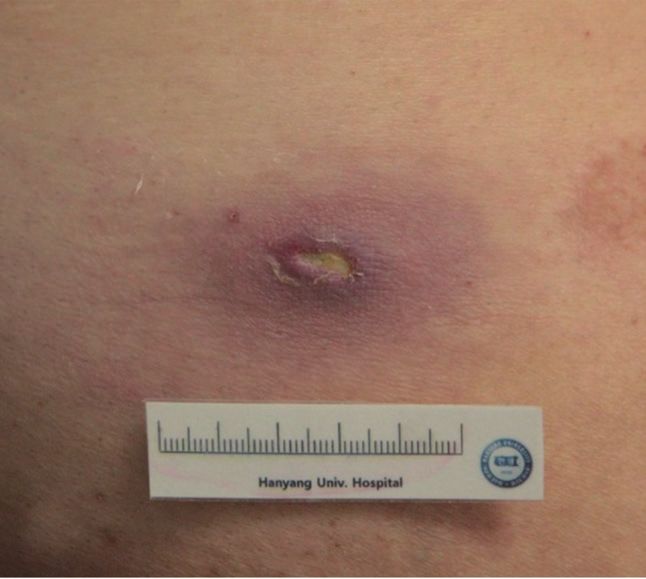
Fig. 1. (A) Cutaneous examination showed two erythematous nodules with small central ulceration on the left buttock and thigh. (B, C)
Incisional biopsies were performed on both erythematous ulcerative nodules along their major axis.
B
A C
Fig. 2. (A) Skin biopsy from the nodule on the buttock showed ulceration, central fibrinoid necrosis and diffuse cellular infiltration in the
dermis (H&E, × 1.25). (B) A high power view revealed the infiltration of inflammatory cells that consisted of lymphocytes, eosinophils and
neutrophils (H&E, × 400). (C) Skin biopsy from upper thigh specimen, several brownish angular fragments were identified, which were
thought to be remnants of larvae (H&E, × 400).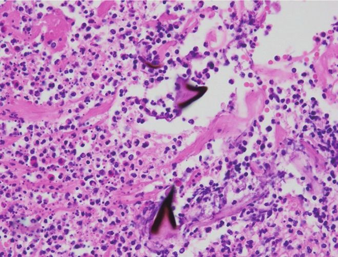
Ko et al.: Myiasis by Cordylobia anthropophaga 201
B C
A D
Fig. 3. (A) Two white and dark-brown colored barrel-shaped larvae were about 10.5 mm in length, and 4.5 mm in width. (B) Their body
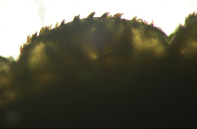
was almost completely covered by conic spines with a brown apex, pointing towards the posterior end. (C) Detail of cuticle spines. (D)
Two copper-colored posterior spiracles, each bearing 3 sinuous spiracular slits which lack a distinct chitinous rim.
B
A C
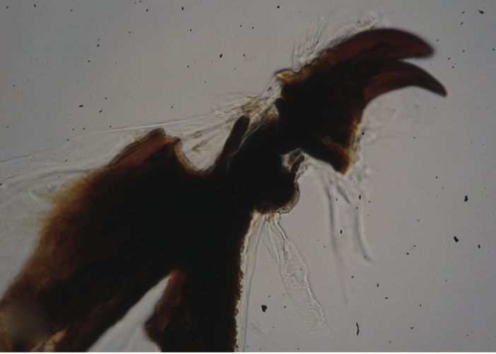
Fig. 4. (A) Anterior end of larva. (B) Paired mouth hooks (toothed, spade-like, oral hooklets) protruded ventrally from anterior end. (C) An-
terior spiracles of larva.
central fibrinoid necrosis and diffuse cellular infiltration of and cellular infiltration extending to subcutis and several deep
lymphocytes, eosinophils and neutrophils in deep dermis (Fig. brownish angular fragments were observed in dermis, which
2A, B). The upper thigh specimen showed gangrenous necrosis were thought to be remnants of larvae (Fig. 2C). Two barrel-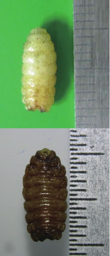
202  Korean J Parasitol Vol. 56, No. 2: 199-203, April 2018

shaped larvae were detected from the each lesion. They were          eral fragments of larvae and surrounding necrosis and granu-
white and dark-brown colored, 10.5 mm long and 4.5 mm                loma formation. For our patient, we believe that removing the
wide (Fig. 3A).                                                      ulcer, probably opening pore, through biopsy procedure
   The larvae were preserved in a solution of 10% KOH, 89%           helped the patient's recovery.
ethanol, 95% ethanol, and 100% ethanol sequentially. The                Furuncular myiasis is common in tropical and subtropical
morphology of their body was almost completely covered by            areas, including central and South America, tropical Africa,
conic spines with a brown apex, pointing towards the posteri-        however, there are few reported cases of furuncular or wound
or end (Fig. 3B, C). Two copper-colored posterior spiracles,         myiasis in Korea. Three patients were Korean [4-6] and the
each bearing 3 sinuous spiracular slits which lack a distinct        other 2 were foreigners who lived in Korea [10,11]. Three pa-
chitinous rim (Fig. 3D). In the anterior end of larva, the           tients were furuncular myiasis caused by C. anthropophaga from
mouth with a pair of spade-like stout hooks and anterior spir-       Cameroon and Uganda [4,6] and D. hominis from Cost Rica
acles were characteristically observed (Fig. 4). Skin lesions were   [5]. Two foreigners had C. anthropophaga from Benin [10] and
markedly improved after the remove of larvae and 2-week an-          D. hominis from South America [11]. These traveling history
tibiotic treatment, and there was no recurrence.                     are consistent with endemic geographic location of C. anthro-
                                                                     pophaga and D. hominis. Actually, C. anthropophaga is most
                        DISCUSSION                                   commonly found in tropical African and D. hominis is most
                                                                     commonly found in Central and South America. Our cases
   C. anthropophaga is known as the skin maggot fly, the mango       also have traveling history to Equatorial Guinea, Central Afri-
fly, the putzi (or putsi) fly, by the French name ver du Cayer       ca. It has been reported in other northeast Asia countries, that
(“worm of Cayer”, an area in present–day Senegal) or, most           is, China and Japan [12-14]. Therefore, although northeast
commonly the tumbu fly [1]. The adult fly is yellow brown,           Asia is not common location of furuncular myiasis, with in-
has brown-black spots on the abdomen, 2 black longitudinal           creasing international travels and works, myiasis may be en-
bands on the thorax, and measure 6 to 12 mm in length [7].           countered more frequently.
   The larvae can penetrate the unbroken skin of the host, who          In conclusion, we report a case of furuncular cutaneous my-
is usually lying on the ground or by the contaminated clothes.       iasis on the left side of the buttock and upper thigh by C. an-
Interestingly, the host usually feels no symptoms at the time of     thropophaga, which has been rarely described in Korea. Consid-
skin penetration by larvae. Therefore, most patients do not          ering the increasing international travel and exchange workers
think maggots as a cause of their skin problems. Within 1 to 2       with foreign countries, physicians should be more concerned
weeks, the larvae develop into the second and third instars,         about clinical manifestations of cutaneous myiasis and mor-
and the 13- to 15-mm sized mature larvae that can emerge             phologic characteristics of diphterous larvae.
from the central pore of the skin lesions [3]. In this report,
morphological characteristics of larvae of C. anthropophaga (ex,                    CONFLICT OF INTEREST
a pair of spade-like hooks, anterior spiracles, and posterior
spiracles) are well observed in Figs. 3 and 4.                         We have no conflict of interest related to this study.
   Early lesions may resemble other reactions due to insect
bite, but furuncular lesions with an intense inflammatory reac-                             REFERENCES
tion in the surrounding tissue rapidly develop [3,8]. The goal
of treatment is removal of the larva and prevention of the sec-       1. McGraw TA, Turiansky GW. Cutaneous myiasis. J Am Acad Der-
ondary infection. Occlusion, larvicides such as ivermectin, or           matol 2008; 58: 907-926.
manual squeezing can be used to remove the larva. Occlusion           2. Robbins K, Khachemoune A. Cutaneous myiasis; a review of the
deprives the larva of oxygen and either kills the larva or induc-        common types of myiasis. In J Dermatol 2010; 49: 1092-1098.
                                                                      3. Ockenhouse CF, Samlaska CP, Benson PM, Roberts LW, Eliasson
es it to move upward in search of air [1]. Manually squeezing
                                                                         A, Malane S, Menich MD. Cutaneous myiasis caused by the Afri-
out the larva is therapeutic option in all forms of furuncular           can tumbu fly (Cordylobia anthropophaga). Arch Dermatol 1990;
myiasis. Sometimes, surgical removal is needed for removing              126: 199-202.
the larvae or their fragments [9]. Indeed, our case showed sev-       4. Park JM, Kim HJ, Choi YJ, Yong TS, Ree HI, Lee MG. A case of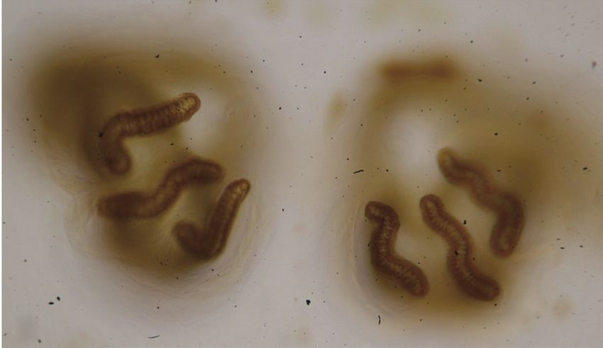
                                                                                 Ko et al.: Myiasis by Cordylobia anthropophaga  203

       furuncular cutaneous myiasis after traveling to Cameroon. Kore-    10. Ko BJ, Cho HK, Lee IY, Yong TS, Whang KU. A case of furuncular
       an J Dermatol 2009; 47: 600-603 (in Korean).                           cutaneous myiasis in a German patient who has traveled to Be-
    5. Youn YH, Kim MR, Oh ST, Cho BK, Lee IY, Park HJ. A case of fu-         nin. Korean J Dermatol 2013; 51: 348-352 (in Korean).
       runcular cutaneous myiasis by Dermatobia hominis. Korean J         11. Shin JY, Kim JH, Kim YC. Furuncular myiasis in a traveler return-
       Dermatol 2015; 53: 570-571 (in Korean).                                ing from South America. Korean J Dermatol 2012; 50: 662-663
    6. Song SM, Kim SW, Goo YK, Hong Y, Ock MS, Cha HJ, Chung                 (in Korean).
       DI. A case of furuncular myiasis due to Cordylobia anthropophaga   12. Deng Y, Liu F, Chen X, Lu S. The first imported cutaneous myia-
       in a Korean traveler returning from Uganda. Korean J Parasitol         sis due to Cordylobia anthropophaga in China. Int J Dermatol
       2017; 55; 327-331.                                                     2013; 52: 120-122.
    7. Veraldi S, Brusasco A, Süss L. Cutaneous myiasis case by larvae    13. Fujisaki R, Makimura K, Hayashi T, Yamamura M, Yamaoka T,
       of Cordylobia anthropophaga (Blanchard). Int J Dermatol 1993;          Shiraishi K, Ishibashi S, Kawakami S, Kurihara T, Nishiya H. Ex-
       32: 184-187.                                                           otic myiasis caused by 19 larvae of Cordylobia anthropophaga in
    8. Chopra A, Probert AJ, Beer WE. Myiasis due to tumbu fly. Lancet        Namibia and identified using molecular methods in Japan.
       1985; 1: 1165.                                                         Trans R Soc Trop Med Hyg 2008; 102: 599-601.
    9. Lane RP, Lowell CR, Griffiths WA, Sonnex TS. Human cutaneous       14. Nagamori K, Katayama T, Kumagai M. A case of cutaneous my-
       myiasis--a review and report of three cases due to Dermatobia          iasis due to Dermatobia hominis in Japan. J Infect Chemother
       hominis. Clin Exp Dermatol 1987; 12: 40-45.                            2007; 13: 255-257.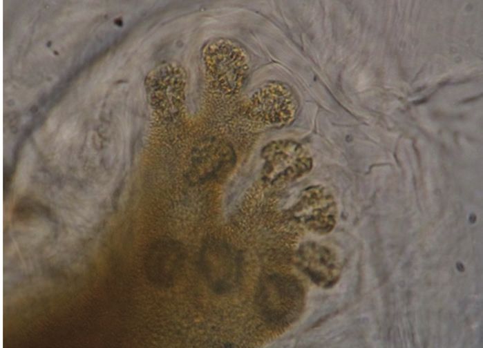
You can also read