A Rare Case of Human Residual Root Myiasis Caused by Clogmia Albipunctata Larvae(Diptera: Psychodidae)
←
→
Page content transcription
If your browser does not render page correctly, please read the page content below

A Rare Case of Human Residual Root Myiasis
Caused by Clogmia Albipunctata Larvae(Diptera:
Psychodidae)
Ying-jie Liu
Department of Pathogenic Biology, School of Basic Medcial Sciences, Henan University, Kaifeng, China
Jin-rui Liu
Xi'an Jiaotong University
Yun Liu
Henan University
Juan Chen ( wfnaca@163.com )
Department of Gynecology and Obstetrics,Kaifeng New District Maternal and Child Health Care Hospital
https://orcid.org/0000-0003-2806-4375
Case Report
Keywords: China, Myiasis, Residual root, Clogmia albipunctata, Larvae
Posted Date: April 5th, 2021
DOI: https://doi.org/10.21203/rs.3.rs-259478/v1
License: This work is licensed under a Creative Commons Attribution 4.0 International License.
Read Full License
Page 1/10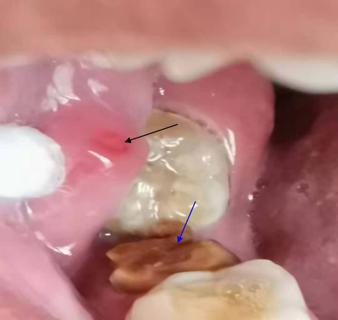
Abstract
Background: Clogmia albipunctata is cosmopolitan in distribution. The adult flies can survive and spread
outdoors during the temperate seasons while continuously breeding in buildings during the winter
months. Because they are non-biting, tiny and quite, most of people do not pay special attention to them.
It is the first case reported that Clogmia albipunctata larvae cause human residual root myiasis.

Case presentation: In December 2020, a 26-year-old woman was referred from Kaifeng New District
Maternal and Child Health Care Hospital to the Department of Pathogenic Biology, Medical College of
Henan University with chief complaint that two active alive larvae were found in the mouth while brushing
her teeth in the morning. The intraoral examination revealed nice oral hygiene and no larvae was found
directly. The right second mandibular molar was a residual root and the mucosa above it was mild
erythematous and edematous and no bleeding on probing was present. While some 50℃ normal saline
was injected in the residual root with syringe, four larvae swarmed out from the residual root. The larvae
were observed by naked eyes and light microscope and the larvae were reared. One adult fly was got 11
days later. They were identified as Clogmia albipunctata larvae. Because the patient was in lactation,
medication was not recommended. Treatment included the removal of all visible larvae followed by
debridement. The patient was followed-up for 1 month and healed. The patient’s residual root myiasis
was associated with sleeping with the mouth open and the smell of rotten food in the residual root
attracted Clogmia albipunctatus to lay eggs in the residual root.

Conclusions: This report implies that even if the oral hygiene is nice in general, the existence of residual
roots maybe results in oral myiasis. The myiasis caused by Clogmia albipunctata larvae should be paid
attention to. It is necessary to treat residual roots in time.

Background
Dental caries result from the chemical dissolution of dental tissues, resulting from the production of
acids generated by bacteria that metabolize carbohydrates from the diet, especially sucrose[1]. Currently
tooth decay is described as a complex sugar dependent biofilm dysbiosis[2, 3].

Dental caries and residual roots are commonly known as ‘worm tooth’ in China though worms do not
cause them. However we did find ‘worms’ in a residual root recently. It belongs to the category of myiasis.

Myiasis is defined as an invasion of live human and vertebrate animals with dipterous larvae, which feed
on the host’s living or dead tissue, ingested food or liquid body substances[4]. Myaisis is regarded as a
disease associated with unhygienic conditions and poor socioeconomic environment[5]. Of note, the case
we present has the following special features. First, the patient is healthy with nice hygienic conditions.
Secend, Myiasis occurs in a residual root which caused by Clogmia albipunctata larvae which was never
reported.

Case Presentation
                                                   Page 2/10
On 26 December 2020, a 26-year-old woman was referred from Kaifeng New District Maternal and Child
Health Care Hospital to the Department of Pathogenic Biology, Medical College of Henan University with
chief complaint that two active alive larvae were found in the mouth while brushing her teeth in the
morning.

The woman was a civil servant living in the suburb of Kaifeng, Henan Province, China. She did not report
suffering from fever, pain or other oral uncomfortable feeling. However, she looked anxious and
frightened. The intraoral examination revealed nice oral hygiene. No larvae was found directly. The right
second mandibular molar was residual root. The mucosa above it showed mild erythematous and
edematous and no bleeding on probing was present (Fig. 1).

While some about 50℃ normal saline was injected in the residual root and on the erythematous and
edematous mucosa with syringe, a total of four larvae swarmed out from the residual root.

The larvae were grasped and removed quickly and gently. They wriggled around actively in water and
crawled quickly on the surface of solid(Fig. 2).

Because the patient was in lactation, medication was not recommended. Treatment included the removal
of all visible larvae followed by debridement. She was advised to irrigate the residual root with
approximately 500ml 50℃ normal saline three times a day before brushing her teeth to remove the
potential eggs and larvae in the residual root. And we advised her to refer to dentist to treat the residual
root as soon as possible.

After careful history taking from the patient, we learned she had the habit of sleeping with the mouth
open. Because she was in lactation, there were many fruits and snacks in the bedroom which attracted
some small flies. It was deduced that while the patient sleeping with mouth open, the flies laid eggs in the
residual root. We advised her to clean the room to eliminate the breeding environment of the flies and
spray with insecticides to exterminate the flies. And we advised her to sleep without the mouth open as
far as possible to avoid the flies laying eggs in the residual root again. The patient was followed for one
month. No more larvae was found again and the erythematous and edematous mucosa was healed.

The larvae were observed by naked eyes and light microscope(Motic BA210,MOTIC CHINA GROUP CO.,
LTD. ). The larva is about 7–8 mm long and 1 mm wide. The body is cylindrical. The body of the larva
consists of a head segment, 3 thoracic segments and 8 abdominal segments.

It is grayish dorsally and white ventrally while its head and tail are dark brown(Fig. 3A). Dorsal aspects of
the body segments were covered with 26 saddles shaped dark chitinous plates. The body is densely
covered with long black back-warding setae dorsally and laterally(Fig. 3B). Caudally, the siphon is cone-
shape. There are two dorsal anal processes and two ventral anal processes with a tuft of hairs at the
end(Fig. 3C). Its head is triangular. The mouthparts are of chewing type. Two breathing tubes appear
extending along the length of the body starting with a pair of anterior spiracles at the prothorax and end
by a pair of posterior spiracles, at the tip of the terminal segment (Fig. 3D).

                                                   Page 3/10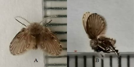
Adult morphological identification is considered the ‘gold standard’ for the identification of Diptera
species[6]. The four alive larvae were reared at 25°C. One Clogmia albipunctata was got 11 days later.

The grayish brown body is about 4mm long. Its antennae has 13 nodes. Its wings are covered with gray
brown minute hairs. Its wings have two black spots each side and there are some white spots on the
wings. (Fig. 4)

From previous morphological characters and comparing all the morphological characters to several
literatures[7–9], it was indentified as the fourth instar larval stage larva of Clogmia albipunctata.

Discussion
Oral myiasis occurs mainly in the tropics and associated with inadequate public and personal hygiene,
senility, alcoholism, mental debility, mouth breathing[10], incompetent lip, cerebral palsy[11], suppurating
lesions, severe halitosis, gingival disease, trauma[12] and so on.

Of note, in this case the patient lived in a decent house with nice surrounding environment and personal
hygiene. However, the food blocked in the residual root is difficult to clean. The patient had the habit of
sleeping with the mouth open. The smell of the rotten food in the residual root attracted Clogmia
albipunctatus to lay eggs in the residual roots. And the rotten food in the residual root provided an ideal
media for the larvae to settle.

The treatments of myiasis included manual removal of larvae and debridement, application of antibiotic
therapy, asphyxiating substances and ivermectin. Because the patient was in lactation, we used the
treatment of manual removal of larvae and debridement.

The main species reported to cause oral myiasis are Cochliomyia hominivorax, Chrysomya bezziana,
Musca domestica, Sarcophaga, Lucilia sericata, Lucilia cuprina, Musca nebulo, Oestrus ovis,
Calliphoridae, Dermatobia hominis, Hypoderma bovis, Hypoderma tarandi and Wohlfahrtia magnifica[13].

Clogmia albipunctata is a primitive Nematocera of the family Psychodidae, subfamily Psychodinae. It is
cosmopolitan in distribution. The adult flies can survive and spread outdoors during the temperate
seasons while continuously breeding in buildings during the winter months. Their larvae are
coprophagous and saprophagous. They feed on decaying organic matter and the feces of the vertebrate.
They are present in moist places such as bathrooms, toilets, hotels and hospitals[14]. They are all around
us all the year round. Because they are non-biting, tiny and quite, most of people do not pay special
attention to them. Clogmia albipunctata can elicit inhalant allergy as a result of inhaling fragments of
their disintegrated body parts and play a significant role as a potential mechanical vector of
pathogens[15]. The larvae of Clogmia albipunctata can cause myiasis.

Larvae of Clogmia albipunctata had been reported to cause human nasopharyngeal myiasis[16],
intestinal myiasis[7, 9] and urinary myiasis[8]. The case of human residual root myiasis caused by
Clogmia albipunctata larvae had not been reported before.
                                                   Page 4/10
Conclusions
Poor hygiene is the most important risk factor and is present in almost all myiasis cases. Therefore, some
people with nice hygiene feel that it is impossible for them to suffer from myiasis. However, we can learn
from this case a healthy person with better hygiene maybe suffers from myiasis under some conditions.
In addition, some people do not pay enough attention to dental hygiene in China. If they have not
symptoms, they will not treat the residual roots in time. Residual root may become the focus of systemic
infection and then cause systemic diseases. Moreover myiasis can occur in residual roots. Therefore, it is
necessary to treat the residual root in time. In the mean time, the human residual root myiasis caused by
Clogmia albipunctata larvae should be paid attention to prevent.
Abbreviations
Not applicable.
Declarations
Ethical Approval and consent to participate
This study was approved by the Human Research Ethics Committee of the Henan University in China and
the committee’s reference number is [HUSOM2021-003]. The patient received an explanation about the
scope of the study, such as objectives, procedures, and potential risks, and signed an informed consent
for the use of the patient’s clinical samples.
Consent for publication
Written informed consent was obtained from the patient for publication of this case study. A copy of the
written consent is available for review by the editor of this journal.
Availability of data and materials
Not applicable.
Competing interests
The authors declare that they have no competing interests.
Funding
The authors received no funding for this work.
Authors’ contributions
Page 5/10Ying-jie Liu conceived and designed the experiments and drafted the manuscript. Juan Chen participated
in the case collection and revised the manuscript. Jin-rui Liu and Yun Liu participated in the microscopic
examination of the larvae and raised the larvae. All authors read and approved the final manuscript.
Acknowledgements
Not applicable.
Footnotes
Not applicable.
References
1. Søren Jepsen, Juan Blanco, Wolfgang Buchalla, Joana C. Carvalho, Thomas Dietrich, Christof Dörfer,
Kenneth A. Eaton, Elena Figuero, Jo E. Frencken, Filippo Graziani, Susan M. Higham, Thomas Kocher,
Marisa Maltz, Alberto Ortiz-Vigon, Julian Schmoeckel, Anton Sculean, Livia M.A. Tenuta, Monique H.
Veen, Vita Machiulskiene. Prevention and control of dental caries and periodontal diseases at
individual and population level: consensus report of group 3 of joint EFP/ORCA workshop on the
boundaries between caries and periodontal diseases. Journal of Clinical Periodontology. 2017; 44
Suppl 18:85-93.
2. Sheiham A, James W P T. Diet and Dental Caries: The Pivotal Role of Free Sugars Reemphasized. J.
Dental Res. 2015; 94: 1341-7.
3. R A Giacaman. Sugars and beyond. The role of sugars and the other nutrients and their potential
impact on caries. Oral Dis. 2018; 24: 1185-97.
4. Farrag H M M, Huseein E A M, Almatary A M, Othman R A. Morphological and initial molecular
characterization of Clogmia albipunctatus larvae(Diptera: Psychodidae) causing urinary myiasis in
Egypt. PLoS Negl Trop Dis. 2019; 13: e0007887.
5. J. Blejter, D. Fischer, S. G. Golombek. Tracheostomy wound myiasis in a child: case report and review
of the literature. Case Rep Pediatr. 2012; doi:10.1155/2012/317862.
6. Giangaspero A, Barlaam A, Pane S, Marchili M R, Onetti Muda A, Putignani L, Hall M J R. Accidental
Nasal Myiasis Caused by Megaselia rufipes(Diptera: Phoridae) in a Child. Journal of Medical
Entomology. 2020; doi:10.1093/jme/tjaa184.
7. Mokhtar Aida Syafinaz, Braima Kamil Ali Obeid, Peng Chin How, Jeffery John, Mohd Zain Siti
Nursheena, Rohela Mahmud, Lau Yee Ling, Jamaiah Ibrahim, Wilson John-James, Abdul-Aziz
Noraishah Mydin. Intestinal Myiasis in a Malaysian Patient Caused by Larvae of Clogmia
albipunctatus (Diptera: Psychodidae). Journal of Medical Entomology, 2016; 53: 957-60.
8. Hjaija D, Sawalha S S, Amr Z S, Katbeh-Bader A, Hassoon R A H. Urinary Myiasis Caused by Clogmia
albipunctata from the Palestinian Territories. Bulletin de la Société de pathologie exotique. 2018;
111: 148-51.
Page 6/109. Nadia Ali El-Dib, Mona Ibrahim Ali, Doaa Ahmed Hamdy,Wegdan Mohamed Abd El Wahab.Human
intestinal myiasis caused by Clogmia albipunctata larvae (Diptera: Psychodidae): First report in
Egypt. Journal of infection and public health. 2020; 13: 661-3.
10. Zachariah Jane Emily, Sehgal Khushboo, Dixit Uma B, Bhatia Rupinder. Oral myiasis: a case
report. Special Care in Dentistry. 2014; 34: 51-3.
11. Vanmathi Vasanthakumar, Parasuraman Varalakshmi, Ramya Vanmathi. Oral myiasis of maxilla
(palatal gingiva). Contemporary Clinical Dentistry. 2020; 11: 162-4.
12. Shenoi Ramakrishna, Kolte Vrinda, Ingole Pranav, Rajguru Jignesh, Karmarkar Jui, Kolte Sunil,
Patankar Kunal. Management of oral myiasis caused by Chrysomya bezziana- A case series. Annals
of Maxillofacial Surgery. 2020; 10: 521-4.
13. Fabio Francesconi, Omar Lupi. Myiasis. Clin Microbiol Rev.2012; 25: 79-105.
14. Onder Z, İnci A, Yıldırım A, Ciloğlu A, Düzlü O. Molecular Characterization of Myiasis-Causing Moth
Flies(Diptera: Psychodidae). Turkiye Parazitol Derg. 2018; 42: 223-8.
15. M. Faulde, M. Spiesberger. Hospital infestations by the moth fly, Clogmia albipunctata(Diptera:
Psychodinae), in Germany. Journal of Hospital Infection. 2012; 81: 134-6.
16. N. Mohammed, K.G.V. Smith. Nasopharyngeal myiasis in man caused by larvae of Clogmia
(=Telmetoscopus) albipunctatus Williston (Psychodidae, Dipt.). Transactions of the Royal Society of
Tropical Medicine and Hygiene. 1976; 70: 91.
Figures
Page 7/10Figure 1
The residual root (blue arrow) and the mild erythematous and edematous mucosa (black arrow).
Page 8/10Figure 2
The four larvae wriggled around actively in a plastic cup with water (red arrows).
Figure 3
The body of the alive Clogmia albipunctata fourth instar larvae A: Full-larva; B: Middle segments showing
dorsal plates and dorsal and lateral setae(×40); C: Caudal part showing siphon and end processes and
anus in lateral view(×40); D: The triangular head (blue arrow) and 2 breathing tubes extending along the
length of the body (red arrows) (×40)
Page 9/10Figure 4
One Clogmia albipunctata was got 11 days later. A: The Clogmia albipunctata (alive) in vertical view B:
The Clogmia albipunctata (dead) in lateral view
Page 10/10You can also read