A Virtual Lecture with Hands-on Simulation of the Gastric Ultrasound Examination - Michelle Horton BSN, RN, CCRN Ginamarie Martucciello BSN, RN ...
←
→
Page content transcription
If your browser does not render page correctly, please read the page content below
A Virtual Lecture with Hands-on Simulation
  of the Gastric Ultrasound Examination

         Michelle Horton BSN, RN, CCRN
      Ginamarie Martucciello BSN, RN, CCRN

              Principal Investigator:
      Michael McLaughlin, DNP, CRNA-APN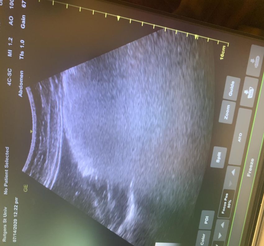
School of Nursing

       Please take a moment to fill out a
             Pre-Intervention Test
             found at the link below
https://rutgers.ca1.qualtrics.com/jfe/form/SV_8
               8oXUbQ3wCRpyh7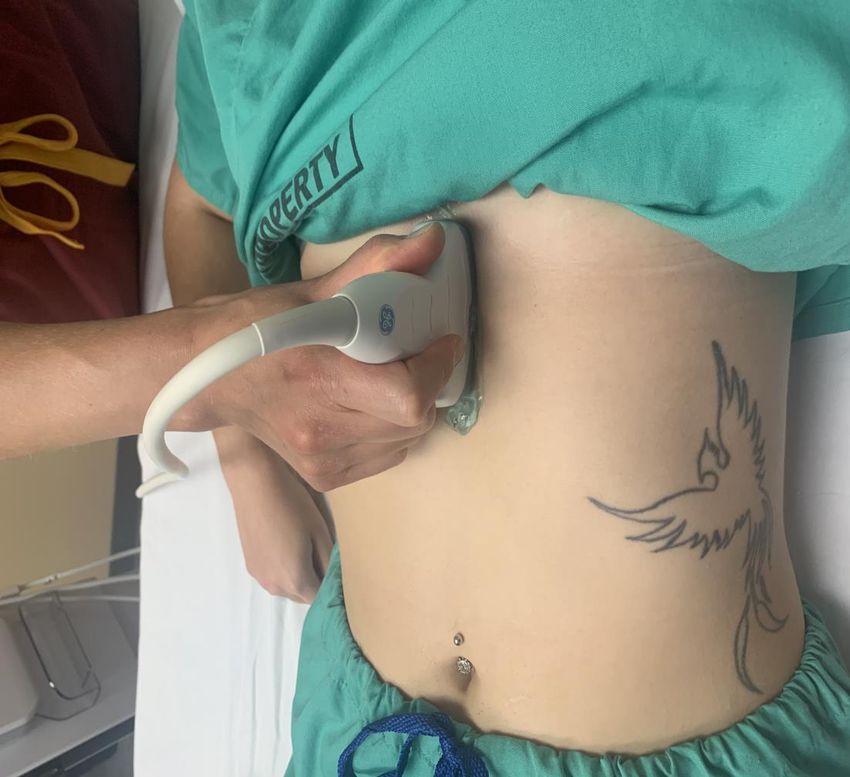
School of Nursing

Aims & Objectives
• To increase the anesthesia provider's confidence in the
  identification of the gastric antrum's contents with point-of-
  care gastric ultrasound
• To increase the anesthesia provider's competency in the
  identification of the gastric antrum's contents with point-of-
  care gastric ultrasound
   o To create a forum that addresses the evidence, indications, benefits,
     use, and interpretation of gastric ultrasound.
   o To provide examples of various prandial statuses with gastric ultrasound
     photos and videos.
   o To provide a hands-on demonstration of gastric ultrasound and the
     various prandial status’ that can be encountered.
   o To disseminate an evidence-based decision tree to aid in data
     interpretation of the gastric ultrasound results to encourage use in the
     clinical setting and promote longevity of the training.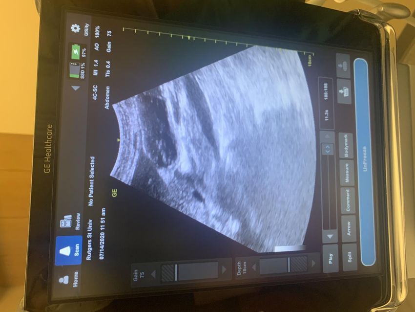
School of Nursing

 Significance
Pulmonary Aspiration is An Anesthesia-Related Complication

• Incidence varies between
School of Nursing

Background
• Classically, prevention strategies rely on asking last time
  patient ate and following fasting guidelines
• Current Assessment Tool: ASA NPO Guidelines is antiquated
  and has several shortcomings
   – Does not take into account a variety of patient factors:
       • Emergent and urgent surgeries
       • Communication and comprehension issues (LOC, AMS, drugs, language
         barrier)
       • Cognitive impairment
       • Pediatric population
       • Patient’s may not be truthful
       • Medical conditions that delay gastric emptying
School of Nursing

A New Way to Evaluate a Patient’s Aspiration Risk
 • Point-of-Care Gastric Ultrasound helps determine the volume
   and gastric content material in the patient’s stomach
 • Gastric ultrasound is:
     – Simple: Findings are easily recognizable & scanning technique can be
       quickly learned and performed
     – Fast: Takes less than 5 minutes
     – Non-invasive
     – Accurate & Reliable: Provides diagnostic data (qualitative and
       quantitative), high specificity and sensitivity
     – Point-of-Care: Performed at bedside, Focused/Limited in scope
     – Real-time

  Helps anesthesia providers answer the question on whether
         the patient is at risk for pulmonary aspiration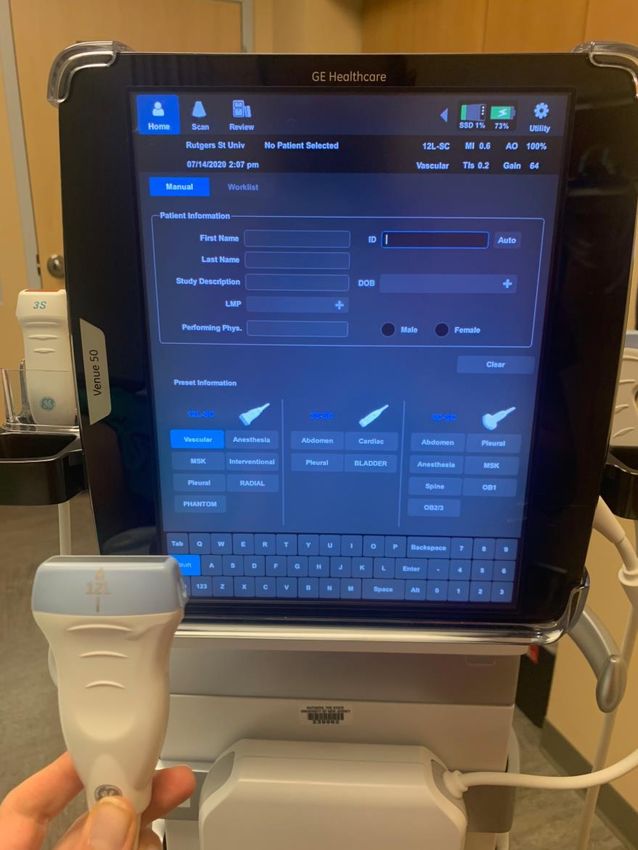
School of Nursing

Evidence: Validity, Reliability, and
Interpretability of Gastric Ultrasound

School of Nursing

Evidence: Impact and Benefits of Gastric
Ultrasound in Changing Anesthetic Management

School of Nursing

Limitations and Barriers
• Inaccurate in abnormal underlying gastric anatomy
   – Previous gastric resection/bypass gastric band
   – Fundoplication
   – Large hiatus hernia
• Antrum difficult to find/assess in 2-3% of normal individuals
• Challenges with:
   – Morbid obesity: older machines may not have adequate depth
   – Pregnancy: antrum may be displaced deep to the liver
   – Unable to position patients in RLD position

School of Nursing

Indications
• Lack of adherence to fasting instructions
   – Emergency or Urgent procedure (no planned fasting)
   – Miscommunication
   – Questionable/Borderline adherence to fasting instructions
• Inability to obtain fasting history
   – Depressed level of consciousness
   – Language barrier
   – Cognitive dysfunction
• Co-morbidities that delay gastric emptying
   –   Pregnancy/Active labor
   –   Diabetes
   –   Severe liver or kidney dysfunction
   –   Neuromuscular disorders
   –   Recent trauma
   –   Pain and chronic opioid use
   –   Gastric dysmotility
School of Nursing

Performing the Scan
• Equipment
   – Ultrasound
   – Adults: Curvilinear, low-frequency (2-5MHz) transducer, abdominal
     setting
   – Children/
School of Nursing Curvilinear Probe, Linear Probe, Abdominal Setting Vascular Setting
School of Nursing
Performing the Scan:
Steps: Scanning Technique
Start supine:
      1.   Probe indicator towards the head
      2.   Place probe just below xiphoid process
      3.   Sagittal plane (Runs mouth to tail)
      4.   Sweep transducer left to right along subcostal margin
      5.   Identify gastric antrum using standard anatomical landmarks
           •    Interpret scans between contractions or when antrum relaxed
      6.   Position to right lateral decubitus
Repeat steps in right lateral decubitus position
School of Nursing Supine Right Lateral Decubitus
School of Nursing

  Performing the scan: Anatomy

• Stomach has 3 anatomical parts:
   1. Fundus- superiorly located
   2. Body- most of the mass
   3. Antrum- inferiorly located, prior to pyloric sphincter: where the exam is
      focused on
• Gastric Antrum
   –   Primary area of interest in assessing gastric contents
   –   Holds food until released to small intestine
   –   Consistently located in epigastrium
   –   Most amendable to examination
   –   Accurately reflects the content of the entire stomach
   –   Less air content that interferes with scan
   –   Usually 3-4cm deep
School of Nursing Landmarks on Ultrasound • Inferior/Posterior to left lobe of the liver (usually at 9 o'clock) • Anterior to pancreas • Anterior to Aorta/IVC
School of Nursing
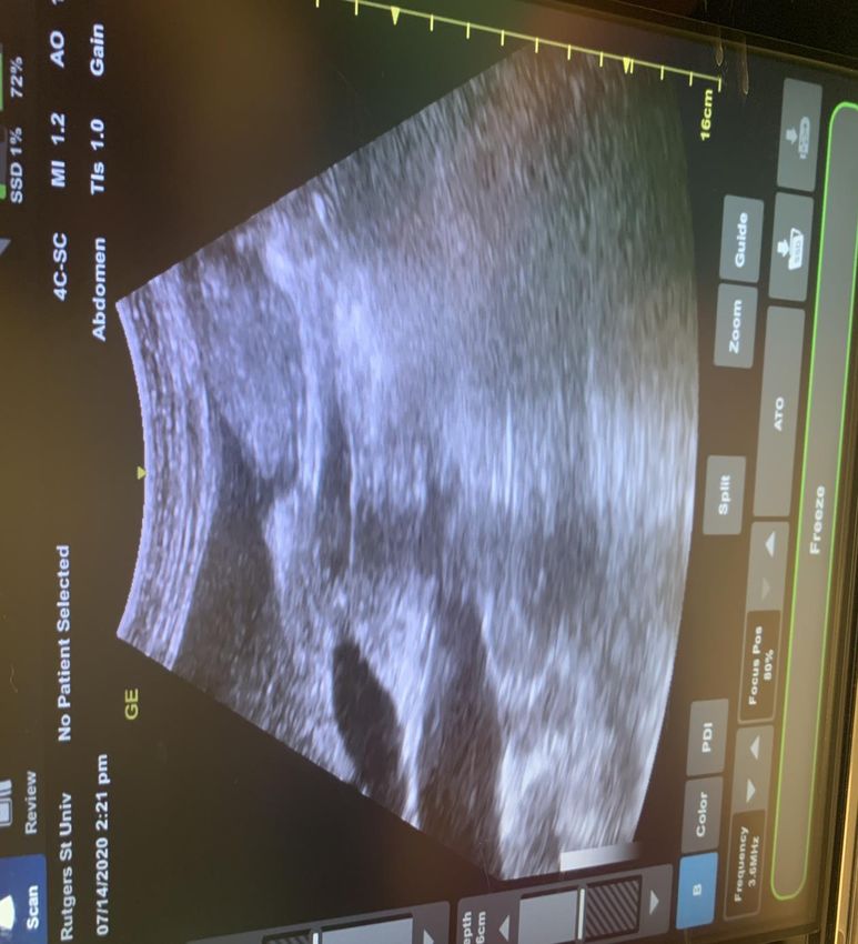
School of Nursing

Types of Gastric Content
•   Empty
•   Solid
•   Milk/Suspensions
•   Clear Fluid
School of Nursing

  Empty
• No content in both
  supine and RLD
• Antrum flat &
  collapsed
• Small, 2-3cm in
  diameter
• Round, Ovoid
  shape
• “Bulls eye” or “Target”
• Ring is thick
  muscularis propriae
School of Nursing

Empty Antrum
School of Nursing

Solid
• Antrum distended
• Thin muscle wall

• Early
   – Contents of high/mixed echogenicity
   – “Frosted glass”
        • Mixing of air and solid along anterior wall
        • Blurring of posterior wall and deeper structures
• Late (1-2 hours following a solid meal)
   – Heterogeneous, particulate content
        • Solid food
   – Homogenous, hyperechoic content
        • Characteristic of dairy products or particulate fluids
School of Nursing
School of Nursing
School of Nursing

Fluid
• Liquids are anechoic or hypoechoic
    – All fluids have similar appearance (gastric secretions to clear fluids e.g.,
      water, tea, apple juice)
•   Antrum round & distended
•   Thin muscular wall
•   Size of antrum is proportional to gastric volume
•   ”Starry Night”
    – Multiple air bubbles on hypoechoic background
    – Seen shortly after ingestion of clear fluids or effervescent drinks
• NOTE: volume assessment can differentiate a low (normal)
  quantity of baseline gastric secretions from a higher (non-
  fasted) volume
School of Nursing
School of Nursing
School of Nursing
Qualitative Assessment
• Assessed by visualization on ultrasound
• 3-Point Grading System
– Grade 0: empty stomach → LOW ASPIRATION RISK
• Negligible fluid
• Empty in both supine and RLD position
– Grade 1
• Small, negligible fluid volume above baseline
• Gastric fluid only seen in RLD
– Grade 2: Volume >1.5mL/kg or Particulate matter → HIGH ASPIRATION
RISK
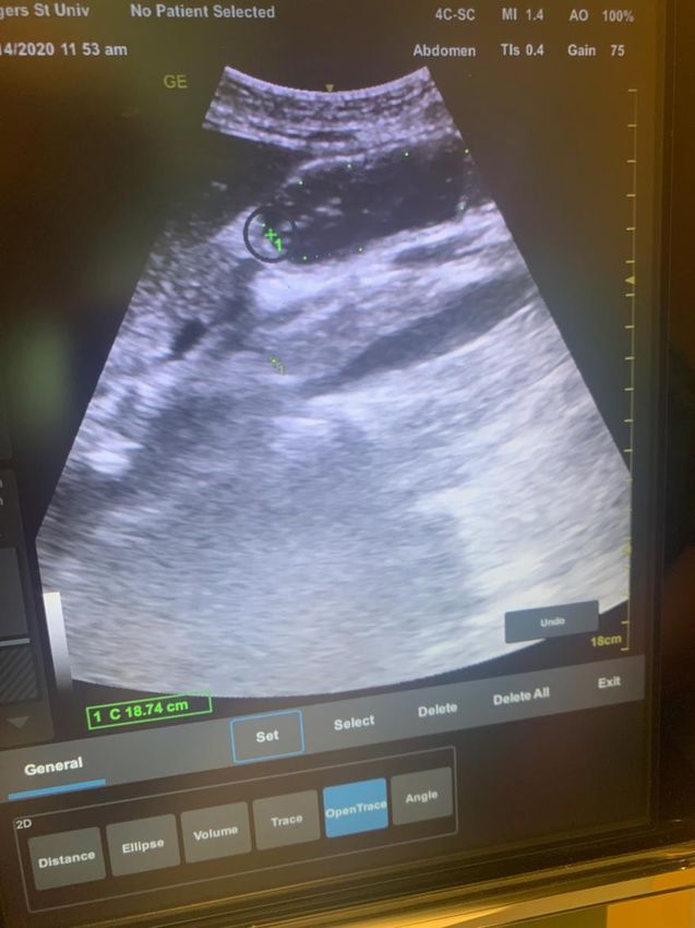
• Particulate matter seen in both supine and RLD positionSchool of Nursing Quantitative Assessment: CSA of Antrum • Only for assessment of clear fluid NOT solid content – Is amount of liquid in stomach Grade I or II? – Any particulate matter detection is an automatic Grade II • Antral CSA has a linear correlation with the gastric volume To measure: 1. Identify antrum at the level of aorta in RLD 2. Freeze screen with antrum at rest (between peristaltic contractions 3. Outline antrum with area mode (include full thickness of the gastric wall from serosa to serosa) 4. Press calculate on ultrasound 5. Apply to predictive model
School of Nursing

Measuring the Antrum CSA
School of Nursing

Formula for Quantitative Assessment
 Gastric Volume (mL) = 27 + (14.6 x RLD CSA) - 1.28 x age (years)
School of Nursing

Clinical Decision Making
School of Nursing

Scanning Tips
•   Always identify your landmarks: liver and aorta/IVC
•   Focus on area below below liver tip and in front of vessels
•   If you can’t identify in supine, change to RLD
•   Ask the patient to take a slow deep breath
    – Moves transverse colon down
• Increased fluid = increased viewing of deeper structures
• Fan and rotate your probe
• Remember: in 2-3% of patients the antrum may not be
  identifiable
• Remember these helpful image terms:
    – Bulls Eye = empty stomach
    – Frosted Glass = solid food in antrum
    – Starry Night = liquid content in antrum
School of Nursing
FAQs
• Do I have to scan in supine, or can I just scan in RLD?
– Scanning in supine is helping because 1) if solid or thick fluid is
observed then the stomach is a Grade II and the exam is complete, and
2) scanning in both positions allows for a qualitative evaluative of
volume
• Is 1.5mL/kg in reference to IBW or TBW?
– Total Body Weight
• Why does age matter when predicting the volume of fluid in
the antrum?
– Older patients tend to have a higher CSA in their antrum, which is
hypothesized to be do to a more compliant gastric wall.
• If I can not locate the antrum, can I assume the stomach is
empty?
– The exam should be considered inconclusive if one is unable to find the
stomach. In 2-3% of patients the antrum may be be located. It could be
posterior to the colon and therefore not able to be appreciated by the
ultrasound.School of Nursing Practice Scans
School of Nursing
School of Nursing

LIQUID

         WHY?
         • Distended, thin
           walls.
         • Hypoechoic/
           anechoic content
         • Enhanced view of
           deeper vascular
           structures
School of Nursing
School of Nursing

EARLY SOLID

              WHY?
              • Distended, thin
                wall
              • Consumption of
                food/air with
                eating → Mixed
                echogenicity aka
                “Frosted glass”
                appearance
              • Blurring of
                posterior wall/
                deeper structures
School of Nursing
School of Nursing

EMPTY

        WHY?
        • Small
        • Flat/compressed
        • Can see muscle
          layers
        • Round/ovoid
        • “Bulls eye”
School of Nursing
School of Nursing

EMPTY

        WHY?
        • Rugae with small
          amount of baseline
          secretions
          (anechoic center)
School of Nursing
School of Nursing

LATE SOLID   WHY?
             • Distended, thin wall
             • Round,
               heterogenous mass
               in center
School of Nursing
School of Nursing

CLEAR LIQUID
               WHY?
               • Distended, thin
                 walls.
               • Hypoechoic/
                 anechoic content
               • Enhanced view of
                 deeper vascular
                 structures
School of Nursing
School of Nursing

EMPTY
        WHY?
        • Small
        • Constricted
        • Can see muscle
          layers
        • Flat/compressed
        • Round/ovoid
School of Nursing
School of Nursing

EMPTY
        WHY?
        • Small
        • Constricted
        • Can see muscle
          layers
        • Flat/compressed
        • Round/ovoid
        • “Bulls eye”
School of Nursing
School of Nursing

EARLY SOLID
              WHY?
              • Distended, thin wall
              • Consumption of food/air
                with eating → Mixed
                echogenicity aka
                “Frosted glass”
                appearance
              • Blurring of posterior
                wall/ deeper structures
School of Nursing Practice Dexterity of US Probe
School of Nursing

Questions?
School of Nursing

      Please take a moment to fill out a
          Post-Intervention Test
           found at the link below

              (Link posted in chat):
https://rutgers.ca1.qualtrics.com/jfe/form/SV_0
               SRJIWmVzcz9A33
School of Nursing
References
Adler, A. C., Greeley, W. J., Conlin, F., & Feldman, J. M. (2016). Perioperative anesthesiology ultrasonographic
evaluation (PAUSE): A guided approach to perioperative bedside ultrasound. Journal of Cardiothoracic and
Vascular Anesthesia, 30(2), 521-529. doi: 10.1053/j.jvca.2015
Alakkad, H., Kruisselbrink, R., Chin, K. J., Niazi, A. U., Abbas, S., Chan, V. W., & Perlas, A. (2015). Point-of-care
ultrasound defines gastric content and changes the anesthetic management of elective surgical
patients who have not followed fasting instructions: A prospective case series. Canadian Journal of
Anesthesia, 62(11), 1188-1195. doi: 10.1007/s12630-015-0449-1.
American Society of Anesthesiologists. (2017). Practice guidelines for preoperative fasting and the use of
pharmacologic agents to reduce the risk of pulmonary aspiration: An updated report. Anesthesiology,
113(3), 376-393. doi: https://doi.org/10.1097/ALN.0000000000001452.
Bisinotto, F. M. B., Pansani, P. L., Silveira, L. A. M., Naves, A. A., Peixoto, A. C. A., Lima, H. M., & Martins, L. B. (2017).
Qualitative and quantitative ultrasound assessment of gastric content. Revista da Associação Médica
Brasileira, 63(2), 134-141. https://doi.org/10.1590/1806-9282.63.02.134.
Bouvet, L., & Chassard, D. (2014). The value of ultrasound for the preoperative evaluation of gastric contents. French
Annals of Anesthesia and Resuscitation, 33(4), 240-247. https://doi.org/10.1016/j.annfar.2014.01.021.
Bouvet, L., Mazoit, J. X., Chassard, D., Allaouchiche, B., Boselli, E., & Benhamou, D. (2011). Clinical assessment of the
ultrasonographic measurement of antral area for estimating preoperative gastric content and volume.
Anesthesiology, 114(5):1086-92.
Bouvet, L., Miquel, A., Chassard, D., Boselli, E., Allaouchiche, B., & Benhamou, D. (2009). Could a single standardized
ultrasonographic measurement of antral area be of interest for assessing gastric contents? A preliminary
report. European Journal of Anesthesiology, 26(12, 1015-1019. doi:10.1097/EJA.0b013e32833161fd.School of Nursing
References
Cubillos, J., Tse, C., Chan, V. W., Perlas, A. (2012). Bedside ultrasound assessment of gastric content: An
observational study. Canadian Journal of Anesthesia, 59(4), 416-423. doi: 10.1007/s12630-011-
9661-9.
El-Boghdadly, K., Kruisselbrink, R., Chan, V. W. S., & Perlas, A. (2016). Images in anesthesiology: Gastric
ultrasound. Anesthesiology, 125(3):595. doi: https://doi.org/10.1097/ALN.0000000000001043.
EFalyar, C. R., & Kantzavelos, L. (2018). Clinical application of point-of-care ultrasound gastric examination in
the management of an ASA class 3E patient: A case report. AANA Journal, 86(5), 379-382.
Fujigaki, T, Fukusaki, M., Nakamura, H., Shibata, O., & Sumikawa K. (1993). Quantitative evaluation of gastric contents
using ultrasound. Journal of Clinical Anesthesia, 5(6), 451-455.
Gagey, A. C., de Queiroz Siqueira, M., Desgranges, F. P., Combet, S., Naulin, C., Chassard, D., & Bouvet, L. (2016).
Ultrasound assessment of the gastric contents for the guidance of the anesthetic strategy in infants with
hypertrophic pyloric stenosis: A prospective cohort study. British Journal of Anesthesia, 116(5), 649-654.
doi: 10.1093/bja/aew070.
Gagey, A. C., de Queiroz Siqueira, M., Monard, C., Combet, S., Cogniat, B., Desgranges, F. P., Robinson, P.,
Chassard, D., & Bouvet, L. (2018). The effect of pre-operative gastric ultrasound examination on the
choice of general anesthetic induction technique for non-elective pediatric surgery. A prospective
cohort study. Anesthesia, 73(3), 304-312. doi: 10.1111/anae.14179.
Gola, W., Domagala, M., & Cugowki, A. (2018). Ultrasound assessment of gastric emptying and the risk of aspiration
of gastric contents in the perioperative period. Anesthesiology Intensive Therapy, 50(4), 297-302.
doi:10.5603/AIT.a2018.0029.
Hamada, S. R., Garcon, P., Ronot, M., Kerever, S., Paugam-Burtz, C., & Mantz J. (2014). Ultrasound
assessment of gastric volume in critically ill patients. Intensive Care Medicine, 40(7), 965-972. doi:
10.1007/s00134-014-3320-x.
Jacoby, J., Smith, G., Eberhardt, M., & Heller, M. (2003). Bedside ultrasound to determine prandial status. American
Journal of Emergency Medicine, 21(3), 216-219.School of Nursing
References
Kline, J., Selai, B., Ardigo, M., & Pugh., M. (2017). Accuracy in evaluating gastric ultrasound images before and
after brief training. Anesthesiology News, Special Edition, 91-95.
Koenig, S. J., Lakticova, V., & Mayo, P. H. (2011). Utility of ultrasonography for detection of gastric fluid during
urgent endotracheal intubation. Intensive Care Medicine, 37(4), 627-631. doi: 10.1007/s00134-010-
2125-9.
Kruisselbrink, R., Arzola, C., Endersby, R., Tse, C., Chan, V., & Perlas, A. (2014). Intra- and interrater reliability of
ultrasound assessment of gastric volume. Anesthesiology, 121(1), 46-51. doi:
https://doi.org/10.1097/ALN.0000000000000193.
Kruisselbrink, R., Arzola, C., Jackson, T., Okrainec, A., Chan, V., & Perlas, A. (2017). Ultrasound assessment of
gastric volume in severely obese individuals: A validation study. British Journal of Anesthesia, 118(1),
77-82. doi: 10.1093/bja/aew400.
Kruisselbrink, R., Gharapetian, A., Chaparro, L. E., Ami, N., Richler, D., Chan, V. W. S., & Perlas A. (2019). Diagnostic
accuracy of point-of-care gastric ultrasound. Anesthesia & Analgesia, 128(1), 89-95. doi:
10.1213/ANE.0000000000003372.
Perlas, A. (n.d.). Gastric Ultrasound. USRA. http://www.usra.ca/regional-anesthesia/specific-
blocks/pocus/gastric.php
Perlas, A., Arzola, C., & Van de Putte, P. (2018). Point-of-care gastric ultrasound and aspiration risk assessment: A
narrative review. Canadian Journal of Anesthesia, 65(4), 437-448. doi: 10.1007/s12630-017-1031-9.
Perlas, A., Chan, V. W., Lupu, C. M., Mitsakakis, N., & Hanbidge, A. (2009). Ultrasound assessment of gastric
content and volume. Anesthesiology, 111(1), 82-89. doi: 10.1097/ALN.0b013e3181a97250.
Perlas, A., Davis, L., Khan, M., Mitsakakis, N., & Chan, V. W. (2011). Gastric sonography in the fasted surgical
patient: A prospective descriptive study. Anesthesia & Analgesia, 113(1), 93-97. doi:
10.1213/ANE.0b013e31821b98c0.School of Nursing
References
Perlas, A., Mitsakakis, N., Liu, L., Cino, M., Haldipur, N., Davis, L., Cubillos, J., & Chan V. (2013). Validation of a
mathematical model for ultrasound assessment of gastric volume by gastroscopic examination.
Anesthesia & Analgesia, 116(2), 357-363. doi: 10.1213/ANE.0b013e318274fc19.
Tampo, A., Suzuki, A., Ijiri, E., Kunisawa, T., & Iwasaki, H. (2013). Preanesthetic gastric assessment with
sonography for a patient with a full stomach. Journal of Clinical Anesthesia, 25(2), 164-165. doi:
10.1016/j.jclinane.2012.10.004.
Van de Putte, P. (2013). Bedside gastric ultrasonography to guide anesthetic management in a nonfasted
emergency patient. Journal of Clinical Anesthesia, 25(2), 165-166.
https://doi.org/10.1016/j.jclinane.2012.10.005.
Van de Putte, P., & Perlas, A. (2014). Ultrasound assessment of gastric content and volume. British Journal of
Anesthesia, 113(1), 12–22. https://doi.org/10.1093/bja/aeu151.
Van de Putte, P., Van Hoonacker, J., & Perlas A. (2018). Gastric ultrasound to guide anesthetic management
in elective surgical patients non-compliant with fasting instructions: A retrospective cohort study.
Minerva Anestesiologica, 84(7), 787-795. doi: 10.23736/S0375-9393.17.12305-9.
Wu, C., Chen, Y., Wang, M., & Pinelis, E. (2017). National trends in admission for aspiration pneumonia in the
United States. Annals of the American Thoracic Society, 14(6), 874-87.You can also read