ABSTRACT BOOK Journée des doctorants - 7 Juillet 2021 - Institut de Chimie Physique
←
→
Page content transcription
If your browser does not render page correctly, please read the page content below
ABSTRACT
  BOOK
 Journée des doctorants – 7 Juillet 2021
       Institut de Chimie Physique
      Salle Magat - Visioconférence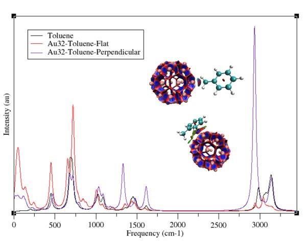
ORAL COMMUNICATIONS

In-vitro analysis by UPLC-MS of a protein complex involved in DNA repair
    Anouchka GATIN1, Isabelle BILLAULT1, Patricia DUCHAMBON2, Guillaume VAN DER REST1 et
                                   Cécile SICARD-ROSELLI1
           1
            Université Paris-Saclay, CNRS, Institut de Chimie Physique UMR 8000, 91405 Orsay Cedex
       2
           CNRS UMR9187, INSERM U1196, Institut Curie, Université Paris-Saclay, 91405 Orsay Cedex

        In-vivo, DNA is continually altered and repaired. In one of the repair pathway, NER (nucleotide
excision repair), the lesion recognition step is performed by XPC 1 (Xeroderma Pigmentosum group C protein).
XPC is a major component of a protein complex also containing human centrin 2 (CEN2). In-cellulo studies
have shown that CEN2, a predominantly cytoplasmic protein, is delocalized to the nucleus when DNA is
damaged. However, CEN2 is not able to pass the nuclear membrane on its own2, its peptide sequence does
not gives it this property. So, how to explain its delocalization when DNA is altered? It has been proved that
XPC and CEN2 are co-localized in the nucleus after a UV exposure. XPC being able to sail from nucleus to
cytoplasm3, what are the role and nature of the CEN2-XPC interaction in the CEN2 delocalization?
        The reactivity of both proteins of interest has been studied isolated and then together following
exposure to ionizing radiation (γ), allowing the formation of oxidizing radicals in-situ. After an enzymatic
digestion step, UPLC-MS and UPLC-MS-MS experiments highlighted the formation of a covalent binding
between XPC and CEN2 following oxidation. Precisely, we identified theses covalent crosslinks as tyrosine-
tyrosine bridges. The excellent chromatographic resolution enables us to emphasize the plurality of di-tyrosine
isomers. New structures of di-tyrosine have been identified4 and are being characterized. As XPC is necessary
for the delocalization of CEN2 to the nucleus, it is important to question about the role and the significance
of this crosslink between both partners.

Figure 1: Schematic representation of DNA lesion recognition process by CEN2-XPC complex in NER. CEN2 is delocalized from the cytoplasm
to the nucleus.

1
  Sugasawa et al., Molecular Cell 2 (1998) 223-232
2
  Klein et al., Cell Science (2009) 3312-3321
3
  Hoogstraten et al., J. Cell Sci. 121 (2008) 2850-2859
4
  Gatin A. et al., Free Radic. Biol. Med. 16 (2021) 461-470
AUTO-OXIDATION OF TETRAHYDROBIOPTERIN AND ITS DERIVATIVES:
                          KINETICS MONITORING BY HPLC-MS/MS

                                            Ayoub BOULGHOBRA

Tetrahydrobiopterin (BH4) is an essential cofactor involved in numerous enzymatic reactions. It is quantified
in CSF samples and used as a biomarker in order to diagnose inborn errors of dopamine and serotonin
metabolism. BH4 is a reduced biopterin that tends to auto-oxidation. In this work, we developed a high-
performance liquid chromatography (HPLC) coupled to tandem mass spectrometry (MS/MS) method that
allowed us to observe the evolution of seven pterins found in BH4 auto-oxidation pathway namely, BH4,
quinonoid dihydrobiopterin (q-BH2), dihydrobiopterin (BH2), dihydropterin (PH2), dihydroxanthopterin (XH2),
biopterin (B) and pterin (P) in addition to neopterin (N) which was used as internal standard. These
compounds were identified thanks to their specific MS/MS transitions and to their retention factors. With this
LC-MS/MS method, we performed BH4 auto-oxidation kinetic study at 10°C using a pH 2.8 ammonium
formate as a reference buffer. We observed that, as shown below, BH4 produce q-BH2 in the first step. The
latter is then isomerized into 7,8-BH2 which is oxidized into XH2 and B. We determined that, in this buffer,
BH4 decreases following a bi-exponential model corresponding to a first-order reaction. The stability-
indicative method that was set up will be used to compare the kinetics obtained at other pH and in other
buffers, with the aim of identifying the ideal conditions for CSF storage.

Scheme: BH4 autoxidation pathway in 0.05 M; pH 2.8 ammonium formate
Investigation on the adsorption geometry of substituted aromatic compounds on the surface of Gold
Nanoparticles: Quantum Chemical Topology and Vibrational Study
Rika Tandiana, Van-Oanh Nguyen-Thi, Cécile Sicard-Roselli, Carine Clavaguera
Institut de Chimie Physique, Université Paris-Saclay rika.tandiana@universite-paris-saclay.fr
Gold Nanoparticles (GNPs), owing to their unique optical properties and versatile preparation strategy, have been
demonstrated to exhibit promising applications in diverse fields, which include bio-sensors, catalysts, nanomedicines,
radiotherapy, and still many others.1,2 Yet, the interfacial interaction of GNPs with their environment (ligand and
solvent) remains elusive. The difference in experimental SERS spectra between closely related amino acids (tyrosine
and phenylalanine) interacting on a metal surface (silver or gold nanoparticles), that arose due to difference in the
orientation of the amino acid,3,4 demonstrates the importance of understanding the interaction in greater details. The
presence of multiple functional groups introduces additional complexity for such interactions. Therefore, we have
systematically investigated the adsorption of small organic molecules on the surface of GNPs of varying size (0.8 ~ 2
nm in diameter) with varied functional groups. In this study, we have focused on the interaction between Au32 and a
series of substituted aromatic compounds that includes benzene, aniline, phenol, toluene, benzoic acid,
acetophenone, methyl benzoate, and thiophenol.
DFT geometry optimization suggests that a benzene ring preferentially lies flat on the surface of the metal in all the
studies systems. This perpendicular orientation is possible for some of other the functional groups, albeit with lower
metal-ligand interaction energies. Correlation between interaction energy and the charge transfer was not observed,
regardless of the choice of the electronic population scheme. Charge decomposition analysis also suggested the back
donation of electrons from GNP to organic molecules. A thorough quantum topological analysis identified multiple
non-covalent interactions and revealed that the nature of the interaction includes dispersive interactions, and dative
interaction between the benzene ring and GNP. Lastly, analysis of the vibrational IR spectra shows significant changes
between an aromatic ligand parallel to the surface of the GNP compared to one where the ligand is perpendicular to
the GNP surface. This distinct IR signal should be experimentally observable and could help quantify the presence of
the various configurations during experiments.
Figure 1 – The calculated IR spectra of Au32 interacting with toluene with flat or perpendicular configuration
References
1. Kuncic, Z. & Lacombe, S. Nanoparticle radio-enhancement: principles, progress and application to cancer
treatment. Physics in Medicine & Biology 63, 02TR01 (2018).
2. Fen-Ying Kong et al. Unique Roles of Gold Nanoparticles in Drug Delivery, Targeting and Imaging Applications.
Molecules 22, 1445 (2017).
3. Moskovits, M. & Suh, J. S. Surface selection rules for surface-enhanced Raman spectroscopy:
calculations and application to the surface-enhanced Raman spectrum of phthalazine on silver. J. Phys. Chem. 88,
5526–5530 (1984).
4. Madzharova, F., Heiner, Z. & Kneipp, J. Surface Enhanced Hyper-Raman Scattering of the Amino Acids Tryptophan,
Histidine, Phenylalanine, and Tyrosine. J. Phys. Chem. C 121, 1235–1242 (2017).Lucie ARBERET
De la chromatographie analytique à la chromatographie préparative : optimisation d’une
séparation bidimensionnelle hors ligne pour la purification des composants d’un colorant
traditionnel mésoaméricain.
La caractérisation des matières colorantes du Codex Borbonicus, manuscrit aztèque du 16ème
siècle conservé à la Bibliothèque de l’Assemblée nationale, a mis en évidence l’utilisation d’un
colorant traditionnel mésoaméricain extrait de l’arbuste Justicia spicigera pour l’obtention de
peintures brunes. La présence, unique à ce jour, de ce colorant dans un codex mésoaméricain, par
ailleurs traditionnellement employé dans le domaine de la teinture de textiles pour l'obtention de
teintes bleues, et le manque de connaissances quant à sa composition chimique, ont motivé
l’analyse approfondie de celui-ci.
Des essais chromatographiques préliminaires ont permis de constater la présence de deux
composés contribuant majoritairement à la coloration de l’extrait obtenu par macération des feuilles
de Justicia spicigera. En vue de leur caractérisation structurale complète, le développement d’une
méthode de purification de ces composés cibles par chromatographie en phase liquide préparative
a été réalisé.
Tout d’abord, les conditions de séparation analytiques ont été optimisées en Chromatographie
Liquide à Polarité de Phases Inversée avec détection UV-Visible et SM. Afin de faciliter le transfert
de la méthode analytique optimisée vers le système préparatif, le choix des phases stationnaires
testées en analytique a été dicté par leur disponibilité à l’échelle préparative. Ainsi, les performances
de 5 phases stationnaires dans 4 systèmes d’élution (variation de modificateur organique et de pH)
ont été comparées vis-à-vis de la séparation des deux composés d’intérêt et des composés non-
désirés avoisinants. La complexité de l’extrait et le choix limité de phases stationnaires ne
permettant pas d’obtenir les composés d’intérêt avec une pureté satisfaisante dans une seule
dimension de séparation, leur isolement en deux dimensions hors ligne sur la même phase
stationnaire a été envisagé. Cela a été permis par l’observation d’une certaine orthogonalité des
systèmes d’élution testés, notamment lors de variation du pH de la phase mobile.
Par la suite, les conditions d’élution optimisées pour chacune des dimensions de séparation ont
été transférées vers le système préparatif permettant l’isolement des composés cibles. Des
techniques d’analyse structurale (spectroscopies IR, RMN du proton et du carbone et
spectrométrie de masse SMn en infusion directe) sont actuellement en cours afin de permettre la
caractérisation des composés colorés d’intérêt isolés.Karwan Ali OMAR Supervisor: Aurélien de la Lande “Influence of the irradiating biomolecule environment on the energy deposition of fast ionic particle” When swift ionic particles penetrate in biomolecules, in a few attoseconds, they deposit some of their energy by Colombian interaction between ion charge and electron cloud of biomolecule. Accurately quantifying the deposited energy is essential in many fields of science for instance astrophysics, nuclear material, nuclear medicine. The mechanism of energy transfer initiates when a positively charged particle encloses the electron cloud of the target martial, drags and strongly polarizes the electrons clouds, the electron density accumulates on the impacting molecular fragment and partially reduces on surrounding molecular fragments. After the projectile left the target, the flow of electrons come back and emit fraction of it to surrounding. The cooperative effect of the surrounding fragments on the deposited energy is crucial to validate the additive properties of the deposited energy in the empirical models. To estimate this effect, we irradiated a supramolecular system and isolated fragments of it by swift proton with difference kinetic energy. The simulations have been done by implemented Real-time time-dependent density functional theory (RT-TD- DFT) which successfully calculates the deposited energy in time real as promising model. We reveal the quantity of non-additive energy deposition that come from the cooperativity of surrounding fragments, is significant, it varies from 5% to 12% with respect to the kinetic energy of the proton in our system.
‘Functional Nanoinks Based on Metal Nanoparticles for Printed Electronics’
                                        A. Amanova 1, L. Assaud, 2 H. Remita 1
                                 1
                                  Institut de Chimie Physique, Université Paris-Saclay
                  2
                   Institut de Chimie Moléculaire et des Matériaux d'Orsay, Université Paris-Saclay
Key words: Conductive inks, flexible printed electronics, inkjet printing, metal nanoparticles
Conductive inks are recent progress in the electronics industry and widely used for the fabrication of flexible printed
electronics. Silver is the commonly used metal for conductive inks due to its excellent electrical conductivity (6.3 x 10 7
Ω-1 m-1) and stability to oxidation. However, the high price of silver and low resistance to electrochemical migration
limits wide industrial applications. In this regard, copper can be a good alternative metal for conductive inks because of
its equivalent conductivity to silver (5.96 x 10⁷ Ω -1 m-1), vast abundance, low price, and strong resistance to
electrochemical migration. In the current work, we are developing inks based on silver and copper nanoparticles
stabilized by polymers and synthesized by chemical, radiolytic, photochemical methods. The prepared inks have been
deposited on the plasma-treated PET and glass substrates by the aerospray method. The inkjet printing method will be
elaborated for printing electrical circuits after adjusting the viscosity, surface tension of inks using various additives.
Sintering with thermal and photonic treatments was performed to increase the electrical conductivity through creation
of percolation path. The 4 point-probe method was employed to measure the electrical conductivity after sintering. The
obtained values of electrical conductivity (1.66 x 10⁵ Ω -1 m-1) of our inks are comparable to commercial conductive inks
in the market. This holds promise to apply our conductive inks in printed electronics such as flexible displays and
sensors.

Kamyshny, A., & Magdassi, S. (2019). Conductive nanomaterials for 2D and 3D printed flexible electronics. Chemical
Society Reviews, 48(6), 1712–1740. https://doi.org/10.1039/c8cs00738a

Fernandes, I. J., Aroche, A. F., Schuck, A., Lamberty, P., Peter, C. R., Hasenkamp, W., & Rocha, T. L. (2020). Silver
nanoparticle conductive inks: synthesis, characterization, and fabrication of inkjet-printed flexible electrodes. Scientific
Reports, 10(1). https://doi.org/10.1038/s41598-020-65698-3

Oliva-Puigdomènech, A., De Roo, J., Van Avermaet, H., De Buysser, K., & Hens, Z. (2020). Scalable
Approaches to Copper Nanocrystal Synthesis under Ambient Conditions for Printed Electronics. ACS
Applied Nano Materials, 3(4), 3523–3531. https://doi.org/10.1021/acsanm.0c0024
DeMon conference: Ab initio derivation of flavin hyperfine interactions in
the protein magnetosensor cryptochrome
Jean Deviers
Some biological functions, such as avian magnetically-oriented navigation or photosynthesis, arise from
quantum processes. In the case of avian magnetoreception, several possible mechanisms exist to rationalise this
phenomenon, but none has yet emerged as the definitive one.
The performance of a magnetic compass can be evaluated by simulating the spin dynamics of an intermediate
radical pair or triad, which notably requires a well-chosen and finely parametrised Hamiltonian.
One crucial parameter of this Hamiltonian, the electron-nucleus hyperfine couplings of the flavin (one of the
proposed radicals), has been previously calculated on one oxidation state of a close molecular homologue. We
however expect that considering the exact molecule in its relevant oxidation states, chemical environment and
thermally-allowed conformations, would equip us with significantly better tensors and a more realistic
Hamiltonian.
A minimal electronic and steric protein environment for the flavin could be delineated. A set of tensors
describing the shape and strength of electron-nucleus hyperfine couplings was then obtained on such a cluster,
in biologically relevant geometries generated along a Molecular Dynamics run. The HFC tensors calculations
made use of a new module for Electron Paramagnetic Resonance (EPR) module, and were greatly accelerated
by the use of density fitting and of the mixed scheme for ERIs evaluation.
Further refinement could consist in including the electronic environment beyond the minimal cluster, using the
QM/MM capabilities of DeMon2k.
The amplitude and shape of the variation of individual HFC tensor elements was obtained, and the geometrical
and electronic causes of these fluctuations could be decorrelated to an extent. These realistic, fluctuating
tensors constitute accurate parameters of a flavin-protein complex and shall be used in spin dynamics
simulations to test various mechanisms of avian magnetoreception.Dina AL ABYAD Localisation et assemblage de protéines chimères activatrices de Nox2 (p47-p67-Rac) sur les membranes de neutrophiles et PLB985 Lors d’une infection, les polynucléaires neutrophiles phagocytent l’agent pathogène et le détruisent grâce à une production massive d’espèces réactives oxygénées(ERO). La source principale de ces ERO est l’anion superoxyde, généré par un complexe enzymatique, la NADPH oxydase (NOX). Ce dernier est formé de six protéines, deux membranaires (gp91phox et p22phox) et quatre cytosoliques régulatrices (p47phox, p67phox, p40phox et Rac) qui peuvent s’assembler soit à la membrane plasmique, soit à la membrane phagosomale pour former un complexe actif. Le but de ce projet est d’étudier une étape de la régulation de l’enzyme : son assemblage. Cette étude a été faite à partir d’une protéine de fusion de trois protéines cytosoliques nommée le trimera sur lequel a été effectué des mutations. Les mutations réalisées sur des domaines d’interaction avec les phospholipides anioniques ont pu mettre en évidence un rôle important de ces interactions dans la formation du complexe actif. Ces interactions pourraient être à l’origine des différentes localisations de la NADPH oxydase (membrane plasmique ou membrane du phagosome). Mots-clés : anion superoxyde; NADPH oxydase ; phagosome ; assemblage ; activité ; trimère During an infection, neutrophils phagocytose pathogens and destroy them through massive production of reactive oxygen species (ROS). The main source of these EROs is the superoxide anion, generated by an enzyme complex, the NADPH oxidase (NOX). The latter consists of six proteins, two catalytic membrane proteins (gp91phox and p22phox) and four regulatory cytosolic ones (p47phox, p67phox, p40phox and Rac) which can be assembled either to the plasma membrane or to the phagosomal membrane to form an active complex. The aim of this project is to study the particular step of the enzyme regulation, its assembly. This study has been realized from a fusion of the cytosolic proteins, named trimera on which mutation has been performed. Mutations of the interaction domains with phospholipids highlight an important role of these interactions on the complex formation. These interactions could be responsible of the different membran localizations of the NADPH oxidase (plasma membrane or phagosomal membrane). Keywords: superoxide anion; NADPH oxidase; phagosome; assembly; activity; trimera
Ferroelectric heterostructures for photoelectrocatalytic hydrogen evolution
                            Qian Xu, Christophe Colbeau-Justin, Mohamed Nawfal Ghazzal
                       Institut de Chimie Physique, UMR 8000 CNRS, Université Paris-Saclay, 91405 Orsay, France

Photoelectrocatalysis as a powerful tool can be used for energy conversion and environmental redemption1. However,
current photoelectrocatalytic efficiency of the semiconductors is largely limited by the effective separation of
photogenerated charge carriers2. The introduction of a built-in electric field induced polarization of ferroelectric
materials is an efficient way to modulate the migration behavior of charge carriers3,4. In this work, a core-shell
TiO2@BaTiO3 heterostructure has been proposed for higher photoelectrocatalytic performance by ferroelectric effect
under external electric field. To better understand the effect of ferroelectric polarization on the photoelectrocatalysis
efficiency of the heterostructures, we also constructed BaTiO3 with different crystalline phases. The obtained
TiO2@BaTiO3 heterostructures were characterized by XRD, SEM, EDX, UV-vis spectrums, and time resolved
microwave conductivity (TRMC) to ascertain the structure and electronic properties of the composites. The difference
in photoelectrocatalytic reactivity of these samples was investigated through photoelectrochemical (PEC) test and H 2
evolution. This work will provide an efficient strategy to design high performance photoelectrochemical systems.

References:
(1) Jing, L.; Zhou, W.; Tian, G.; Fu, H. Surface Tuning for Oxide-Based Nanomaterials as Efficient Photocatalysts. Chem. Soc. Rev.
2013, 42, 9509-9549.
(2) Ding, C.; Shi, J.; Wang, Z.; Li, C. Photoelectrocatalytic Water Splitting: Significance of Cocatalysts, Electrolyte, and Interfaces.
ACS Catalysis 2016, 7, 675-688.
(3) Wu, F.; Yu, Y.; Yang, H.; German, L. N.; Li, Z.; Chen, J.; Yang, W.; Huang, L.; Shi, W.; Wang, L.; Wang, X. Simultaneous
Enhancement of Charge Separation and Hole Transportation in a TiO2 -SrTiO3 Core-Shell Nanowire Photoelectrochemical System.
Adv. Mater. 2017, 29, 1701432.
(4) Zhang, M.; Li, F.; Benetti, D.; Nechache, R.; Wei, Q.; Qi, X.; Rosei, F. Ferroelectric Polarization-Enhanced Charge Separation
in Quantum Dots Sensitized Semiconductor Hybrid for Photoelectrochemical Hydrogen Production. Nano Energy 2021, 81, 105626.
On the role of radical-SAM enzyme Viperin by advanced computational
approaches
Angela Parise,a,b Tiziana Marino,a Nino Russo,a and Aurelien de la Landeb
a
Dipartimento di Chimica e Tecnologie Chimiche, Università della Calabria, Via Pietro Bucci, 87036 Arcavacata di
Rende, CS, Italy; b Université Paris-Saclay, CNRS, Institut de Chimie Physique UMR8000, Orsay, France
Viperin is an enzyme found in mammals, including humans. It catalyses the synthesis a molecule capable of
interfering with viruses replication. Its effect has been verified to inhibit a broad range of viruses. A recent
study1 revealed that viperin catalyses conversion reaction of cytidine triphosphate (CTP) to 3'-deoxy-3',4'-
Didehydro-CTP (ddh-CTP). The viperin central domain contains the catalytic pocket that consist in a CxxxCxxC
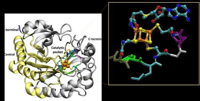
motif, characteristic of the radical S-adenosylmethionine enzymes and a [4Fe-4S] cluster (Figure 1). In this
contribution we will show the structural characteristics of viperin in the presence and absence of the CTP
substrate through Molecular Dynamics simulations. Then, we will show the use of different computational
techniques (QM, QM/MM and QM/MMpol) to describe the ionization potential of the Fe-S cluster. Specifically
we analyse the possible spin configurations for the Fe-S cluster through the use of the constrained
constrained Density Functional Theory (cDFT) and how the inclusion of the surrounding first and second
coordination shell of the cluster influences the description of the ionization energy, in the diabatic
description of the system.
Figure 1. Cartoon diagram of viperin where N-terminal domain is in light blue, central domain in yellow, C-terminal
domain in grey. [4Fe-4S] cluster shown as orange and yellow spheres, S-Adenosylmethionine and cysteines bond to
cluster are in ball and stycks, residues X of CX3CX2C are in tube colored by residue. In the square zoom on viperin
binding site. (PDB code 6Q2P)
[1] Gizzi, A. S.; Grove, T. L.; Arnold, J. J.; Jose, J.; Jangra, R. K.; Garforth, S. J.; Du, Q.; Cahill, S. M.; Dulyaninova, N. G.;
Love, J. D.; Chandran, K.; Bresnick, A. R.; Cameron, C. E.; Almo, S. C. A Naturally Occurring Antiviral Ribonucleotide
Encoded by the Human Genome. Nature 2018, 558 (7711), 610–614.POSTERS
Relaxation dynamics through a conical intersection: Quantum and
                    quantum-classical studies

     CARLOTTA PIERONI1,2 *, EMANUELE MARSILI3, DAVID LAUVERGNAT2, and GIOVANNI
                GRANUCCI1, FEDERICA AGOSTINI2, MAURIZIO PERSICO1

              1 Universitàdi Pisa, Dipartimento di Chimica e Chimica Industriale, via G. Moruzzi
                                               13, 56124 Pisa, Italy
           2 Université Paris-Saclay, CNRS, Institut de Chimie Physique UMR8000, 91405 Orsay,

                                                      France
           3 Department of Chemistry, Durham University, South Road, Durham DH1 3LE, United

                                                     Kingdom
                                      *carlotta.pieroni@phd.unipi.it

Trajectory-based approaches to nonadiabatic dynamics are powerful tools for predicting the fate of a
molecule after photo-excitation [1] or the products of a collision reaction. The approximations, in the treatment
of nuclear dynamics and in the description of electron-nuclear coupling, make them computationally efficient,
at least compared to a numerically-exact solution of the time-dependent Schrodinger equation. Clearly, the
computational efficiency comes at the price of losing accuracy or missing critical features, such as tunnelling
and zero-point energy, interferences, quantum decoherence, to name a few. Related to this point we study
the relaxation process through a conical intersection of a photo-excited retinal chromophore model. The
analysis is based on a twoelectronic-state two dimensional Hamiltonian developed by Hahn and Stock [2] to
reproduce, with a minimal model, the main features of the 11-cis to all-trans isomerization of the retinal of
rhodopsin. In particular, we focus on the performance of various trajectory-based schemes to nonadiabatic
dynamics: trajectory surface hopping (TSH) [3], trajectory surface hopping including energy decoherence
corrections (TSH-EDC) [4], Ehrenfest dynamics (EH) [5], and the coupledtrajectory mixed quantum-classical
(CT-MQC) [6] scheme derived from the exact factorization of the time-dependent electron-nuclear
wavefunction. We compare quantum-classical results to numerically-exact quantum vibronic wavepacket
dynamics. The purpose of the work is to investigate, by analyzing electronic and nuclear observables, how
the sampling of initial conditions for the trajectories affects the subsequent dynamics [7].

References:
[1] S. K. Min, F. Agostini, I. Tavernelli, and E. K. U. Gross, “Ab initio nonadiabatic dynamics with coupled trajectories:
A rigorous approach to quantum (de)coherence,” J. Phys. Chem. Lett. 8, 3048–3055 (2017). [2] S. Hahn and G.
Stock, “Quantum-mechanical modeling of the femtosecond isomerization in rhodopsin,” J. Phys. Chem. B 104, 1146–
1149 (2000).
[3] J. C. Tully, “Molecular dynamics with electronic transitions,” J. Chem. Phys. 93, 1061 (1990). [4] G. Granucci and M.
Persico, “Critical appraisal of the fewest switches algorithm for surface hopping,” J. Chem. Phys. 126, 134114 (2007).
[5] J. C. Tully, “Mixed quantum-classical dynamics,” Faraday Discuss.110, 407 (1998)
[6] F. Agostini, S. K. Min, A. Abedi, and E. K. U. Gross, “Quantum-classical non-adiabatic dynamics: Coupled- vs.
    independent-trajectory methods,” J. Chem. Theory Comput.12, 2127–2143 (2016)
[7] C. Pieroni, E. Marsili, D. Lauvergnat, and F. Agostini, “Relaxation dynamics through a conical intersection:
Quantum and quantum-classical studies” J. Chem. Phys. 154, 2021
Dounia ZAMIATI
Novel live-cell imaging method based on FRET-FLIM to explore Membrane Contact Sites
Abstract
The Endoplasmic Reticulum (ER) is a voluminous cellular compartment, taking up more than 40% of the total cell
volume. It is also very dynamic, as it can explore up to 97% of the cell in less than fifteen minutes. The ER’s membrane
continuously forms contact zones with the other cellular compartments’ membranes. Various signaling pathways,
which govern basic cellular activities and coordinate cell actions, can be observed at these Membrane Contact Sites
(MCS). They play an important role in calcium regulation or lipid homeostasis, as well as organelle trafficking. The
study and understanding of these MCS is a relatively recent undertaking, and much about them is still unknown.
The dynamic study of these MCS is still a challenge. The Golden Standard uses electron microscopy, which limits the
study to fixed - dead - cells. Photonic microscopy can generate images and videos from live cells, but only with a spatial
resolution of 250 nm. Therefore, it is impossible to observe the MCS, using only basic photonic microscopy, when the
distance between the ER and another compartment is established to be between 5 and 20 nm.
My internship’s main goal is to participate in establishing a new live-cell imaging method, based on FRET-FLIM, to
explore these MCS. One of the objectives of this study is to investigate if the proteins known to tether the ER to another
membrane accomplish this purpose alone, or if they form clusters. Preliminary data demonstrates protein-protein
interactions occurring between members of the E-syt, VAP and ORP families, via a combination of FRET Efficiency
measurements.
Membrane Contact Sites are places where proteins aggregate to complete various biological tasks necessary to the
smooth running of the cells. Our data showed that these proteins often work in concert, forming complexes or favoring
the same microdomains. This novel imaging method lays a strong foundation to the study of signaling pathways and
proximity between proteins at these MCS. Moreover, it could be further developed to function on videos in order to
explore MCS in space and time, during various biological processes.Théo BEGUIN
Assessment of molecular origins of photobleaching of fluorescent
proteins
Fluorescent proteins (FPs) are used as genetically encoded probes to study various biochemical events by cell
imaging. During the last decades, a lot of different FPs have been engineered with different colours,
photophysical properties, sensitivities and biochemical behaviour to get optical markers optimized for
different imaging technics. However, the photostability, one of the most common wanted parameters for FPs,
remains difficult to optimize. This difficulty comes from the global misunderstanding of the phenomenon of
photobleaching, the process by a FP loses irreversibly its emissive abilities after prolongated excitation of its
chromophore. Here we report a global study of the photobleaching of the Citrine, a Yellow FP very used
despite its poor photostability. Thanks to a homemade irradiation setup, samples of Citrine have been
photobleached and its absorption and emission spectra have been monitored to analyse the kinetics of the
phenomenon. The influence of parameters like irradiation power, wavelength selection of the irradiating light,
concentration, pH and O2 presence in the samples has been studied to better understand the mechanisms
involved in photobleaching. SDS-PAGE electrophoresis and mass spectrometry have been carried on the
photoproducts in order to try to characterise them. We identified two types of photobleaching pathways: The
first is the reaction of the excited state of Citrine’s chromophore with dissolved O2 which cause cytotoxicity
and drive to oxidation of numerous residues of the FP. It also causes the stabilisation of a protonated non-
fluorescent state of the chromophore. The second reaction involved is the self-reaction of the chromophore
with light, leading to cleavage of the protein and loss of its absorbing and emissive properties.Cellulose Nanocrystals in Spherical Titania-sols Microdroplet: From Dynamic Self-assembly to
Nanostructured Microsphere Synthesis
Cong Wang,1 Erwan Paineau,2 Hynd Remita,1 Mohamed Nawfal Ghazzal1*
1
Institut de Chimie Physique, UMR 8000 CNRS, Université Paris-Saclay 91405 Orsay, France
2
Laboratoire de Physique du Solide, UMR 8502 CNRS, Université Paris-Saclay 91405 Orsay, France
Cellulose nanocrystals (CNCs) are spindle-like rods derived from a variety of natural resources via strong acid
hydrolysis,1 which can spontaneously organize into a cholesteric structure, called chiral (Ch) nematic structure.2 Self-
organization of CNCs into well-ordered Ch nematic liquid crystals are ubiquitous in nature,3 inspiring widespread
interest in the development of green functional materials with versatile applications, such as biomimetic nanomaterials,
soft nanotechnology, optoelectronics and photocatalysis technology.4 The transfer of such natural Ch nematic
nanostructures into artificial functional materials on the nano- or micro-scale, however, is extremely challenging.5
Typically, studies have focused on obtaining long-range ordered Ch nematic structure in planar functional films by using
evaporation-induced self-assembly (EISA) technology. Only a few attempts have been dedicated to investigating the Ch
nematic structure of CNCs aqueous suspension in spherical microdroplets, describing the evolution of CNCs as a simple
isotropic-to-anisotropic phase transition. The direct observation of the self-organization of CNCs in spherical
microdroplet is challenging, in particular with regard to the involvement of inorganic precursors (such as metal chlorides
or alkoxides, they are not stable in aqueous solution). In this study, we encapsulated CNCs in spherical titania-sols
microdroplets using an inverse micro-emulsion method. We monitored the self-assembly process from seconds to hours
with the aim to determine the key steps that drive the self-organization of CNCs from isotropic to Ch nematic phase in
such aqueous sol. Polarized optical microscopy (POM) provides a unique perspective to witness the evolution process
of CNCs tactoids from nucleation, growth and migration to equilibration in spherical microdroplet. Simultaneously,
growth mechanisms as well as the induced structural defects during different evolution stages have been identified
during the process. Moreover, the impact of temperature on the Ch nematic structure was investigated and a proof-of-
concept of the transfer of such structure into solid TiOx/C microspheres by hydrothermal method was demonstrated.
The hybrid TiOx/C microspheres exhibited high absorption in the visible range and promising application in
photocatalytic H2 generation. This study therefore provides insights into the self-assembly process of CNCs in spherical
confinement, favoring further comprehension of growth mechanism as well as opening up a new approach to transfer
the Ch nematic structure into spherical nanostructured materials. Furthermore, it is also relevant for a wide range of
disciplines to synthesize advanced hybrid materials with rod-shaped particles or liquid crystals.
Reference
1. Vanderfleet, O.M., and Cranston, E.D. (2020). Production routes to tailor the performance of cellulose
nanocrystals. Nat. Rev. Mater., 1-21.
2. Kose, O., Tran, A., Lewis, L., Hamad, W.Y., and MacLachlan, M.J. (2019). Unwinding a spiral of cellulose
nanocrystals for stimuli-responsive stretchable optics. Nat. Commun. 10, 1-7.
3. Wilts, B.D., Whitney, H.M., Glover, B.J., Steiner, U., and Vignolini, S. (2014). Natural Helicoidal Structures:
Morphology, Self-assembly and Optical Properties. Materials Today: Proceedings 1, 177-185.
4. Wang, L., Urbas, A.M., and Li, Q. (2020). Nature-Inspired Emerging Chiral Liquid Crystal Nanostructures:
From Molecular Self-Assembly to DNA Mesophase and Nanocolloids. Adv. Mater. 32, e1801335.
5. Liu, Y., Agthe, M., Salajkova, M., Gordeyeva, K., Guccini, V., Fall, A., Salazar-Alvarez, G., Schutz, C., and
Bergstrom, L. (2018). Assembly of cellulose nanocrystals in a levitating drop probed by time-resolved small
angle X-ray scattering. Nanoscale 10, 18113-18118.Feven-Alemu KORSAYE Coupling density based descriptors with Real Time DFT for the description of electronic density evolution induced by irradiation The aim of this project is to setup computational approaches enabling to predict and analyze the evolution in time of the electron density in molecular systems after light irradiation. To this end, we will focus on the analysis of the electron density reorganization and excited states evolution after the perturbation, taking into account phenomena occurring in attosecond timescale. This will enable actually to follow the electronic excited states formation and their evolution in time. The project will be carried out by the means of Density Functional Theory (DFT) and Time Dependent-Density Functional Theory (TDDFT), both in the Linear Response (LR-TDDFT) and Real Time (RT-TDDFT) approaches. We will focus first on defining the accuracy and the possible methodological artifacts of RT-TDDFT using adapted density based descriptors, and next on the application of the so developed methods to molecules of biological relevance focusing on those known to generate Charge Transfer (CT) or Charge Separated (CS) states. For this latter, the development and use of diagnostic tools to comprehend the reliability of the obtained RT-TDDFT results will indeed be of particular relevance.
You can also read