ECMM/ISHAM recommendations for clinical management of COVID- 19 associated mucormycosis in low- and middle-income countries - DukeSpace
←
→
Page content transcription
If your browser does not render page correctly, please read the page content below

Received: 8 June 2021 | Accepted: 11 June 2021
DOI: 10.1111/myc.13335
ORIGINAL ARTICLE
ECMM/ISHAM recommendations for clinical management of
COVID-19 associated mucormycosis in low-and middle-income
countries
Shivaprakash M. Rudramurthy1 | Martin Hoenigl2,3 | Jacques F. Meis4 |
Oliver A. Cornely5,6,7 | Valliappan Muthu8 | Jean Pierre Gangneux9 |
10 1
John Perfect | Arunaloke Chakrabarti | ECMM and ISHAM
1
Department of Medical Microbiology, Postgraduate Institute of Medical Education and Research, Chandigarh, India
2
Division of Infectious Diseases, Department of Internal Medicine, Medical University of Graz, Graz, Austria
3
Division of Infectious Diseases and Global Public Health, University of California San Diego, San Diego, California, USA
4
Department of Medical Microbiology and Infectious Diseases, Center of Expertise in Mycology, Radboud University Medical Center/Canisius Wilhelmina
Hospital, Nijmegen, the Netherlands
5
Department of Internal Medicine, Excellence Center for Medical Mycology (ECMM), Faculty of Medicine and University Hospital Cologne, University of
Cologne, Cologne, Germany
6
Chair Translational Research, Cologne Excellence Cluster on Cellular Stress Responses in Aging-A ssociated Diseases (CECAD), Faculty of Medicine, University
of Cologne, Cologne, Germany
7
German Centre for Infection Research (DZIF), Partner Site Bonn-Cologne, Cologne, Germany
8
Department of Pulmonary Medicine, Postgraduate Institute of Medical Education and Research, Chandigarh, India
9
CHU Rennes, Inserm, EHESP, Irset (Institut de recherché en santé, environnement et travail) –UMR_S 1085, Université de Rennes, Rennes, France
10
Division of Infectious Diseases, Department of Medicine, Duke University Medical Center, Durham, North Carolina, USA
Correspondence
Arunaloke Chakrabarti, Department of Abstract
Medical Microbiology, Postgraduate
Reports are increasing on the emergence of COVID-19–associated mucormycosis
Institute of Medical Education and
Research, Chandigarh, Chandigarh (CAM) globally, driven particularly by low- and middle-income countries. The recent
160012, India.
unprecedented surge of CAM in India has drawn worldwide attention. More than
Email: arunaloke@hotmail.com
28,252 mucormycosis cases are counted and India is the first country where mu-
cormycosis has been declared a notifiable disease. However, misconception of man-
agement, diagnosing and treating this infection continue to occur. Thus, European
Confederation of Medical Mycology (ECMM) and the International Society for Human
and Animal Mycology (ISHAM) felt the need to address clinical management of CAM
in low- and middle-income countries. This article provides a comprehensive docu-
ment to help clinicians in managing this infection. Uncontrolled diabetes mellitus and
inappropriate (high dose or not indicated) corticosteroid use are the major predispos-
ing factors for this surge. High counts of Mucorales spores in both the indoor and
outdoor environments, and the immunosuppressive impact of COVID-19 patients as
well as immunotherapy are possible additional factors. Furthermore, a hyperglycae-
mic state leads to an increased expression of glucose regulated protein (GRP- 78) in
endothelial cells that may help the entry of Mucorales into tissues. Rhino-orbital mu-
cormycosis is the most common presentation followed by pulmonary mucormycosis.
1028 | © 2021 Wiley-VCH GmbH. wileyonlinelibrary.com/journal/myc Mycoses. 2021;64:1028–1037.
RUDRAMURTHY et al. | 1029
Recommendations are focused on the early suspicion of the disease and confirmation
of diagnosis. Regarding management, glycaemic control, elimination of corticosteroid
therapy, extensive surgical debridement and antifungal therapy are the standards for
proper care. Due to limited availability of amphotericin B formulations during the pre-
sent epidemic, alternative antifungal therapies are also discussed.
KEYWORDS
corticosteroids, COVID-19, diabetes, infection, Mucorales, mucormycosis, SARS-CoV-2
1 | I NTRO D U C TI O N spp., Cunninghamella spp. and Saksenaea spp. Present data suggest
that Rhizopus arrhizus is the predominant agent causing CAM in
The current pandemic of Coronavirus disease (COVID-19) caused India.10 The distribution of different species varies amongst differ-
by the novel Coronavirus SARS-CoV-2 has already affected more ent geographical regions. For instance, Rhizopus arrhizus is the most
than 174 million and has killed 3.8 million people across the world. common species in India15,16 and France17 but Cunninghamella spp.is
Mortality is causally related to the severe pneumonia caused by the most common in Spain.18
SARS-CoV-2 along with an aberrant host immune response in the Mucorales are thermo-tolerant fungi with a ubiquitous distribu-
form of a cytokine storm. Secondary infections due to bacteria tion. They are found on organic substrates such as decaying fruit and
and fungi increase the mortality rate.1,2 Among fungal infections, vegetable matter, crop debris, bread, compost piles, animal excreta
COVID-19–associated pulmonary aspergillosis (CAPA) carries a high and soil within indoor and outdoor environments.19 An ecological
mortality, with an all-cause mortality ranging between 33%–8 0%, study conducted in India revealed the presence of many species of
as the diagnosis of CAPA is difficult.3-5 Due to overlapping clinical Mucorales in soil. 20 Mucorales are also present in indoor environ-
features of CAPA and COVID-19–associated acute respiratory dis- ments such as air conditioning filters. 21,22 A recent study from North
tress syndrome (ARDS), the European Confederation of Medical India reported large numbers of Mucorales spores in both hospitals
Mycology (ECMM) and International Society for Human and Animal and outdoor air. 21 The mean spore count of Mucormycetes in outdoor
Mycology (ISHAM) jointly defined diagnostic criteria and manage- samples ranged between 0.73 to 8.60 cfu/m3 across different sea-
3
ment protocol of patients with CAPA. Mucormycosis did not draw sons. Within the hospital, the mean spore count was slightly higher
similar attention, as the disease was considered rare. Recent reviews in an airconditioned area (0.88–1.72 cfu/m3) compared to a non-
on worldwide cases highlighted the importance of COVID-19– airconditioned area (0.68–1.12 cfu/m3). 21 R arrhizus was the predom-
6-9
associated mucormycosis (CAM) globally. Particularly the recent inant Mucorales isolated in both indoor and outdoor air. 21
unprecedented surge of CAM in India has drawn worldwide atten- Rhino-orbito-cerebral mucormycosis (ROCM) and pulmonary
tion.10,11 During the last year, a 2.1 time increase of mucormycosis mucormycosis are generally observed in patients with uncontrolled
prevalence was reported due to COVID-19 in a multicentre study diabetes or having immunosuppressive conditions such as patients
in India.10 However, this year those numbers have been exploding: receiving corticosteroid treatment, cancer chemotherapy or im-
Already more than 28,252 mucormycosis cases have been reported munotherapy, haematological stem cell transplants, those with
and mucormycosis has been declared a notifiable disease in India.12 prolonged neutropenia or solid organ transplants.13,15 In contrast
Although the ECMM and the Mycoses Study Group (MSG) together cutaneous or subcutaneous mucormycosis is generally observed
developed a global guideline for diagnosis and management of mu- after trauma.15
13
cormycosis, we felt the need to address the specific issues related
to CAM such as any misconceptions on the management, diagnosing
and managing this infection that more frequently occur in low and 3 | PATH O G E N E S I S O F C A M
middle-income countries (LMIC). Experts from ISHAM and ECMM
prepared recommendations to help clinicians manage this infection. The sporangiospores of Mucorales vary in size depending on spe-
cies (range 3–11 µm). These spores are released from the sporan-
gium and dispersed into the air, where a vulnerable host population
2 | A E TI O LO G Y A N D R I S K FAC TO R S may acquire an infection. Inhalation is the major route of acquiring
mucormycosis. The relatively larger spores of R arrhizus get trapped
Mucormycosis is caused by few genera of fungi in the order Mucorales in the nasal epithelium and sinuses and thus may lead to ROCM,
of the phylum Mucormycota. The phylum Mucormycota comprises 55 whereas the relatively smaller spores of Cunninghamella spp. can
genera with 261 species of which 38 are known to be human patho- reach the lower respiratory tract leading to pulmonary mucormy-
gens.14 Common species causing mucormycosis include Rhizopus cosis.15 However, available evidence suggests that any pathogenic
spp., Rhizomucor spp., Mucor spp., Lichtheimia spp., Apophysomyces Mucorales species can produce any type of clinical presentation. 23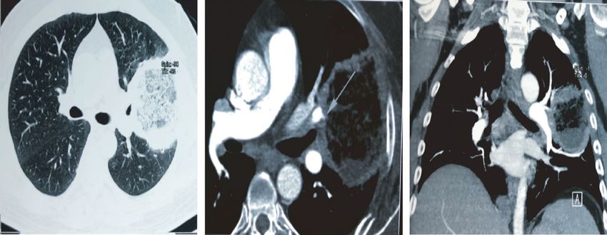
1030 | RUDRAMURTHY et al.
3.1 | Uncontrolled diabetes in CAM is well established, but the dose and the duration of steroid therapy
required to increase the risk of different infections is not as well
The major underlying disease noted in mucormycosis cases in LMICs defined.35
such as India, Iran or Mexico is uncontrolled diabetes with or with-
out ketoacidosis.14,21,22,24 Among COVID-19 patients, uncontrolled
diabetes had been reported at 7%–21%. However, the prevalence 3.3 | Iron and hyperferritinaemia
in India is above 30%, with 2% of patients developing diabetic ke-
toacidosis. Acute or stress-induced hyperglycaemia has been noted Iron overload and deferoxamine therapy are well-known risk fac-
in 50% of hospitalised COVID-19 patients. 25,26 SARS-CoV-2 itself tors for mucormycosis.36 The free iron captured by siderophores of
can induce acute diabetes and diabetic ketoacidosis by damaging Rhizopus species helps in its growth.36 Severe COVID-19 is a hyper-
pancreatic islets cells, which have a high expression of angioten- ferritinaemic state due to hyperinflammation.37 In severe COVID-19
sin converting enzyme-2 receptors, as has been noted with SARS- patients, ferritin level rises 1.5 to 5 times higher than in non-severe
CoV-1, and indirectly by damaging small blood vessels supplying cases (average ferritin concentration of >800 μg/L).37 High IL-6
pancreatic beta cells. 27,28 Increased resistance to insulin due to the concentrations in COVID-19 patients have been correlated to dis-
profound inflammatory reaction, may also play some role in the in- ease severity.38 IL-6 directly stimulates ferritin production and in-
duction of hyperglycaemia.8,29 In addition, a few cases of euglycemic creases the synthesis of hepcidin which in turn sequesters iron in
diabetic ketoacidosis have been reported in COVID-19 cases, espe- enterocytes and macrophages thus preventing them to efflux from
cially those receiving sodium-glucose co-transporter-2 inhibitors these cells leading to increased intracellular iron load.39 This excess
30,31
(SGLT2). intracellular iron generates reactive oxygen species (ROS) causing
Type 2 diabetes mellitus itself is an immunocompromised state damage to the tissue and free iron is released in the circulation and
which leads to dysregulated, dysfunctional innate and adaptive im- available to Mucorales.8,9,38
mune cells making the host susceptible to infections by Mucorales.32
Due to increased glycosylation, IL-10 production by lymphocytes
and macrophages is significantly reduced. Diabetes mellitus also re- 3.4 | Endothelial damage
duces polymorphonuclear leukocyte mobilisation and chemotaxis.32
Hyperglycaemia and acidosis can induce phagocytic cell dysfunction Comparison of autopsy lung specimens obtained from expired pa-
leading to increased risk of Mucorales infections. tients due to COVID-19 and those with acute respiratory distress
syndrome (ARDS) secondary to influenza A (H1N1) infection, has
revealed severe vascular endothelial injury along with the presence
3.2 | Role of corticosteroids in CAM of intracellular virus and disrupted cell membranes in COVID-19
deceased patients. Pulmonary vessels show widespread thrombo-
In a multicentre study from India, inappropriate corticosteroid use sis along with significantly higher (9 times, p < .001) alveolar capil-
was noted in 63.3% patients.10 The situation was aggravated fur- lary microthrombi in COVID-19 patients compared to patients with
ther during the second wave of COVID-19, when steroids were used influenza.40 Thrombosis can also occur in small veins supplying the
indiscriminately and inappropriately to overcome the challenge of pancreas, thereby damaging it and causing insulin deficiency lead-
the oxygen acquisition crisis and resulting oxygen desaturation in ing to diabetes mellitus.41 As endothelial adhesion and penetration
patients. The over-the-counter availability and the practice of self- is an early step in establishing mucormycosis,36 endothelial damage
medication with corticosteroids complicates the situation even fur- observed in severe COVID-19 disease may play an important patho-
ther. The use of corticosteroids in the treatment of COVID-19 may genic role.8
act as a double-edged sword. Firstly, corticosteroids can increase
blood glucose levels by acting as a substrate for oxidative stress me-
tabolism with lipolysis, proteolysis and hepatic glucose production. 3.5 | Overexpression of GRP78
They also increase insulin resistance in up to 60%–8 0% of patients
depending on the dose and type used.33 Secondly, they affect vir- Hyperglycaemia is a stress condition that induces the overexpres-
tually all immune cells. Some major immune suppressive actions of sion of the glucose regulated protein (GRP78) which is present in the
corticosteroids are given as: (i) antagonism of macrophage matura- lumen of the endoplasmic reticulum and expressed in mammalian
tion and differentiation, (ii) decrease of interleukin-1, interleukin-6, cells.42 The CotH protein kinase belonging to the spore coating pro-
tumour necrosis factor, proinflammatory prostaglandins and leu- tein family in Rhizopus acts as the ligand for GRP78, which helps the
kotrienes production by macrophages, (iii) suppression of micro- fungus to adhere and invade endothelial and nasal epithelial cells.43
bicidal activity of activated macrophages.34 They also suppress Sabrili et al demonstrated significantly higher serum GRP78 levels
neutrophil adhesion to endothelial cells and impair lysosomal en- in COVID-19 patients compared to a COVID-19 negative control
zyme release, respiratory burst and chemotaxis to the site of infec- group. These findings suggest amplified pathogenetic role of GRP78
tion.34 Increased risk of infection following glucocorticoid therapy in CAM.44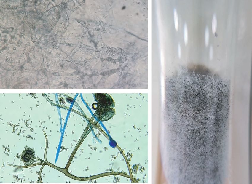
RUDRAMURTHY et al. | 1031
Therefore, the pathogenesis of CAM is a complex issue, in which without pain has also been well described. An eschar may be seen in
environment, vulnerable patients, corticosteroid use and COVID-19 the nasal septum, palate, eyelid, face or orbital areas. Cranial nerve
play synergistic roles in the disease pathogenesis (Figure 1). palsies are also common. With invasion of the brain the patient may
present with features of cerebral oedema such as coma, and vascular
invasion may lead to cerebral infarcts. Another common presenta-
4 | W H E N A N D H OW TO C LI N I C A LLY tion includes sinus cavernous thrombosis a loss of vision.
S U S PEC T C A M
During the present epidemic nearly 90% of cases presented as ROCM 4.2 | Pulmonary mucormycosis
10
and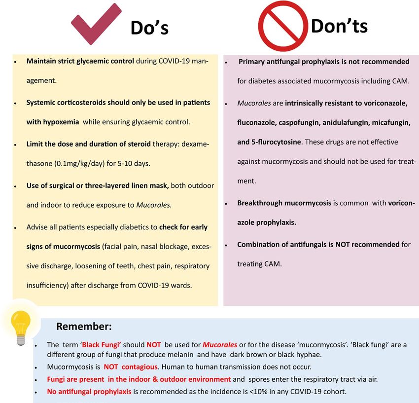
1032 | RUDRAMURTHY et al.
(A) (B) F I G U R E 2 Left facial and orbital
swelling with chemosis in a case of rhino-
orbital mucormycosis (A), and a large
necrotic ulcer on the left half of palate in
a patient with left maxillary mucormycosis
(B). Coronal view of magnetic resonance
imaging of paranasal sinuses showing
bilateral maxillary and ethmoidal sinusitis
(C), more involvement on right side,
contrast study showed post-contrast
enhancement (D)
(C) (D)
(A) (B) (C)
F I G U R E 3 Computed Tomography (CT) thorax with CT pulmonary angiography showing reverse halo in left upper lobe (A), CT pulmonary
angiography showed vessel occlusion sign (B & C)
imaging (MRI) of brain and paranasal sinuses. MRI has an advantage pulmonary aspergillosis), reverse halo sign, multiple nodules and
over CT as it can determine the extent of fungal invasion whereas CT pleural effusions13 (Figure 3).
is better in identifying the bony erosion, which is noted in advanced
stages of infection.13 Radiological features of ROCM are mucosal
thickening and opacification of the sinuses, oedema, inflammation 5.2 | Mycological diagnosis
or infarction of the brain (Figure 2C,D).
Radiological features of pulmonary mucormycosis are non- A definitive diagnosis can be made by microbiological and/or histo-
specific and difficult to interpret as many features overlap with other pathological examination of tissues obtained from different lesions.
fungal pneumonias such as pulmonary aspergillosis. The reversed Nasal samples from ROCM, broncho alveolar lavage (BAL) samples,
halo sign is commonly associated with pulmonary mucormycosis.9 mini-BAL, non-bronchoscopic lavage, transbronchial biopsy from
Other features include signs of pleural effusions and multiple nod- pulmonary mucormycosis may provide an early clue of infection. The
ules. Lung CT may be confused with COVID-19 related shadows. In samples are first examined with KOH or KOH calcofluor-white, which
such cases, mucormycosis should be suspected when there is a thick- demonstrate characteristic broad aseptate or pauci-septate hyphae
walled lung cavity (need to differentiate from COVID-19–associated (measuring 6-16 or even up to 25 µm) with folding of the hyphae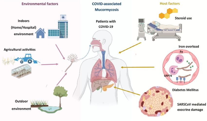
RUDRAMURTHY et al. | 1033
giving a ribbon-like appearance (Figure 4A). Right-angle branch- successful management. Controlling or eliminating the underlying
ing can be often noted especially on histopathological examination. predisposing factors such as diabetes, ketoacidosis, corticosteroids
Demonstration of this kind of hyphae and isolation of Mucorales from intake, immunomodulators and any immunosuppressant agents are
endoscopically collected debrided tissue/biopsy or a CT guided bi- crucial and help in preventing progression of the disease. Invasive
opsy from a lung lesion confirms the diagnosis of mucormycosis. For mucormycosis is a medical emergency condition and frontline anti-
successful isolation of Mucorales, tissue or biopsy samples should not fungal therapy with amphotericin B (optimally lipid-based formu-
be homogenised or grinded but instead it should be cut into small bits lations) should be initiated at the earliest, as a delay of ≥6 days in
using scissors before inoculating culture media. Homogenisation dam- certain patients is associated with twofold increase in mortality rate at
ages the fragile cell wall of Mucorales making the fungi non-viable re- 12 weeks.50 Management of mucormycosis is a team effort depend-
sulting in no growth on the media. In culture, Mucorales can easily be ing on presentation and extent of involvement. The team generally
identified as they typically produce cottony growth (Figure 4B), and constitutes an infectious diseases specialist, microbiologist, ENT sur-
with histopathology stains, angioinvasion and occlusion of the ves- geon, general surgeon, maxillofacial surgeon, intensivist, ophthalmol-
sels with thrombi, necrosis and haemorrhagic infarction can be noted. ogist, neurologist, histopathologist, radiologist and pharmacologist.51
PCR-based molecular techniques have been shown to have high sensi-
tivity especially from fresh tissue sections.45-47 A real-time PCR-based
kit is commercially available for the detection of Mucorales DNA in 6.1 | Surgical management
various clinical specimens.48,49 Identification of Mucorales to species
level is based on microscopic features (Figure 4C), PCR followed by Early and aggressive surgical resection and debridement of the af-
sequencing and Matrix-Assisted Laser Desorption/Ionization-Time fected tissues is necessary for local control of mucormycosis.13
13
of Flight (MALDI-TOF). Though no biomarker is available for the di- In ROCM infection complete debridement of the external infected
agnosis of mucormycosis, repeated negative galactomannan antigen, tissues including bones as well as internal tissues by endoscopic de-
1,3-ß-D-glucan, and Aspergillus specific PCR results in a patient with bridement or orbital exenteration results in higher survival rates.13 In
strong suspicion of invasive mould infections of the lungs may suggest cases of recurrence, repeated resection and debridement is necessary.
pulmonary mucormycosis.13 In pulmonary mucormycosis, resection of the affected lung (if localised
or single lobe is involved) may be beneficial to the patient by preventing
him/her from undergoing emergency surgery for controlling bleeding
6 | TR E ATM E NT A PPROAC H E S FO R C A M in a later course of disease further increasing the chance of survival.
Standard approaches for the management of CAM are similar to the
management of mucormycosis in non-COVID-19 patients, which are 6.2 | Antifungal therapy
outlined in the ECMM-MSG global guideline.13
For instance, early diagnosis of mucormycosis based on clinical, Amphotericin B (if available as lipid-based formulations) is the drug
microbiological, histopathological or radiological features is key for of choice for first line therapy of mucormycosis. Among azoles,
(A) (B)
(C)
F I G U R E 4 Broad aseptate hyphae
on potassium hydroxide wet mount (A),
classical cottony appearance of Rhizopus
arrhizus colony (B) Microscopic picture of
Rhizopus arrhizus (C)1034 | RUDRAMURTHY et al. F I G U R E 5 Treatment algorithm for CAM prepared by the Fungal Infections Study Forum (modified)51 [CVC, central venous catheter; PICC, peripherally inserted central catheter; TDM, therapeutic drug monitoring] posaconazole and isavuconazole are effective, whereas itraconazole considered. Intravenous therapy followed by itraconazole suspen- has shown in vitro activity against Mucorales. Of all the different sion is the preferred formulations. If only itraconazole capsules are injectable amphotericin B formulations (liposomal amphotericin B, available, they should be taken with acidic beverages such as cola. amphotericin B deoxycholate, amphotericin B lipid complex, ampho- Concomitant use of proton-pump inhibitors decreases the absorp- tericin B colloidal dispersion) available, liposomal amphotericin B is tion of this drug. Therapeutic drug monitoring should be done after strongly recommended at a dose of 5 mg/kg per day in 200 ml of 5% 5 days of treatment with itraconazole. dextrose over 2–3 h for 3–6 weeks. Although a few reports on the use of iron chelators, especially The current epidemic of mucormycosis in India has led to lim- deferasirox have shown improvement along with antifungal therapy, ited availability or even non-availability (in certain regions) of am- the evidence is not robust. Deferasirox may be considered in those photericin B, posaconazole or isavuconazole, making the situation patients who have diabetes as a risk factor but should probably be imperative to use alternative antifungals. In the current Indian sce- avoided in patients with a haematological malignancy.13 nario where, antifungal drugs are not easily available, we agree with the recommendation of Fungal Infection Study Forum at Figure 5. However, we recommend to follow the global guideline on mucor- 7 | PR E V E NTI O N A N D M I S CO N C E P TI O N S mycosis after the availability of antifungal drugs.13 Itraconazole is generally not recommended for the management of mucormycosis. Steps to prevent or reduce the occurrence of COVID-associated mu- In fact, there are only few case reports available using itraconazole cormycosis and misconception on the terminology and acquisition of either alone or in combination with amphotericin B for mucormy- infection is depicted in Figure 6. cosis.52-54 However, in vitro antifungal susceptibility testing has consistently shown itraconazole is active against Mucorales55,56 but trial has not been done to evaluate its effect in vivo. In a recent 8 | CO N C LU S I O N multicentric study from India, it has been shown that the minimum inhibitory concentration values of itraconazole against all species An urgent attention is essential to curb the epidemic of CAM in of Mucorales are less than the epidemiological cut-off value (2 µg/ low- and middle-income countries, especially in India. Recent mul- ml) defined for R arrhizus by Espinel Ingroff et al.55,56 Hence when ticentre study in India has confirmed that proper glycaemic control amphotericin B, isavuconazole and posaconazole are not available, and appropriate steroid therapy behaviour are essential while man- itraconazole therapy, 200 mg thrice a day for 3–6 weeks may be aging COVID-19 patients. Early diagnosis is important for better
RUDRAMURTHY et al. | 1035
F I G U R E 6 Figure depicting prevention
and misconception regarding COVID-19–
associated mucormycosis
outcome of CAM patients. Clinicians while discharging from COVID Foundation), German Federal Ministry of Research and Education,
wards, should advice the diabetic patients on the early symptoms Immunic, Janssen, Medicines Company, Melinta Therapeutics; per-
and signs of CAM. Mucormycosis management requires comprehen- sonal fees from Allecra Therapeutics, Al-Jazeera Pharmaceuticals,
sive facility of accurate diagnosis, competent surgical and medical Biocon, Biosys, CoRe Consulting, Entasis, Grupo Biotoscana,
team. Administration and pharma companies should work together IQVIA, Matinas, Menarini, Molecular Partners, MSG-ERC, Mylan,
to overcome the deficit of antifungal drugs at the earliest period of Nabriva, Noxxon, Octapharma, Paratek, PSI, Roche Diagnostics,
time. During the antifungal drug deficit period clinicians may man- Seres, Shionogi; others from Wiley (Blackwell); outside the submit-
age the patients according to the guideline suggested here. The epi- ted work. JPG received research fundings from Gilead and Pfizer.
demic has taught us those fungal diseases should not be neglected; Other authors expressed no conflict of interest in preparation of the
training on managing fungal infections among laboratory personnel manuscript.
and clinicians are essential in each country especially in countries
having high rate of fungal infection. Research is essential in fungal AU T H O R S C O N T R I B U T I O N S
diseases especially those that disproportionately affect the large AC, MH, OAC, JFM initiated the discussion for the recommendation,
population of low-and middle-income countries. MH and JPG provided the recommendation on behalf of European
Confederation of Medical Mycology, AC, JFM, OAC, JP provided the
AC K N OW L E D G E M E N T S recommendation on behalf of International Society for Human and
We acknowledge Dr Atul Patel, Sterling Hospital, Ahmedabad, India Animal Mycology, SMR and VM wrote the draft, all authors reviewed
for providing the clinical and radiological images. We also thank Dr the manuscript.
Shreya Singh, Department of Medical Microbiology, Postgraduate
Institute of Medical Education and Research, Chandigarh for prepar- DATA AVA I L A B I L I T Y S TAT E M E N T
ing the illustration (Figures 1 and 6). No new data is generated.
C O N FL I C T O F I N T E R E S T ORCID
MH received research funding from Pfizer, Astellas, Gilead, Scynexis, Shivaprakash M. Rudramurthy https://orcid.
MSD and NIH. OAC reports grants and personal fees from Actelion, org/0000-0002-9097-9253
Amplyx, Astellas, Basilea, Cidara, Da Volterra, F2G, Gilead, MedPace, Jean Pierre Gangneux https://orcid.org/0000-0002-4974-5607
Merck/MSD, Pfizer, Scynexis; grants from DFG (German Research Arunaloke Chakrabarti https://orcid.org/0000-0003-1555-38071036 | RUDRAMURTHY et al.
20. Prakash H, Ghosh A, Rudramurthy S, et al. The environmental
REFERENCES source of emerging Apophysomyces variabilis infection in India. Med
1. Feldman C, Anderson R. The role of co-infections and secondary Mycol. 2016;54(6):567-575.
infections in patients with COVID-19. Pneumonia. 2021;13(1):5. 21. Prakash H, Singh S, Rudramurthy SM, et al. An aero mycological
https://doi.org/10.1186/s41479-021-0 0083-w analysis of Mucormycetes in indoor and outdoor environments of
2. Al-Hatmi AMS, Mohsin J, Al-Huraizi A, Khamis F. COVID-19 associ- northern India. Med Mycol. 2020;58(1):118-123.
ated invasive candidiasis. J Infect. 2021;82(2):e45-e 46. https://doi. 22. Caetano LA, Faria T, Springer J, et al. Antifungal-resistant Mucorales
org/10.1016/j.jinf.2020.08.005 in different indoor environments. Mycology. 2019;10(2):75-83.
3. Koehler P, Bassetti M, Chakrabarti A, et al. Defining and managing https://doi.org/10.1080/21501203.2018.1551251
COVID-19-associated pulmonary aspergillosis: the 2020 ECMM/ 23. Rammaert B, Lanternier F, Zahar JR, et al. Healthcare-associated
ISHAM consensus criteria for research and clinical guidance. Lancet mucormycosis. Clin Infect Dis. 2012;54(Suppl 1):S44-S54.
Infect Dis. 2021;21(6):e149-e162. https://doi.org/10.1016/S1473 24. Skiada A, Pavleas I, Drogari-Apiranthitou M. Epidemiology and di-
-3 099(20)30847-1 agnosis of mucormycosis: an update. J Fungi. 2020;6(4):265. https://
4. Blaize M, Mayaux J, Nabet C, et al. Fatal invasive aspergillosis and doi.org/10.3390/jof604 0265
coronavirus disease in an immunocompetent patient. Emerg Infect 25. Ceriello A. Hyperglycemia and COVID-19: What was known and
Dis. 2020;26(7):1636-1637. what is really new? Diabetes Res Clin Pract. 2020;167:108383.
5. Salmanton-García J, Sprute R, Stemler J, et al. COVID-19–associated https://doi.org/10.1016/j.diabres.2020.108383
pulmonary aspergillosis, March–August 2020. Emerg Infect Dis. 26. Li X, Xu S, Yu M, et al. Risk factors for severity and mortality in
2021;27(4):1077-1086. adult COVID-19 inpatients in Wuhan. J Allergy Clin Immunol.
6. Garg D, Muthu V, Sehgal IS, et al. Coronavirus Disease (Covid-19) 2020;146(1):110-118. https://doi.org/10.1016/j.jaci.2020.04.006
Associated Mucormycosis (CAM): case report and systematic re- 27. Yang JK, Lin SS, Ji XJ, Guo LM. Binding of SARS coronavirus to its
view of literature. Mycopathologia. 2021;186(2):289-298. https:// receptor damages islets and causes acute diabetes. Acta Diabetol.
doi.org/10.1007/s11046-021-0 0528-2 2010;47(3):193-199.
7. Singh AK, Singh R, Joshi SR, Misra A. Mucormycosis in COVID-19: 28. Müller JA, Groß R, Conzelmann C, et al. SARS-CoV-2 infects and
a systematic review of cases reported worldwide and in India. replicates in cells of the human endocrine and exocrine pancreas.
Diabetes Metab Syndr Clin Res Rev. 2021;15(4):102146. Nat Metab. 2021;3(2):149-165. https://doi.org/10.1038/s42255-
8. John TM, Jacob CN, Kontoyiannis DP. When uncontrolled diabetes 021-0 0347-1
mellitus and severe covid-19 converge: the perfect storm for mu- 29. Kothandaraman N, Rengaraj A, Xue BO, et al. COVID-19
cormycosis. J Fungi. 2021;7(4):298. https://doi.org/10.3390/jof70 Endocrinopathy with hindsight from SARS. Am J Physiol -Endocrinol
40298 Metab. 2021;320(1):e139-e150.
9. Hoenigl M, Seidel D, Carvalho A, et al. The emergence of COVID-19 30. Oriot P, Hermans MP. Euglycemic diabetic ketoacidosis in a patient
associated mucormycosis: analysis of cases from 18 countries. with type 1 diabetes and SARS-CoV-2 pneumonia: case report
Bioxriv. 2021. https://doi.org/10.2139/ssrn.3844587 and review of the literature. Acta Clin Belgica Int J Clin Lab Med.
10. Patel A, Agarwal R, Rudramurthy SM, et al. Multicenter epidemi- 2020;16:1-5. https://doi.org/10.1080/17843286.2020.1780390
ologic study of coronavirus disease-associated mucormycosis. 31. Vitale RJ, Valtis YK, McDonnell ME, et al. Euglycemic diabetic ke-
India. Emerg Infect Dis. 2021;27(9). https://doi.org/10.3201/eid27 toacidosis with COVID-19 infection in patients with type 2 diabe-
09.210934. tes taking SGLT2 inhibitors. AACE Clin Case Rep. 2021;7(1):10-13.
11. Mishra N, Mutya VSS, Thomas A, et al. A case series of inva- https://doi.org/10.1016/j.aace.2020.11.019
sive mucormycosis in patients with COVID-19 infection. Int J 32. Morales-Franco B, Nava-V illalba M, Medina-Guerrero EO, et al.
Otorhinolaryngol Head Neck Surg. 2021;7(5):867. https://doi. Host-pathogen molecular factors contribute to the pathogene-
org/10.18203/issn.2454-5929.ijohns20211583 sis of Rhizopus spp. in Diabetes Mellitus. Curr Trop Med Reports.
12. Statement from Health Minister, Government of India to press. 2021;8(1):6-17.
https://www.tribun eindia.com/news/nation/28-252-b lack-f ungu 33. Tamez-Pérez HE. Steroid hyperglycemia: prevalence, early detec-
s-c ases-in-india-265262 (last accessed, 8 May 2021) tion and therapeutic recommendations: a narrative review. World J
13. Cornely OA, Alastruey-Izquierdo A, Arenz D, et al. Global guideline Diabetes. 2015;6(8):1073-1081.
for the diagnosis and management of mucormycosis: an initiative 34. Boumpas DT, Chrousos GP, Wilder RL, et al. Glucocorticoid therapy
of the European Confederation of Medical Mycology in coop- for immune-mediated diseases: basic and clinical correlates. Ann
eration with the Mycoses Study Group Education and Research Intern Med. 1993;119(12):1198-1208.
Consortium. Lancet Infect Dis. 2019;19(12):e405-e 421. 35. Youssef J, Novosad SA, Winthrop KL. Infection risk and safety of
14. Walther G, Wagner L, Kurzai O. Updates on the taxonomy of corticosteroid use. Rheum Dis Clin North Am. 2016;42(1):157-176.
mucorales with an emphasis on clinically important taxa. J Fungi. https://doi.org/10.1016/j.rdc.2015.08.004
2019;5(4):106. https://doi.org/10.3390/jof504 0106 36. Ibrahim AS, Spellberg B, Walsh TJ, Kontoyiannis DP. Pathogenesis
15. Prakash H, Chakrabarti A. Global epidemiology of mucormycosis. J of mucormycosis. Clin Infect Dis. 2012;54(Suppl 1):S16-S22.
Fungi. 2019;5(1):26. https://doi.org/10.3390/jof5010026 37. Gómez-Pastora J, Weigand M, Kim J, et al. Hyperferritinemia in
16. Prakash H, Chakrabarti A. Epidemiology of mucormycosis in India. critically ill COVID-19 patients –Is ferritin the product of inflamma-
Microorganisms. 2021;9(3):1-12. tion or a pathogenic mediator? Clin Chim Acta. 2020;509:249-251.
17. Lanternier F, Dannaoui E, Morizot G, et al. A global analysis of mu- 38. Perricone C, Bartoloni E, Bursi R, et al. COVID-19 as part of the
cormycosis in France: the RetroZygo study (2005–2007). Clin Infect hyperferritinemic syndromes: the role of iron depletion therapy.
Dis. 2012;54(Suppl 1):S35-S 43. Immunol Res. 2020;68(4):213-224.
18. Guinea J, Escribano P, Vena A, et al. Increasing incidence of mucor- 39. Edeas M, Saleh J, Peyssonnaux C. Iron: innocent bystander
mycosis in a large Spanish hospital from 2007 to 2015: Epidemiology or vicious culprit in COVID-19 pathogenesis? Int J Infect Dis.
and microbiological characterization of the isolates. PLoS One. 2020;97:303-3 05. https://doi.org/10.1016/j.ijid.2020.05.110
2017;12(6):1-10. https://doi.org/10.1371/journal.pone.0229347 40. Ackermann M, Verleden SE, Kuehnel M, et al. Pulmonary vascular
19. Richardson M. The ecology of the zygomycetes and its impact on endothelialitis, thrombosis, and angiogenesis in Covid-19. N Engl J
environmental exposure. Clin Microbiol Infect. 2009;15(Suppl 5):S2- Med. 2020;383(2):120-128.
S9. https://doi.org/10.1111/j.1469-0691.2009.02972.xRUDRAMURTHY et al. | 1037
41. Lim S, Bae JH, Kown HS, Nauck MA. COCID-19 and diabetes mel- iated-Mucormycosis/images/Treatment-C AM-20-5-21.pdf (last
litus: from pathophysiology to clinical management. Nature Rev accessed 8 June 2021)
Endocrinol. 2021;17:11-3 0. 52. Ganesh R, Manikumar S, Vasanthi T. Rhinocerebral mucormycosis
42. Liu M, Spellberg B, Phan QT, et al. The endothelial cell receptor in an adolescent with type 1 diabetes mellitus: case report. Ann
GRP78 is required for mucormycosis pathogenesis in diabetic mice. Trop Paediatr. 2008;28(4):297-3 00.
J Clin Invest. 2010;120(6):1914-1924. 53. Quinio D, Karam A, Leroy JP, et al. Zygomycosis caused by
43. Gebremariam T, Liu M, Luo G, et al. CotH3 mediates fungal invasion Cunninghamella bertholletiae in a kidney transplant recipient. Med
of host cells during mucormycosis. J Clin Invest. 2014;124(1):237-250. Mycol. 2004;42(2):177-180.
44. Sabirli R, Koseler A, Goren T, et al. High GRP78 levels in Covid-19 54. Venkatachalam VP, Anand N. Paranasal mucormycosis: Unusual
infection: a case-control study. Life Sci. 2021;265:118781. https:// presentation in otherwise healthy child. Indian J Otolaryngol Head
doi.org/10.1016/j.lfs.2020.118781 Neck Surg. 2007;59(3):264-266.
45. Zaman K, Rudramurthy SM, Das A, et al. Molecular diagnosis of 55. Patel A, Kaur H, Xess I, et al. A multicentre observational study on
rhino-orbito-cerebral mucormycosis from fresh tissue samples. J the epidemiology, risk factors, management and outcomes of mu-
Med Microbiol. 2017;66(8):1124-1129. cormycosis in India. Clin Microbiol Infect. 2020;26(7):944.e9-944.
46. Jillwin J, Rudramurthy SM, Singh S, et al. Molecular identification of e15. https://doi.org/10.1016/j.cmi.2019.11.021
pathogenic fungi in formalin-fixed and paraffin-embedded tissues. J 56. Espinel-I ngroff A, Chakrabarti A, Chowdhary A, et al.
Med Microbiol. 2020;70(2). https://doi.org/10.1099/jmm.0.001282 Multicenter evaluation of MIC distributions for epidemiologic
47. Pandey M, Xess I, Singh G, et al. Conventional PCR as a reliable method cutoff value definition to detect amphotericin B, posacon-
for diagnosing invasive mucormycosis in resource-limited settings. J azole, and itraconazole resistance among the most clinically
Med Microbiol. 2021;70(5). https://doi.org/10.1099/jmm.0.001370 relevant species of Mucorales. Antimicrob Agents Chemother.
48. Skiada A, Lass-Floerl C, Klimko N, et al. Challenges in the diagnosis 2015;59(3):1745-1750.
and treatment of mucormycosis. Med Mycol. 2018;56:S93-S101.
49. Guegan H, Iriart X, Bougnoux ME, et al. Evaluation of MucorGenius®
mucorales PCR assay for the diagnosis of pulmonary mucormycosis. J
How to cite this article: Rudramurthy SM, Hoenigl M, Meis JF,
Infect. 2020;81(2):311-317. https://doi.org/10.1016/j.jinf.2020.05.051
50. Chamilos G, Lewis RE, Kontoyiannis DP. Delaying amphotericin
et al; ECMM and ISHAM. ECMM/ISHAM recommendations
B-based frontline therapy significantly increases mortality among for clinical management of COVID-19 associated
patients with hematologic malignancy who have zygomycosis. Clin mucormycosis in low-and middle-income countries. Mycoses.
Infect Dis. 2008;47(4):503-509. 2021;64:1028–1037. https://doi.org/10.1111/myc.13335
51. Fungal Infections Study Forum. Treatment of Covid associated
mucormycosis. 2021. http://www.fisftrust.org/covid-19/AsscoYou can also read