Acute gastroesophageal intussusception in a juvenile Australian shepherd dog: endoscopic treatment and long-term follow-up
←
→
Page content transcription
If your browser does not render page correctly, please read the page content below
Shibly et al. BMC Veterinary Research 2014, 10:109
http://www.biomedcentral.com/1746-6148/10/109
CASE REPORT Open Access
Acute gastroesophageal intussusception in a
juvenile Australian shepherd dog: endoscopic
treatment and long-term follow-up
Sarina Shibly1*, Sandy Karl1, Katharina M Hittmair2 and Reinhard A Hirt1
Abstract
Background: Canine gastroesophageal intussusception (GEI) is a rare and potentially fatal disease usually affecting
puppies or young dogs < 3 months of age and of medium to large breeds. Surgical intervention has been
advocated as the therapy of choice by most authors. Endoscopic treatment may offer an advantageous or
alternative method of treatment.
Case presentation: GEI was diagnosed in a nine-week-old Australian Shepherd dog with an acute onset of
vomiting and regurgitation and compatible radiographic findings on thoracic radiography. Treatment consisted of
endoscopic gastric repositioning and placement of a percutaneous endoscopic gastrostomy (PEG) tube to prevent
repeated dislocation of the stomach, and to allow for nutritional supplementation During a follow- up period of
eight months, thoracic radiographs were obtained showing persistent esophageal dilatation in the absence of
compatible clinical signs.
Conclusion: Endoscopic intervention is an effective, alternative in selected canine GEI- cases, allowing for rapid,
minimally invasive confirmation of diagnosis and therapy. After initial treatment, radiographic long-term follow-up
seems prudent even in asymptomatic patients.
Keywords: Esophageal obstruction, Canine, Eso2phagoscopy, PEG tube, Gastroesophageal intussusception
Background junction (sliding hernia) or by cranial displacement of
In dogs, esophageal dysfunction is usually not difficult to the stomach adjacent to the esophagus [8,9].
diagnose, as patients show typical clinical signs such as Canine GEI is a rare disease usually affecting puppies
regurgitation, dysphagia, odynophagia, repeated swallo- of medium to large breeds younger than three months
wing attempts and excessive salivation. However, identi- of age [3]. Males and German Shepherd dogs seem to be
fication of a definitive diagnosis of esophageal disease overrepresented, the latter possibly because of their pre-
which allows specific therapy can be challenging and disposition for congenital megaesophagus [4-6]. The pa-
require laboratory testing, radiography and endoscopy [1,2]. tient reported here is a female and belongs to a breed in
GEI is a condition in which the stomach (in total or in which GEI, to the authors’ knowledge, has not been pre-
part) is translocated into the intrathoracic esophageal viously described.
lumen [3-7]. Additionally, adjacent organs (e.g. duode-
num, spleen, pancreas, omentum) may also invaginate Case presentation
[3-5]. In contrast to GEI, hiatal hernias are characterized A nine-week-old, female Australian Shepherd dog weig-
either by cranial malpositioning of the gastroesophageal hing five kg was presented to the Emergency Department
of the University of Veterinary Medicine Vienna, Austria,
with a three-day history of profuse vomiting and regurgi-
* Correspondence: sarina.shibly@vetmeduni.ac.at tation. No clinical signs had been noticed before the acute
1
Department for Small Animals and Horses, Clinic of Internal Medicine and onset of disease. The puppy had been purchased from a
Infectious Diseases, University of Veterinary Medicine Vienna, Veterinaerplatz
1, Vienna A-1210, Austria breeder one week prior to presentation. The primary
Full list of author information is available at the end of the article veterinarian suspected a hiatal hernia based on thoracic
© 2014 Shibly et al.; licensee BioMed Central Ltd. This is an Open Access article distributed under the terms of the Creative
Commons Attribution License (http://creativecommons.org/licenses/by/2.0), which permits unrestricted use, distribution, and
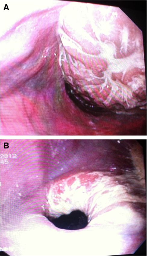
reproduction in any medium, provided the original work is properly credited.Shibly et al. BMC Veterinary Research 2014, 10:109 Page 2 of 6 http://www.biomedcentral.com/1746-6148/10/109 and abdominal positive contrast radiographs. The puppy chloride (6 ml/kg/h) to balance dehydration and hypo- had been fed solid food for a couple of weeks without any kalemia, ranitidine (2 mg/kg IV BID, Ulsal Injectable, difficulty, had been healthy from birth, and was the largest Gebro Pharma GmbH, Fieberbrunn, A) as gastric pro- puppy of the litter. tectant, maropitant (1 mg/kg IV SID, Cerenia Injectable, Initial physical examination revealed lethargy, a body Pfizer Animal Health Austria GmbH, Vienna, A) as anti- condition score of four out of nine, reduced skin turgor, emetic, and amoxicillin-clavulanic acid (22 mg/kg IV increased vesicular lung sounds and abdominal tender- BID, Clavamox Injectable, 550 mg, Sandoz GmbH, ness on palpation. Other parameters were within normal Vienna, A) to prevent possible aspiration pneumonia. limits. A fecal test for parvoviral antigen (IDEXX SNAP® Esophageal endoscopy was performed under general test) was negative. anesthesia (butorphanol 0.1 mg/kg IV, Butomidor Inject- Clinicopathologic findings on blood examination were able, 10 mg/ml, Richter Pharma AG, Wels, A; propofol metabolic alkalosis (pH 7.51 [reference value 7.351-7.463], 5 mg/kg IV, Propofol „Fresenius“ 1% with MCT Inject- HCO3 38.6 mmol/L [reference value 18-24 mmol/L]), able, Fresenius Kabi Austria GmbH, Graz, A; and inhala- hypokalemia (2.9 mmol/L [reference value 3.6-5.6 mmol/L]), tional isoflurane). both likely attributable to the vomiting, and a mild, stress- Inspection of the esophagus with a flexible video- induced hyperglycemia (132 mg/dl [reference value 55- endoscope (Olympus GIF 165) revealed accumulation of 100 mg/dl]). intraluminal fluid, food particles and contrast media in the Thoracic radiographs in left lateral recumbency showed cranial and midsection of the esophagus, while the caudal a contrast-filled esophagus, due to a barium contrast study third of the esophagus appeared distended and obstruc- performed by the referring veterinarian 4 hours pre- ted by an intraluminal mass consistent with the sto- viously. A diverticulum of the esophagus in the cranial mach (Figure 2A). Uncomplicated gastric reposition was mediastinum was suspected with severe esophageal dis- achieved by advancement of the endoscope against the tention in the caudal portion of the mediastinum. An gastric mucosa. Closure of the gastric cardia appeared in- intraluminal soft-tissue mass with traces of barium con- complete. The caudal esophagus remained dilated with trast and rugal folds was visible in the caudal thorax. The the mucosa macroscopically intact (Figure 2A). To pre- trachea was displaced ventrally, and the stomach silhou- vent repeated dislocation of the stomach and to allow for ette was not visible in the cranial abdomen. There was no nutrition bypassing the esophagus, a percutaneous endo- evidence of lung consolidation or infiltration, although scopic gastrostomy (PEG) tube (mushroom/Pezzar style esophageal obstruction caused by gastroesophageal intus- silicone catheter, Surgivet, Smiths Medical, Dublin, OH, susception (GEI) was strongly suspected (Figure 1). USA) was placed in the left abdominal wall. Inspection of Initial treatment for dehydration, and electrolyte im- the cranial and middle parts of the esophageal lumen after balances from the regurgitation and vomiting consisted removal of its contents failed to demonstrate the presence of intravenous saline supplemented with potassium of the radiographically suspected cranial diverticulum. Figure 1 Thoracic radiograph in left lateral recumbency of the dog on the day of presentation. The esophagus is filled with barium contrast and is dilated. The stomach (S) is invaginated into the esophagus. There is a suspected diverticulum of the esophagus cranial to the heart (arrow).
Shibly et al. BMC Veterinary Research 2014, 10:109                                                                         Page 3 of 6
http://www.biomedcentral.com/1746-6148/10/109

                                                                           Thoracic radiographs in left lateral recumbency taken
                                                                         36 hours after gastric repositioning showed resolution of
                                                                         the gastroesophageal intussusception. The esophagus
                                                                         still appeared dilated, and an interstitial and mild alveo-
                                                                         lar lung pattern was identified and thought to be from
                                                                         aspiration pneumonia (Figure 3).
                                                                           Thirty-six hours after endoscopy, enteral nutrition was
                                                                         initiated via PEG tube using a bland diet. The dog was
                                                                         clinically normal, playful, tolerated oral water and sucral-
                                                                         fate well. Over the next days, blood values returned to
                                                                         normal and oral feeding from an elevated position was
                                                                         gradually introduced. Although neither vomiting nor re-
                                                                         gurgitation were observed, thoracic radiographs in left
                                                                         lateral recumbency nine days after initial presentation
                                                                         showed persisting caudal esophageal dilation.
                                                                           The patient was discharged nine days after admission.
                                                                         The owner was instructed to offer incrementally increas-
                                                                         ing small portions of food formed to meatballs from an
                                                                         elevated position five times daily and was taught how to
                                                                         use the PEG tube to maintain the dog’s nutrition. Raniti-
                                                                         dine, sucralfate and amoxicillin-clavulanic acid were pre-
                                                                         scribed as oral medications. The dog remained clinically
                                                                         unremarkable, gained weight and size quickly, and the
                                                                         PEG tube was removed two weeks after discharge as oral
                                                                         feedings covered the patient’s nutritional demands.
                                                                           Thoracic radiographs in left lateral recumbency re-
                                                                         peated at five weeks, four months and eight months after
                                                                         discharge revealed persistent esophageal dilatation, ab-
                                                                         sence of abnormal lung patterns, and the patient con-
                                                                         tinues to tolerate commercially available dog food and
                                                                         treats without difficulty. Thoracic radiographs in left la-
                                                                         teral recumbency eight months after initial presentation
 Figure 2 Esophageal lumen. (A) Obstruction by intussuscepted            show the esophagus is still dilated with a slight dis-
 stomach. (B) Incomplete cardiac closure following gastric Reposition.   tension cranial to the heart. There is a striped luminal
                                                                         pattern in the caudal section consistent with a narrower
                                                                         lumen of the esophagus. The stomach is filled with con-
Based on endoscopic findings the final diagnosis was GEI                 trast medium (Figure 4).
with secondary esophageal dilation.
   After an uneventful recovery from anesthesia, the pa-                 Discussion
tient’s condition improved. Partial parenteral nutrition, a              GEI has been reported in different species, including
metoclopramide constant rate infusion (CRI, 0.01 mg/kg/h,                adult and pediatric humans [10,11], dogs [3,7,12-19],
Paspertin 10 mg Injectable, Abbott Products GmbH,                        domestic cats [4,20,21] and an adult leopard [22]. While
Hannover, D) to prevent vomiting and a lidocaine CRI                     the chronic recurrent form causes intermittent gas-
(0.05 mg/kg/h, Xylanaest purum 1% Injectable, Gebro                      trointestinal signs, patients with acute persistent GEI
Pharma GmbH, Fieberbrunn, A) as analgetic and radical                    present with an acute onset of clinical signs from
scavenger were added to the therapeutic regimen. Twelve                  esophageal obstruction, occasionally accompanied by
hours after endoscopy, lidocaine was progressively re-                   respiratory distress caused by the intrathoracic mass ef-
duced and discontinued. A mucosal protectant (sucralfate,                fect and/or aspiration pneumonia [19,23]. Additional
0.1 g/kg PO TID, Ulcogant oral suspension 1 g/5 ml,                      clinical signs of acute disease include regurgitation,
Merck S.L., Mollet Del Valles, E) was administered, along                vomiting, drooling, dysphagia and abdominal discom-
with small amounts of water. Feeding via the PEG tube                    fort [8,13]. Reduced venous return, endotoxic shock and
was withheld for another 24 hours to prevent possible                    release of inflammatory mediators can lead to cardio-
mucosal irritation caused by gastroesophageal reflux or                  vascular impairment and rapid death [17]. The case re-
vomiting.                                                                ported here can be classified as acute GEI, as no
Shibly et al. BMC Veterinary Research 2014, 10:109 Page 4 of 6 http://www.biomedcentral.com/1746-6148/10/109 Figure 3 Thoracic radiograph in left lateral recumbency 36 hours after gastric repositioning. The esophagus is dilated (arrowheads) and there are no traces of contrast agent left. A mild interstitial and alveolar lung pattern indicates aspiration pneumonia. A PEG tube is visible in the abdomen. abnormalities had been noticed until three days prior to obstruction, acid bile peptic disease and pregnancy presentation. [10,11]. In cats, chronic intermittent GEI predominates GEI occurs with reverse gastric peristalsis in combin- [4,20,21], while dogs tend to develop the acute form of ation with a sudden sustained increase in abdominal the condition [3,12,14,17,18,23]. In dogs, formation of pressure [3]. The pathogenesis of this condition has not GEI has been linked to increased intraabdominal pres- been fully elucidated and is likely multifactorial [6]. Ab- sure from vomiting or blunt trauma [6], negative intra- normalities including esophageal motility disorders, hia- thoracic pressure caused by respiratory disease [6,24] as tal enlargement and lower esophageal sphincter failure well as preexisting esophageal disease, especially megae- presumably predispose to GEI [8,9]. sophagus [3,6-8]. In human medicine, adults are affected more often Canine GEI usually affects puppies of medium and than children, and risk factors include eating disorders, large breeds younger than three months of age [3] with alcohol abuse, sudden sustained exertion, small-bowel males and German Shepherd dogs being overrepresented Figure 4 Thoracic radiograph with esophageal contrast study in left lateral recumbency eight months after diagnosis. The esophagus is dilated and filled with barium contrast and there is a slight distension or diverticulum is located cranial to the heart. There is a striped pattern in the caudal esophagus, and the stomach is filled with contrast agent.
Shibly et al. BMC Veterinary Research 2014, 10:109                                                                              Page 5 of 6
http://www.biomedcentral.com/1746-6148/10/109

[4-6]. The patient reported here is a female and belongs       In the management of GEI, endoscopy allows for defini-
to a breed in which GEI, to the authors’ knowledge, has        tive diagnosis of the condition, evaluation of the integrity
not been described before.                                     of the upper gastrointestinal tract lining, repositioning of
   It may be speculated that the patient described in this     the prolapsed stomach as well as placement of a PEG
report developed GEI as a result of an acute vomiting          tube serving as gastropexy-device and enabling enteral
episode (e.g. caused by stress due to relocation or pos-       nutrition bypassing a possibly impaired esophagus [7].
sible dietary changes). This is supported by breeder’s ac-     One limitation of the endoscopic approach is the inabil-
count that neither regurgitation nor dysphagia had been        ity to assess and correct concurrent problems such as
observed before the acute onset of signs at nine weeks         hiatal abnormalities. Furthermore, with PEG tube place-
of age. Furthermore, the puppy reportedly had been the         ment only a unilateral gastropexy is established, whereas
largest of the litter, which is an unexpected historical       bilateral gastropexy has been recommended by some au-
finding in a dog with congenital esophageal disease. On        thors for management of GEI [15,17]. Interestingly, left
the other hand, radiographic evidence of esophageal            unilateral gastropexy has been advocated by other au-
dilatation which was not identified during endoscopy           thors, since this technique results in positioning of the
persisted over months following gastric repositioning          esophageal hiatus to the left of the midline possibly im-
without causing concurrent clinical signs. This finding        proving the therapeutic success rate [14,23]. Endoscopic
argues against a reversible esophageal dilatation caused       repositioning of the stomach needs to be performed with
by the intussuscepted stomach, and demonstrates that           caution and should not be attempted in cases of adhe-
disappearance of clinical signs does not necessarily equal     sions or evident compromise of the gastric wall to avoid
spontaneous resolution of congenital esophageal disease        iatrogenic perforations [7]. Furthermore, the procedure
[7]. Probably a combination of a congenital esophageal         should be carried out by an experienced clinician to
disorder combined with severe vomiting due to other            minimize complications and duration of anesthesia.
causes triggered GEI in the case described here.                  To the authors’ knowledge, three cases of endoscopic
   Procedures to diagnose GEI include survey or contrast       reductions of GEI have been reported. One in a cat and
radiography, fluoroscopy and endoscopy [4,7]. In the           the other two cases in dogs [7,8,20]. The patient de-
present case, contrast radiography revealed an intralumi-      scribed by McGill and coworkers [7] was a seven-week-
nal soft tissue mass within the caudal thoracic esophageal     old female Siberian husky with esophageal dilatation and
lumen with vertical lamellar accumulations of contrast         GEI and the only case to date using PEG tube place-
medium compatible with gastric folds. The definitive diag-     ment for definitive treatment. The nine-week-old female
nosis of GEI was established using esophagoscopy.              Australian Shepherd described here had an unremark-
   Dogs that suffer from GEI can deteriorate rapidly with      able history up to the acute vomiting episode that had
mortality rates up to 95% reported for complicated cases       started three days prior to presentation. After radio-
[4,5]. Patient stabilization and repositioning of the trans-   graphic evaluation and initial medication, flexible endos-
located viscera needs to be initiated when there are           copy allowed for verification of diagnosis and successful
acute presentations with little delay [3].                     treatment of GEI within only three hours after admission
   Generally, surgical intervention with reduction of the      leading to a rapid clinical recovery. Long-term follow-up
intussusception followed by gastropexy has been advo-          including consecutive thoracic radiographs revealed per-
cated as the treatment of choice by most authors               sisting esophageal dilatation despite complete absence of
[5,14,15,17,18,23]. Potential disadvantages include pro-       compatible clinical signs, indicating that repeated diag-
longed general anesthesia [7] in a potentially hemodyna-       nostic imaging might be reasonable to enable continued
mically compromised, sometimes malnourished patient,           patient care and owner education, as clinical presenta-
the invasiveness and expense of surgery, and the inability     tion may not reflect esophageal abnormalities present
to fully evaluate the condition of the esophageal and gas-     necessitating particular attentiveness.
tric mucosa, which may impact prognosis. Furthermore,
an extended convalescence period, and possible postsurgi-      Conclusion
cal complications such as wound infections or delayed tis-     Acute GEI is a rare and potentially fatal disease and
sue healing need to be considered. These factors, in           should be considered as a differential diagnosis in patients
addition to potential congenital esophageal abnormalities      with acute signs of esophageal obstruction in all dogs.
resulting in a guarded prognosis [7], may influence deci-      Endoscopic treatment of GEI consisting of gastric reposi-
sion-making leading to euthanasia of a patient with a po-      tioning and PEG tube placement appears to be an effect-
tentially controllable condition.                              ive, cost-efficient alternative to conventional surgery.
   Flexible endoscopy is a minimally invasive technique
                                                               Abbreviations
requiring a brief general anesthestic procedure and of-        CRI: Constant rate infusion; GEI: Gastroesophageal intussusception;
fering a number of diagnostic and therapeutic options.         PEG tube: Percutaneous endoscopic gastrostomy tube.
Shibly et al. BMC Veterinary Research 2014, 10:109 Page 6 of 6
http://www.biomedcentral.com/1746-6148/10/109
Competing interests 18. Masloski A, Besso J: What is your diagnosis? gastroesophageal
None of the authors has financial or personal relationships that could intussusception with megaesophagus in a dog. J Am Vet Med Assoc 1998,
inappropriately influence or bias the content of the paper. 212:23–24.
19. Applewhite AA, Cornell KK, Selcer BA: Diagnosis and treatment of
intussusceptions in dogs. Comp Cont Educ Pract 2002, 24:110–127.
Authors’ contributions 20. Van Camp S, Love NE, Kumaresan S: Radiographic diagnosis –
SS and SK were responsible for the study design. SK was responsible for gastroesophageal intussusception in a cat. Vet Radiol Ultrasound 1998,
initial treatment of the dog. Endoscopic intervention was performed by SS. 39:190–192.
KMH was responsible for diagnostic imaging. RAH, SK and SS were 21. Van Geffen C, Saunders JH, Vandevelde B, Van Ham L, Hoybergs Y, Daminet
responsible for clinical examinations and medical care of the patient. SS and S: Idiopathic megaoesophagus and intermittent gastro-oesophageal
SK drafted the manuscript. SS, SK, KMH and RAH were involved in work intussusception in a cat. J Small Anim Pract 2006, 47:471–475.
supervision and writing of the manuscript. All authors read and approved 22. Hettlich BF, Hobson HP, Snakard EP, Johnson JH: Gastroesophageal
the final manuscript. intussusception in a leopard (Panthera pardus). J Zoo Wildl Med 2010,
41:519–521.
Author details 23. Von Werthern CJ, Montavon PM, Fluckinger MA: Gastro-oesophageal
1
Department for Small Animals and Horses, Clinic of Internal Medicine and intussusception in a young German shepherd dog. J Small Anim Pract
Infectious Diseases, University of Veterinary Medicine Vienna, Veterinaerplatz 1996, 37:491–494.
1, Vienna A-1210, Austria. 2Department for Small Animals and Horses, Clinical 24. Arndt JW, Marks SL, Kneller SK: What is your diagnosis? hiatal hernia due
Section of Diagnostic Imaging, University of Veterinary Medicine Vienna, to laryngeal squamous cell carcinoma. J Am Vet Med Assoc 2006,
Veterinaerplatz 1, Vienna A-1210, Austria. 228:693–694.
Received: 20 May 2013 Accepted: 25 March 2014 doi:10.1186/1746-6148-10-109
Published: 7 May 2014 Cite this article as: Shibly et al.: Acute gastroesophageal intussusception
in a juvenile Australian shepherd dog: endoscopic treatment and long-
term follow-up. BMC Veterinary Research 2014 10:109.
References
1. Jergens AE: Diseases of the esophagus. In Textbook of veterinary internal
medicine. 7th edition. Edited by Ettinger SJ. Philadelphia: WB Saunders Co;
2010:1487–1499.
2. Spillmann T: Esophageal diseases – diagnostic and therapeutic approach.
In Proceedings of the world small animal veterinary association. Sydney,
Australia: 2007.
3. Roach W, Hecht S: What is your diagnosis? gastroesophageal
intussusception. J Am Vet Med Assoc 2007, 231:381–382.
4. Martínez NI, Cook W, Troy GC, Waldron D: Intermittent gastroesophageal
intussusception in a cat with idiopathic megaesophagus. J Am Anim
Hosp Assoc 2001, 37:234–237.
5. Pietra M, Gentilini F, Pinna S, Fracassi F, Venturini A, Cipone M: Intermittent
gastroesophageal intussusception in a dog: clinical features,
radiographic and endoscopic findings, and surgical management. Vet Res
Commun 2003, 27:783–786.
6. Rasmussen L: Stomach. In Textbook of small animal surgery. 3rd edition.
Edited by Slatter D. Philadelphia: WB Saunders Co; 2003:631–632.
7. McGill SE, Lenard ZM, See AM, Irwin PJ: Nonsurgical treatment of
gastroesophageal intussusception in a puppy. J Am Anim Hosp Assoc
2009, 45:185–190.1.
8. Guilford WG: Diseases of swallowing. In Strombeck’s Small animal
gastroenterology. 3rd edition. Edited by Guilford WG, Center SA, Strombeck
DR. Philadelphia: WB Saunders Co; 1996:211–235.
9. Tamms TR: Diseases of the esophagus. In Handbook of small animal
gastroenterology. 2nd edition. Edited by Tamms T. St. Louis: WB Saunders
Co; 2003:149–151.
10. Gowen GF, Stoldt HS, Rosato FE: Five risk factors identify patients with
gastroesophageal intussusceptions. Arch Surg 1999, 134:1394–1397.
11. Lukish JR, Eichelberger MR, Henry L, Mohan P, Markle B: Gastroesophageal
intussusception: a new cause of acute esophageal obstruction in
children. J Pediatr Surg 2004, 39:1125–1127.
12. Rowland MG, Robinson M: Gastro-oesophageal intussusception in an
adult dog. J Small Anim Pract 1978, 19:121–125. Submit your next manuscript to BioMed Central
13. Leib MS, Blass CE: Gastroesophageal intussusception in a dog: a review of and take full advantage of:
the literature and a case report. J Am Anim Hosp Assoc 1984, 20:783–790.
14. Clark GN, Spodnick GJ, Rush JE, Keyes ML: Belt loop gastropexy in the
• Convenient online submission
management of gastroesophageal intussusception in a pup. J Am Vet
Med Assoc 1992, 201:739–742. • Thorough peer review
15. Greenfield CL, Quinn MK, Coolman BR: Bilateral incisional gastropexies for • No space constraints or color figure charges
treatment of intermittent gastroesophageal intussusception in a puppy.
• Immediate publication on acceptance
J Am Vet Med Assoc 1997, 228:693–694.
16. Weekley LB, Read R, Wu E, Takeda S, Hsia CC, Johnson RL: • Inclusion in PubMed, CAS, Scopus and Google Scholar
Gastroesophageal intussusception associated with pneumonectomy in a • Research which is freely available for redistribution
dog. Contemp Top Lab Anim Sci 1997, 36:91–93.
17. Graham KL, Buss MS, Dhein CR, Barbee DD, Seitz SE: Gastroesophageal
intussusception in a Labrador retriever. Can Vet J 1998, 39:709–711. Submit your manuscript at
www.biomedcentral.com/submitYou can also read