Evaluation of the sterilization effect of a new mercury-free UVC light source
←
→
Page content transcription
If your browser does not render page correctly, please read the page content below
R ESEARCH         ARTICLE                                                           ScienceAsia 45 (2019): 458–464
doi: 10.2306/scienceasia1513-1874.2019.45.458

Evaluation of the sterilization effect of a new
mercury-free UVC light source
Han Gaoa,b , Tongkuo Yuanb,c , Jian Zhaod , Hongye Liue , Huancai Yina,b,∗
a
     University of Science and Technology of China, Hefei, Anhui 230026 China
b
     CAS Key Lab of Bio-Medical Diagnostics, Suzhou Institute of Biomedical Engineering and Technology,
     Chinese Academy of Sciences, Suzhou, Jiangsu 215163 China
c
     School of Life Sciences, Shanghai University, Shanghai 200433 China
d
     NovelUv Co., Ltd, Shanghai 201108 China
e
     Clinical Laboratory, Changchun Center Hospital, Jilin, Changchun 130051 China
∗
    Corresponding author, e-mail: yinhc@sibet.ac.cn
                                                                                                Received 13 May 2019
                                                                                                 Accepted 29 Oct 2019

ABSTRACT: So far, short-wave UV produced by low-pressure mercury lamps has been widely applied in sterilization,
wastewater treatment, and bioassays, which could be due to its low cost, high conversion efficiency, and small size.
However, metal pollution is caused by the evitable releasing of mercury. In this study, a new mercury-free lamp was
developed, which generated UV light by the electron beam excitation (EBE) of YPO4 :Bi3+ under vacuum conditions.
Such lamps emit light at 241 nm and possessed a photoelectric effect. Excellent sterilization effect was obtained in
Candida albicans, Staphylococcus aureus, Pseudomonas aeruginosa, and Escherichia coli, in the absence of ozone. These
effects positively correlated with the treatment distances and time. Furthermore, oxidative stress was found to play
a major role in the sterilization process of our light sources, since no production of ozone was detected during each
treatment. After all, EBE lamps could be a promising tool for sterilization, and our results provided the theoretical basis
for its microorganism-killing effects.

KEYWORDS: UV, mercury-free lamp, electron beam excitation (EBE) UV lamp, ozone-free, oxidative stress

INTRODUCTION                                                   and no pollution 5, 6 . Despite the long-term devel-
                                                               opment, UVC-producing LEDs are limited by their
Nowadays, UV light sources have been widely ap-                complicated manufacturing process, high cost, low
plied in industrial, agriculture, and medical fields,          photoelectric conversion efficiency, and severe heat
due to their fluorescent, biological, photochemical            radiation 7 . In fact, the thermal emission cathode
and photoelectric effects 1 . In particular, short-wave        produced by LG Company, which are of the most ma-
UV with wavelengths of 200–280 nm has been found               tured manufactory techniques, only exhibits a low
useful in sterilization, wastewater treatment and              photoelectric conversion efficiency at 2%. Hence
biological detection 2 . Among them, low-pressure              an efficient, non-polluting, and long-lifetime UV
mercury lamps producing 254 nm UV light were                   sources are still in demand.
mostly accepted, which could be due to their long                   Based on the recent publications, UV light could
lifetime, high photoelectric conversion efficiency,            be generated by electron beam excitation of phos-
mature manufacturing process, and low cost 3 . Un-             phate, rare earth-doping material and ZnO 8 . With
fortunately, these lamps are being criticized for their        similar mechanisms, Oto et al developed new UV
releasing of mercury, which cause irreversible fatal           lamps by electron beam excitation of AlGaN quan-
damage to living organisms 4 . According to Mina-              tum wells, which exhibited a high power of 100 mW
mata Treaty, mercury-containing UV light sources               at 240 nm 9 . Max Shatalov et al used AlGaN to
will be gradually replaced in 2020. Thus more and              develop a UV light source with a wavelength of
more attentions are being paid on the development              278 nm 10 . Compared with UV LED, EBE-UV light
of mercury-free lamps.                                         sources possessed simple production processes, no
     The first developed light sources are mainly              thermal effect, and high photoelectric conversion
light-emitting diodes (LEDs), which have advan-                efficiency, thus exhibiting a promising application
tages of long life, small size, short start-up time,

www.scienceasia.org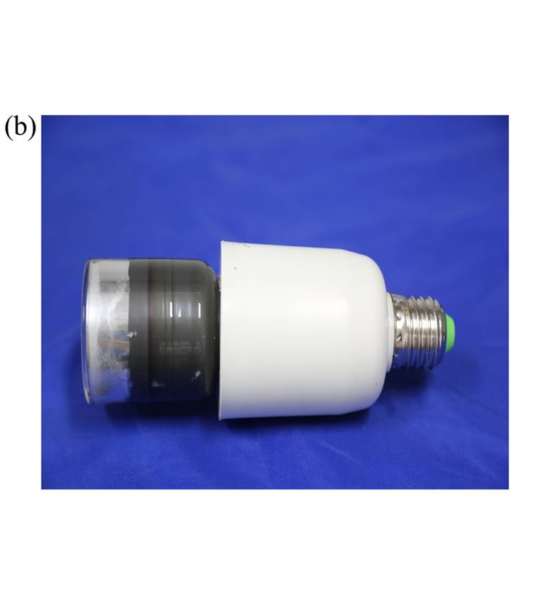
ScienceAsia 45 (2019)                                                                                          459

  (a)                              HV

                                 VACUUM
                                 ELECTRONBEAM
          UVC

                   LUMINESCENT                  ELECTRON
                   MATERIAL                     SOURCE

Fig. 1 (a) Schematic diagram of YPO4 :Bi3+ EBE-UV lamp,    Fig. 2 (a) Schematic diagram of sterilization device;
HV = high voltage; (b) UV lamp body diagram.               (b) physical map of the equipment.

prospect 11, 12 .                                          the electrical and optical powers, respectively. After
     In this study, a small sterilization device was       calculation, η should be 4.01%.
produced with an EBE-UV light source. After char-               The schematic and digital illustrations of the
acterization of photoelectric conversion efficiency        lamp are shown in Fig. 1. Its emission spectra were
and wavelength, the sterilization properties of this       evaluated by a UV spectrometer (Gentec, Florida,
lamp were tested with four microorganisms, includ-         USA) in a wavelength range of 200–360 nm. Ir-
ing Candida albicans, Staphylococcus aureus, Pseu-         radiance power of EBE-UV was measured using a
domonas aeruginosa, and Escherichia coli. After            PH100-SiUV Probe accumulated UV meter (Ushio
then, the main sterilization mechanism of this lamp        Inc., Japan), after the switch was turned on, the
was detected, to provide the basis for its application     UV meter was set as zero first. Then the irradiance
in sterilization.                                          power was randomly detected at 5 different posi-
                                                           tions of the same height to calculate the average
MATERIALS AND METHODS                                      value.
EBE-UV characterization                                    Sterilization device
The EBE-UV light was produced by electron beam             To test the efficiency of UV light source, a small
excitation on YPO4 :Bi3+ , and the cold cathode is         sterilizer with adjustable distance and treatment
protected by vacuum environment 4 . In brief, the          time was designed. The structure is shown in Fig. 2.
electrons hit the matrix of YO4 :Bi3+ , causes energy           The sterilizer consists of two parts: tank and
transfer, and excites the fluorescence. With specific      shell. The tank was made of corrosion-resistant
optical filter, the wavelength of UV would be limited      stainless-steel material (ISO11), with a hole in the
around 241 nm. By DC Ammeter and Voltmeter, the            upper field. The light sources located inside the
input power of the UV lamp is 1.3 W, and output            shell, right on the upper of the tank holes. The light-
power is determined to be 52.13 mW. The photo-             ing time, interval time, and automatic light-off could
electric conversion  rate η is calculated according       be controlled automatically. A transparent viewing
to η = Plight /Pe × 100%, where Plight and Pe are          window is present on the outer shell, to observe

                                                                                                  www.scienceasia.org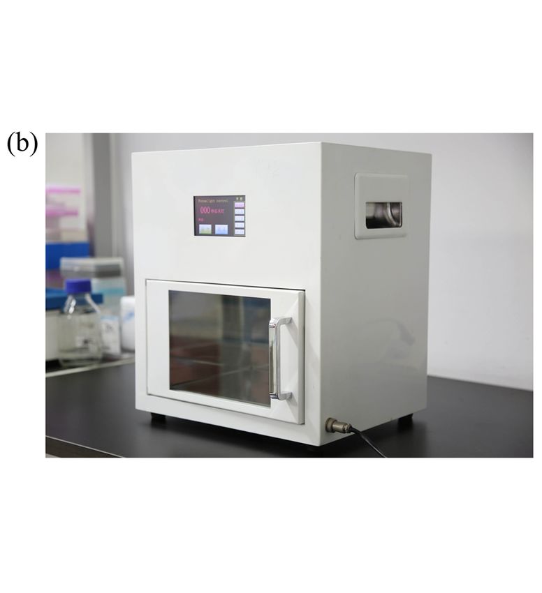
460                                                                                         ScienceAsia 45 (2019)

the entire illumination process. Tin foil was used         tubes (EP), to measure intracellular ROS with com-
to cover the shading treatment on the observation          mercial Kits (Beyotime, China) 17 . 10 µm 2,7-
window, to avoid the light resurrection during UV          dichlorodihydrofluorescein diacetate (DCFH-DA)
treatment. Finally, adjust the distances between the       was added to the treated suspensions and incubated
light sources and the plane of the lowest-end plates,      for 20 min at 37 °C. Then the cells were washed and
to determine the sterilization characteristics.            suspended in 1.0 ml of PBS. The pictures of accu-
                                                           mulated 20 ,70 -dichlorofluorescein (DCF) inside cells
Culture of microorganisms                                  were taken with a digital camera (Olympus Corpo-
C. albicans (ATCC10231), S. aureus (ATCC25923),            ration, Tokyo) and a Zeiss axiophot fluorescence mi-
P. aeruginosa (ATCC9027), and E. coli (ATCC25922)          croscopy (Carl Zeiss, Oberkochen, Germany) with
obtained from American Type Culture Collection,            a green filter. After then, the fluorescence of sus-
were cultured at 37 °C in tryptic soy broth (pH 7.2,       pension was detected with a Multi-Mode microplate
BD Diagnosis Systems, Sparks, MD) 13–15 . All the          Reader (BioTek, US) using an excitation/emission
bacteria and yeast kept on a rotary shaker at              wavelength of 488/525 nm. Relative fluorescence
220 rpm were cultivated for 8 h. The concentration         intensity (RFI) of the intracellular ROS level was
of cells was 109 CFU/ml as assessed by 600 nm              normalized to the total number of microorganism 18 ,
absorption.                                                and then expressed as the fold increase of control
                                                           group.
Evaluation of sterilization effect
                                                                          DCFEX /(1 − Disappearance %)
Microorganisms, including yeast (C. albicans),                    RFI =                                ,
Gram-positive bacteria (S. aureus), and Gram-                                       DCFControl
negative bacteria (E. coli and P. aeruginosa), were        where DCFEX and DCFControl are the detected fluores-
used to evaluate the sterilization effect of EBE-          cence of the treated groups and the control group,
UV. After reaching the set concentrations of 109           respectively.
CFU/ml, the four strains of microorganisms were
diluted by 1:10 000 16 . 100 µl of dilution was cul-       Ozone content detection
tured on the solid nutrient agar medium, and then          The light resource of the UV sterilizer was fixed at
immediately placed in the sterilization device. All        15 cm from the bottom of the plate. Since all the
agar media were treated with UV at 15 cm from the          microorganisms died after 20 min irradiation. The
UV light source at 37 °C. According to the tolerance       ozone production was detected after then. Ozone
of each strain, different time gradients were used         instrument (JSA8–O3, Shenzhen, China) was used
for C. albicans (0.0, 1.0, 2.0, 3.0, and 4.0 min),         to detect the ozone content at 6 random points to
P. aeruginosa (0.0, 1.0, 1.5, 2.0, and 2.5 min),           obtain an average value. The enclosed space of this
S. aureus (0.0, 0.5, 1.0, 1.5, 2.0, 2.5, 3.0, and          device is 15 cm in height, 23 cm in length, 10 cm
3.5 min), and E. coli (0.0, 5.0, 10.0, 15.0, 20.0, 25.0,   in width. The total volume is about 3.45 l. As a
30.0, 35.0, and 40.0 s). Then the survived colonies        positive control, the ozone produced by an ordinary
were counted and compared with untreated control.          254 nm lamp (TUV, 8 W, Philips) was detected in the
The differences were divided by the total colonies,        same condition, and the results indicated a 2.7 ppm
and the results were defined as the disappearance          production of ozone.
rate. The obtained values were plotted against the
irradiation dose (K, (mW.s/cm2 )) of UV, which was         DNA damage analysis
calculated as K = I × T /103 , where I and T are the       After UVC irradiation for indicated time, the four
irradiation intensity (µW/cm2 ) and irradiation time       microorganisms were collected and extracted for
(s), respectively. To prevent cells from reactivation,     their DNA with a commercial kit. The degree of DNA
the processes of UV treatment and cultivating were         damage was determined by the alkaline version
shaded away from light.                                    of the Comet Assay (single-cell gel electrophoresis
                                                           SCGE) as described by Lanier et al 19 . The irradia-
Intracellular ROS assay                                    tion time was selected for each microorganism with
After sterilization, the microorganisms on the plate       the occurrence of significant death. The obtained
were scraped off with 1.0 ml PBS. About 100 µl             comets were visualized with a fluorescent micro-
of the treated suspensions were taken out for              scope (Carl Zeiss, Oberkochen, Germany) at 40 ×
total number counting, and the remaining sus-              magnification (Achroplan and ECPlan-Neofluar ob-
pensions were collected into 1.5 ml eppondorf              jectives) with the red filter (excitation 546 nm and

www.scienceasia.org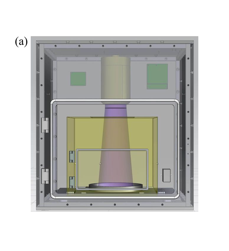
ScienceAsia 45 (2019) 461
It could be seen that the sterilization efficiency
of this EBE-UV light was dose-dependent, and all
the microorganisms could be killed under the ir-
radiation lower than 66.6 mW.s/cm2 . As shown
in Fig. 4a, the most tolerant microorganism was
C. albicans, while E. coli was the most easily killed
bacteria (Fig. 4d). The tolerance of P. aeruginosa
and S. aureus was found to be between C. albicans
and E. coli (Fig. 4bc).
Sterilization mechanism
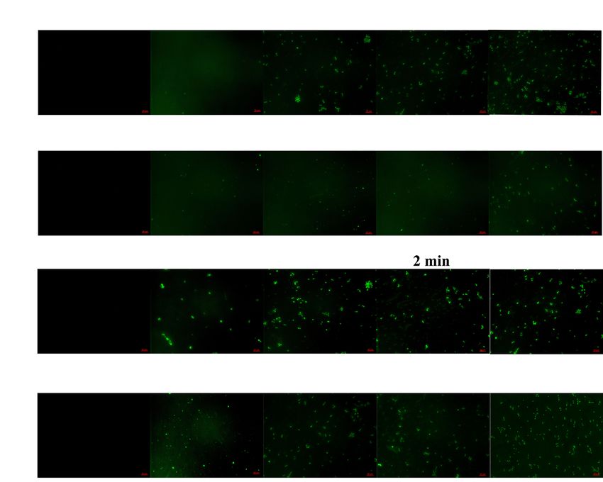
Fig. 3 Emission spectra of YPO4 :Bi3+ EBE-UV lamp. The changes of ROS curve in the four microorgan-
isms after UV treatment were shown in Fig. 5a. As
the irradiation time and dose increase, the levels of
Table 1 Radiation intensity at different distance (DIST)
ROS increase to different degrees for each microor-
from the lighting tube.
ganism. To more intuitively show the effect of irra-
DIST/cm 3 6 9 12 15 diation on cells, the fluorescence intensity of DCF in
Irradiance (µW/cm2 ) 651.1 280.6 139.1 84.2 55.5 cells was photographed by fluorescence microscopy
in the experiment, and the results were shown in
the following Fig. 5b, which became brighter with
emission 590 nm). The obtained images were anal- the increase of treatment time. Surprisingly, no
ysed using AxioVision software version 4.7.2 (Carl production of ozone was detected during the treat-
Zeiss, Oberkochen, Germany). ment of EBE-UV lights, as the data kept below zero
during the detection. In addition, significant DNA
Statistical analysis damages were noticed after UVC irradiation, as DNA
All data represent here are mean ± SD of three inde- migration exhibited an obvious comet style (Fig. 6
pendent experiments with each in triplicate (n = 9). and Table 2).
Statistical analysis was performed using SPSS Statis-
tic 15.0 Software (SPSS Inc., Chicago, Illinois, USA). DISCUSSION
Comparisons were conducted between control and
With the development of science and technology,
treated groups with student’s t-test, *p < 0.05 was
the demands for human health continue to increase.
considered significant.
As an important environmental pollutant, mercury
RESULTS is posing a huge threat to human health due to
their extensive applications in the industry 20 . On
Characterization of EBE-UV light sources
19 January 2013, the United Nations Environment
The diameter of the front-edge of UV light source Programme adopted Minamata Convention, to re-
is 50 mm, and the length of the whole light source duce the releasing of mercury in the world 21 . For
is 155 mm. The emission spectrum is shown in the UV light sources, the convention clearly states
Fig. 3, which indicates a core wavelength of 241 nm that after 2020, low-cost mercury lamps will be
with a half-peak width of 15 nm. The photoelectric unavailable. Hence all countries are developing
conversion efficiency was determined to be 4.01%. high-efficiency and mercury-free UV light sources,
After 1000-h operation, the photoelectric conver- to occupy a huge market including lighting, steril-
sion efficiency will be reduced by about 75%. The ir- ization, and wastewater treatment.
radiance at different distances from the light source In this study, a new sterilization device was
is shown in Table 1, which decreases gradually with constructed with new mercury-free UV sources.
the increase in distance. The lamp has a maximum emission wavelength of
241 nm with a half-width of 15 nm. The pho-
Evaluation of sterilization effect toelectric conversion efficiency was calculated to
To evaluate the microorganism-killing effect of be 4%. After improvement on the matrix, much
deep-UV light, four strains of microorganisms higher photoelectric conversion efficiency of even
(yeast, Gram-positive and Gram-negative bacteria) 12% could be achieved, which is much higher than
were irradiated with 241 nm UV at different doses. LG’s commercial products (data to be published).
www.scienceasia.org462 ScienceAsia 45 (2019)
Fig. 4 The sterilization efficiency at 241 nm was examined by disappearance of (a) C. albicans, (b) P. aeruginosa,
(c) S. aureus, and (d) E. coli. Data are expressed as means ± SD of 3 samples, ***p < 0.001 compared with untreated
control.
(
b) Co
ntr
ol 1mi
n 2mi
n 3mi
n 4mi
n
bi
C.al ans
c
Co
ntr
ol 1mi
n 1.
5mi
n 2mi
n 2.
5mi
n
r
.ae
P a
nos
ugi
Co
ntr
ol 1mi
n 1.
5mi
n 2.
5mi
n
e
S.aurus
Cont
rol 10s 20s 30s 40s
ol
E.c i
Fig. 5 (a) Intracellular ROS concentration after the treatment with 241 nm EBE-UV lamp in yeast, Gram-positive,
and Gram-negative bacteria: ROS value of C. albicans (), P. aeruginosa ( ), S. aureus (Î), and E. coli (È). (b) DCF
fluorescence produced by bacteria with fluorescence microscope.
In the previous reports, the effective radiation 300 mJ/cm2 (3.0 × 10³ mW · s/cm2) to inactivate
power of conventional sterilization lamp (254 nm) 10 bacteria species in platelet concentrates 23 . On
was 150 mJ/cm2 (1.5 × 10³ mW · s/cm2) , which the other hand, Dotson et al used doses from 0.1–
could kill C. albicans up to 10−5 CFU/ml 22 . The dose 0.2 J/cm2 (1.0 × 10³ to 2.0 × 10³ mW · s/cm2) for
used by Mohr H and Gravemann U was even higher, drinking water disinfection 24 . All these irradiation
www.scienceasia.orgScienceAsia 45 (2019) 463
Table 2 Analytical results of comet assay.
L-head L-tail L-comet Head-DNA Tail-DNA Tail Olivetail
Sample
(µm) (µm) (µm) (%) (%) moment moment
C. albicans 0 min 27.0 ± 2.4 4.3 ± 0.6 31.4 ± 3.3 97.53 ± 3.96 2.46 ± 0.85 0.09 ± 0.02 0.32 ± 0.02
C. albicans 1 min 31.6 ± 3.8 37.7 ± 4.0* 69.3 ± 9.8* 73.33 ± 5.95 26.66 ± 3.05* 8.93 ± 2.08* 9.20 ± 2.92*
C. albicans 3 min 27.3 ± 3.6 33.5 ± 5.5* 60.8 ± 4.2 73.54 ± 8.57 26.45 ± 3.43* 10.72 ± 1.99* 4.03 ± 0.12*
P. aeruginosa 0 min 25.7 ± 3.4 3.0 ± 1.1 28.7 ± 3.7 99.95 ± 8.78 0.04 ± 0.02 0.02 ± 0.003 0.01 ± 0.09
P. aeruginosa 1 min 23.9 ± 3.6 21.7 ± 4.8* 45.7 ± 3.3 79.37 ± 6.99 20.62 ± 3.01* 4.33 ± 2.02* 3.93 ± 1.33*
P. aeruginosa 2 min 19.7 ± 3.9 29.8 ± 6.2* 49.5 ± 4.8 38.10 ± 4.22* 61.89 ± 5.78* 17.95 ± 2.04* 9.98 ± 1.96*
S. aureu 0 min 27.1 ± 2.4 3.7 ± 1.6 30.7 ± 7.6 99.93 ± 12.55 0.06 ± 0.45 0.009 ± 0.0003 0.009 ± 0.0003
S. aureu 0.5 min 26.0 ± 8.8 18.7 ± 7.3* 44.7 ± 6.9 84.28 ± 7.98 15.71 ± 4.02* 2.82 ± 1.78* 2.73 ± 1.71*
S. aureu 2 min 25.6 ± 5.8 50.9 ± 7.4* 76.5 ± 9.7* 63.83 ± 4.72 36.16 ± 3.28* 18.08 ± 9.14* 12.81 ± 7.65*
E. coli 0 min 23.2 ± 3.3 4.0 ± 1.4 27.2 ± 4.1 99.97 ± 9.38 0.02 ± 0.01 0.007 ± 0.003 0.003 ± 0.002
E. coli 10 s 19.4 ± 4.8 3.9 ± 2.4 23.4 ± 8.6 99.92 ± 27.72 0.07 ± 0.02 0.002 ± 0.001 0.007 ± 0.006
E. coli 40 s 19.4 ± 4.0 9.6 ± 2.1* 28.9 ± 5.1 74.16 ± 8.23 25.83 ± 7.77* 2.32 ± 1.54* 2.56 ± 1.35*
*
p < 0.05 versus control.
generate ozone 25 . Although ozone can be useful
for sterilization, it has strong oxidizing properties,
which can cause ageing of equipment and is not suit-
able for human environment 26, 27 . In this respect,
the application of new UV light sources could be
advantageous.
CONCLUSION
In this paper, a novel sterilization device was con-
structed with EBE-UV light sources, which gen-
erated UV light with an electron beam to excite
YPO4 :Bi3+ material. This device had a narrow emis-
sion peak (241 ± 25 nm), high photoelectric con-
version efficiency (4.01%), environmental friend-
liness (without mercury) and low cost (around
Fig. 6 Representative comet assay images for the four 0.5 $/mW). This device exhibited an excellent sen-
strains: C. albicans, P. aeruginosa, S. aureus, and E. coli sitization effect at 15 cm from the light source, with
after the treatment with EBE-UV light. oxidative stress as the major mechanism. Hence
it has great application prospects for the surface
sterilization of objects.
doses were much higher than that in this experi-
Acknowledgements: This work was supported by
ment, where the new UV sources killed all the four
grants from National Science and Technology Major
microorganisms after irradiation of 20 min and the
Project of China (2017ZX10302301-003) and National
dose reached 66.6 mW · s/cm2 . Such phenomenon
Key R&D Programme of China (2017YFF0108600 &
could be due to the narrow spectral peak at 241 nm
2017YFC0110400).
compared with broad-spectrum of traditional lamps.
According to literature, DNA damage, ROS ef- REFERENCES
fects, and production of ozone could be the rea-
1. Rozhansky IV, Zakheim DA (2006) Analysis of depen-
sons of UV sterilization. In this experiment, ROS
dence of electroluminescence efficiency of AIInGaN
effects and DNA damages have been found in this LED heterostructures on pumping. Phys Status Solidi
experiment (Fig. 5 and Fig. 6). However, no ozone C 3, 2160–2164.
was detected after the irradiation of UV lights for 2. Gayan E, Serrano MJ, Pagan R, Alvarez I, Condon
20 min. Such phenomenon could be occurred on S (2015) Environmental and biological factors influ-
the fact that the emission spectrum of EBE lights encing the UV-C resistance of Listeria monocytogenes.
does not contain 185 nm, which ionizes the air to Food Microbiol 46, 246–253.
www.scienceasia.org464 ScienceAsia 45 (2019)
3. Chen J, Loeb S, Kim JH (2017) LED revolution: D, Bellopede R, Grimaldi N, Nardone A, Zarrilli R,
fundamentals and prospects for UV disinfection ap- et al (2015) Effect of treatment with an overheated
plications. Environ Sci Water Res Technol 3, 188–202. dry-saturated steam vapour disinfection system on
4. Yoo ST, Hong JH, Kang JS, Park KC (2018) Deep- multidrug and extensively drug-resistant nosocomial
ultraviolet light source with a carbon nanotube cold- pathogens and comparison with sodium hypochlorite
cathode electron beam. J Vac Sci Technol B 36, ID activity. BMC Res Notes 8, ID 551.
02C103. 17. Li JH, Wang G, Zhu HQ, Zhang M, Zheng XH, Di
5. De Volder MF, Tawfick SH, Baughman RH, Hart AJ ZF, Liu XY, Wang X (2014) Antibacterial activity of
(2013) Carbon nanotubes: present and future com- large-area monolayer graphene film manipulated by
mercial applications. Science 339, 535–539. charge transfer. Sci Rep 4, ID 4359.
6. Hirayama H, Maeda N, Fujikawa S, Toyoda S, Kamata 18. Soares SS, Gutierrez-Merino C, Aureliano M (2007)
N (2014) Recent progress and future prospects of Mitochondria as a target for decavanadate toxicity in
AlGaN-based high-efficiency deep-ultraviolet light- sparus aurata heart. Aquat Toxicol 83, 1–9.
emitting diodes. Jpn J Appl Phys 53, ID 100209. 19. Lanier C, Bernard F, Dumez S, Leclercq J, Lemiere
7. Song K, Mohseni M, Taghipour F (2016) Application S, Vandenbulcke F, Nesslany F, Platel A,et al (2016)
of ultraviolet light-emitting diodes (UV-LEDs) for Combined effect of Cd and Pb spiked field soils
water disinfection: a review. Water Res 94, 341–349. on bioaccumulation, DNA damage, and peroxidase
8. Ge CQ, Xie CS, Hu ML, Gui YH, Bai ZK, Zeng DW activities in trifolium repens. Environ Sci Pollut Res
(2007) Structural characteristics and UV-light en- 23, 1755–1767.
hanced gas sensitivity of La-doped ZnO nanoparti- 20. Beck SE, Ryu H, Boczek LA, Cashdollar JL, Jeanis
cles. Mater Sci Eng B 141, 43–48. KM, Rosenblum JS, Lawal OR, Linden KG (2017)
9. Oto T, Banal RG, Kataoka K, Funato M, Kawakami Evaluating UV-C LED disinfection performance and
Y (2010) 100 mW deep-ultraviolet emission from investigating potential dual-wavelength synergy. Wa-
aluminium-nitride-based quantum wells pumped by ter Res 109, 207–216.
an electron beam. Nat Photonics 4, 767–770. 21. Gibb H, O’Leary KG (2014) Mercury exposure and
10. Shatalov M, Sun WH, Lunev A, Hu XH, Dobrinsky health impacts among individuals in the artisanal
A, Bilenko Y, Yang JW, Shur M, et al (2012) AlGaN and small-scale gold mining community: a com-
deep-ultraviolet light-emitting diodes with external prehensive review. Environ Health Perspect 122,
quantum efficiency above 10%. Appl Phys Express 5, 667–672.
ID 082101. 22. Buonanno M, Stanislauskas M, Ponnaiya B, Bigelow
11. Watanabe K, Taniguchi T, Niiyama T, Miya K, AW, Randers-Pehrson G, Xu Y, Shuryak I, Smilenov L,
Taniguchi M (2009) Far-ultraviolet plane-emission et al (2013) 207-nm UV light - a promising tool for
handheld device based on hexagonal boron nitride. safe low-cost reduction of surgical site infections. I:
Nat Photonics 3, 591–594. in vitro studies. PLoS One 8, ID e76968.
12. Ichikawa N, Ikeda K, Honda Y, Taketomi H, Kawai K, 23. Mohr H, Gravemann U, Bayer A, Mueller TH (2009)
Suzuki T (2016) Development of a UV light source Sterilization of platelet concentrates at production
using Pr:LuAG thin film pumped by electron beam. scale by irradiation with short-wave ultraviolet light.
Electron Commun Jpn 99, 33–39. Transfusion 49, 1956–1963.
13. Sousa AM, Machado I, Nicolau A, Pereira MO (2013) 24. Dotson AO, Rodriguez CE, Linden KG (2012) UV
Improvements on colony morphology identification disinfection implementation status in US water treat-
towards bacterial profiling. J Microbiol Methods 95, ment plants. J Am Water Works Assoc 104, 318–324.
327–335. 25. Szeto W, Li JT, Huang HB, Leung DYC (2018)
14. Barboza DD, Martins LCA, Correa TQ (2018) Pho- VUV/TiO2 photocatalytic oxidation process of methyl
todynamic inactivation of Staphylococcus aureus and orange and simultaneous utilization of the lamp-
Escherichia coli using a new bacteriochlorin as pho- generated ozone. Chem Eng Sci 177, 380–390.
tosensitizer. In: Kessel DH, Hasan T (eds) Optical 26. Galante R, Ghisleni D, Paradiso P, Alves VD, Pinto
Methods for Tumor Treatment and Detection - Mecha- TJA, Colaco R, Serro AP (2017) Sterilization of
nisms and Techniques in Photodynamic Therapy 27th, silicone-based hydrogels for biomedical application
California, USA. using ozone gas: comparison with conventional tech-
15. Narita K, Asano K, Morimoto Y, Igarashi T, Hamblin niques. Mater Sci Eng C 78, 389–397.
MR, Dai TH, Nakane A (2018) Disinfection and 27. Wang YZ, Wang HY, Li XS, Liu DX, Jiang YF, Sun
healing effects of 222-nm UVC light on methicillin- ZH (2013) O3 /UV synergistic aging of polyester
resistant Staphylococcus aureus infection in mouse polyurethane film modified by composite UV ab-
wounds. J Photochem Photobiol B 178, 10–18. sorber. J Nanomater 2013, ID 169405.
16. Bagattini M, Buonocore R, Giannouli M, Mattiacci
www.scienceasia.orgYou can also read