Molecular characteristics of Neisseria meningitidis in Qatar
←
→
Page content transcription
If your browser does not render page correctly, please read the page content below
www.nature.com/scientificreports
OPEN Molecular characteristics
of Neisseria meningitidis in Qatar
Manal Mahmoud Hamed1,3*, Fayaz Ahmad Mir1,3, Emad Bashier Ibrahim Elmagboul1,
Abdullatif Al‑Khal1, Muna A. Rahman S. Al. Maslamani1, Anand Sarwottam Deshmukh1,
Hamad Eid Al‑Romaihi2, Mohd. Ahmed M. Sharif Janahi1, Fatma Ben Abid1,
Adila Shaukat Ali Kashaf1, Gulab Sher1, Vinod Kumar Gupta1, Godwin J. Wilson1,
Junais Kadalayi1 & Sanjay H. Doiphode1
The aim of the current study is to review the molecular characteristics of Neisseria meningitidis (N.
meningitidis) in Hamad Medical Corporation, which is the provider of secondary and tertiary care in the
state of Qatar. A total of 39 isolates of N. meningitidis from the period of 2013 to 2018 were revived
and identified by Vitek, and susceptibility on the basis of the E test was retrieved from the patient’s
files. The revived isolates were subjected to multilocus sequence typing. The most common serogroup
(19) of N. meningitidis was W135, of which 12 were isolated from blood and CSF. ST-11 was the most
predominant ST clonal complex causing N. meningitidis cases (61.53%). Clonal complex ST-41/44 was
the second most observed complex (3, 2 of which were related to serogroup B). The most frequent
sequence type was 9596 (8 isolates). Determining the molecular pattern of N. meningitidis in Qatar is
helpful for understanding the strains circulating in Qatar, and the study of the resistance trend of such
strains may be very helpful for empirical treatment of future patients.
Invasive Neisseria meningitidis (N. meningitidis) infection (meningitis, meningococcemia) is associated with
high mortality and morbidity worldwide despite the use of proper antibiotics. Fever, bacteremic pneumonia,
resentations1,2.
pericarditis, septic arthritis, urethritis and ear infection are less frequent p
Capsules of N. meningitidis are important virulent factor that allow the organisms to escape human immuno-
logical defense mechanisms. The organism is known to change its capsular polysaccharide by capsular switching
to evade the immune system; thus, the introduction of vaccine serotypes might result in an increase in serotypes
that are not included in the vaccines by a mechanism such as capsular switching. Genetic changes have also been
observed after the introduction of an outer protein-based vaccine for serogroup B, with an increase in serotypes
C and Y alongside major changes in outer membrane p roteins3. The genetic makeup changes are due to either
the acquisition of foreign DNA from other strains and genera or other mechanisms, such as the recombination
of self-genes. Capsular switching results in a change in the serogroup of the N. meningitidis strain due to a change
in capsular polysaccharides but will reserve the same lineage, as shown by MLST. Capsular switching was proven
during the Hajj season in the Saudi Arabia outbreak caused by serogroup W-1353.
N. meningitidis can be classified by capsular polysaccharides into twelve serogroups (previously thirteen
serogroups, but group D is no longer considered a separate group). Among the twelve capsular polysaccha-
ride serogroups, only five (A, B, C, W-135, and Y) are known to frequently lead to the development of disease
worldwide. Most N. meningitidis found in the throat are untypable or lack a capsule that is required for invasive
infection. Serogroup distribution differs in different parts of the world; serogroup A is the most commonly
isolated serogroup in sub-Saharan Africa4,5, while serogroups B, C, and Y are the most common serogroups
found in Europe and the United States (US). In Asia, serogroup A is the predominant s erogroup5. Most cases
in the US are sporadic, but outbreaks are nonetheless a common occurrence. Epidemics are known to occur in
sub-Saharan Africa (in 25 countries), with the most recent outbreak occurring during the Hajj season in Saudi
Arabia and caused by serogroup W-1356,7.
A recent method of characterization of N. meningitidis is molecular typing methods, which are used to study
the relatedness of outbreak isolates by detecting minimal changes in the genome. Molecular typing tests detect
variations to characterize the clones circulating in a certain geographical area and their closeness to any of the
known international clones. An example of these tests is MLST, which has become the most widely used test
worldwide6–8.
1
Hamad Medical Corporation, Doha, Qatar. 2Ministry of Health, Doha, Qatar. 3These authors contributed equally:
Manal Mahmoud Hamed and Fayaz Ahmad Mir. *email: mhamed@hamad.qa
Scientific Reports | (2021) 11:4812 | https://doi.org/10.1038/s41598-021-84262-1 1
Vol.:(0123456789)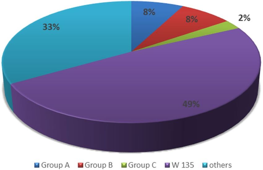
www.nature.com/scientificreports/
MLST, the gold standard for molecular typing, classifies meningococcal strains into different sequence types
(STs) based upon polymorphisms in seven housekeeping genes. This technique helps identify any hyperinvasive
lineages, i.e., meningococcal STs that are independent of serogroups and are disproportionately associated with
disease relative to carriage levels. Seven-locus MLST has been recommended at least for a representative selec-
tion of each country or for enhanced regional surveillance, and this information is essential for the national and
international management of meningococcal disease and the study of meningococcal population biology and
evolution7,8.
In a 2-year study conducted in Qatar (1998–2000) the leading causes of meningitis were revealed to be Strepto-
coccus pneumonia, which resulted in 30% of the cases of meningitis included in the study, Haemophilus influenzae
(24%), and N. meningitidis (5%); however, these findings may not reveal the actual numbers, as partially treated
cases were excluded from the study. The rather low number caused by N. meningitidis was explained at that time
by the adequate number of people who opted to receive the meningococcal vaccine in Qatar before they left for
Hajj or Umrah, as requested by the Kingdom of Saudi Arabia9.
Resistance to antibiotics, especially penicillin, has been reported worldwide due to mutations in the con-
stitution of penicillin-binding protein, PBP 2, and less commonly due to the production of penicillinases. The
occurrence of penicillin resistance in the closely related Neisseria gonorrhea drove researchers to also consider
the possibility of developing resistance in N. meningitidis. Commonly used antibiotics are third-generation
cephalosporins, such as ceftriaxone. Resistance to rifampin and trimethoprim/sulfamethoxazole is rare, but
reports of resistance have been p ublished1,10–12.
Identification of the major serogroups will help in understanding the efficacy of vaccine coverage and the
main circulating serogroup in Qatar. Genetic characterization is essential in understanding the characteristics
and magnitude of circulating clones and provides national authorities in Qatar with valuable data about the
efficacy of the vaccine policy and vaccination schedule and the effectiveness of the available vaccine as well as
suggestions for strategies for better control of N. meningitidis infections and spread13.
To the best of our knowledge, there has been no study of the genetic characteristics of N. meningitidis in Qatar
or any analysis comparing the Qatari invasive strains with international strains. This study represents the first
report of N. meningitidis molecular characteristics in Qatar.
Methods
We subjected our strains to molecular typing techniques for epidemiological purposes to investigate potential
outbreak strains in Qatar. All strains that were stored in our repository (in the period between February 2013
and March 2018) were revived and subjected to the following procedures:
Isolation and culture. All meningococcal isolates, principally invasive isolates in Qatar representing all
known invasive genotypes and serogroups, stored in our cryorepository were tested.
The lyophilized N. meningitidis cryovials were removed from the Hamad General Hospital repositories,
where they were preserved at − 80 °C, and allowed to thaw for 10 min in a biological safety cabinet. The tubes
were then opened, and beads from each cryovial were transferred to chocolate agar plates by using a sterile
needle. The chocolate agar was streaked for bacterial isolation, and the beads were discarded in a yellow sharps
box. The streaked chocolate agar plates were sealed with parafilm and incubated at 37 °C in a microaerophilic
incubator with a 5–10% CO2 supply for 18 h. After an 18-h incubation period, smooth, moist, glistening, and
convex colonies of N. meningitidis had grown. The colonies were subcultured onto a fresh chocolate agar plate
and incubated at 37 °C in a microaerophilic incubator with a 5–10% CO2 supply for another 18 h. The next day,
the N. meningitidis colonies were subjected to DNA extraction.
Antimicrobial susceptibility, serogrouping and all patient details were retrieved from the patient files. The
serogrouping used antisera against the major serogroups (A, B, C, W-135, X, and Y), which cause life-threatening
disease14,15.
DNA extraction and sequencing. DNA was extracted by the PureLink Genomic DNA Mini Kit (Invitro-
gen) according to the manufacturer’s instructions. In brief, N. meningitidis colonies were transferred to 180 μL
Digestion Buffer, 20 μL of Proteinase K was added, and the tube was briefly vortexed. The tube was incubated
at 55 °C with occasional vortexing until lysis was complete. Then, 20 μL of RNase A was added to the lysate,
mixed well by vortexing and incubated at room temperature for 2 min. Two hundred microliters of lysis/binding
buffer was added to the lysate and mixed well by vortexing to obtain a homogeneous solution. Then, 200 μL of
96–100% ethanol was added to the lysate and mixed well by vortexing for 5 s to obtain a homogeneous solution.
The lysate (~ 640 μL) was added to the spin column and centrifuged at 10,000 × g for 1 min. The collection tube
was discarded, and the spin column was transferred into a clean collection tube. Then, 500 μL of wash buffer
1 was added to the column and centrifuged at 10,000 × g for 1 min. The collection tube was discarded, the spin
column was transferred into a clean collection tube, and 500 μL wash buffer 2 was added to the column and cen-
trifuged at maximum speed for 3 min. The collection tube was discarded, and the spin column was transferred to
a sterile 1.5-mL microcentrifuge tube. Then, 50 μL of elution buffer was added to the column, incubated at room
temperature for 1 min and centrifuged at maximum speed for 1 min. The eluted DNA was stored at − 20 °C. All
centrifugations were carried out at room temperature8,16.
Multilocus sequence typing (MLST) and FetA typing. MLST on patient isolates was performed in
HMC laboratories, and MLST and FetA typing were performed as previously described using the primers listed
on the PubMLST website (http://pubmlst.org/neisseria/)8,16. PCR was carried out using Maxima Hot Start Mas-
ter Mix (Thermo Scientific) in 25 μL of reaction with 12.5 μL Master Mix (2X), 1 μL (10 μM) forward primer, 1
Scientific Reports | (2021) 11:4812 | https://doi.org/10.1038/s41598-021-84262-1 2
Vol:.(1234567890)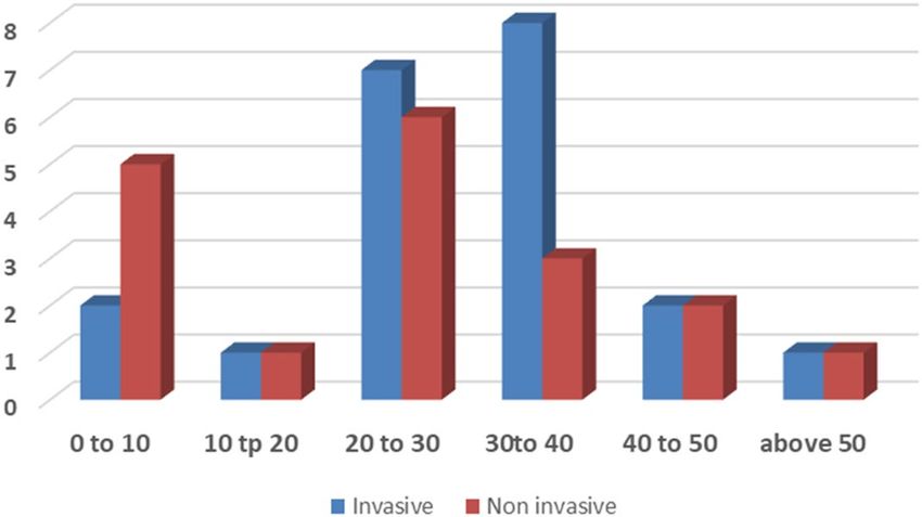
www.nature.com/scientificreports/
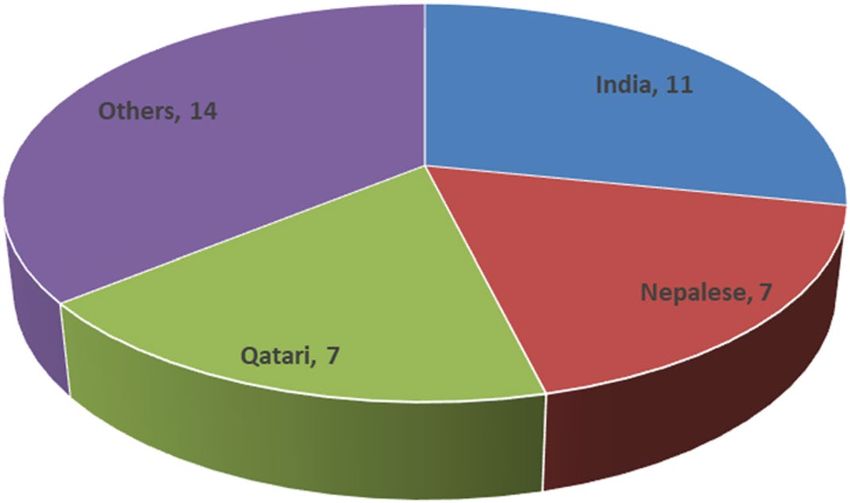
Figure 1. Nationality distribution among the patients.
μL (10 μM) reverse primer, 2 μL DNA template, and 8.5 μL nuclease-free water. The thermal cycling conditions
were as follows: initial denaturation at 94 °C for 2 min; 35 cycles of 94 °C for 1 min, 55 °C for 1 min, and 72 °C for
1 min; and a final extension at 72 °C for 2 min. The amplification was checked by running 5 µl of each reaction
product on a 2% agarose gel with a 100 bp DNA ladder (Invitrogen). PCR products were purified by ExoSAP-IT
reagent (USB, Affilmetrix). In brief, 5 μL of PCR product was mixed with 2 μL of ExoSAP-IT reagent and incu-
bated at 37 °C for 15 min, followed by 80 °C for 15 min. Sequencing was carried out by the BigDye Terminator
ver. 3.1 cycle sequencing kit (Applied Biosystems) in a 5 μL reaction volume consisting of 2 µL of BigDye Termi-
nator 3.1 Ready Reaction Mix, 2 µL of forward/reverse primer (0.6 µM), and 1 µL of purified PCR product. The
thermal cycling conditions were 25 cycles of 96 °C for 10 s, 50 °C for 5 s, and 60 °C for 2 min. After completion of
PCR, 15 μL of sterile dH2O was added to each sample to a final volume of 20 μL. Sequencing reaction products
were purified by a BigDye XTerminator purification kit (Applied Biosystems) according to the manufacturer’s
instructions. Then, 90 µL of SAM solution was mixed with 20 µL of BigDye XTerminator bead solution to pre-
pare the SAM/BigDye XTerminator bead working solution, which was added to each sample. Then, the sample
was mixed on a vortexer for 30 min and centrifuged at 1000 × g for 2 min. The purified sequencing reaction
products were analyzed on an ABI 3500 Genetic Analyzer (Applied Biosystems) according to the manufacturer’s
instructions. Designation of sequence types (ST) and clonal complexes (CC) were retrieved by comparison on
the PubMLST website (http://pubmlst.org/neisseria/)8,16.
Ethical approval. I confirm that all methods were carried out in accordance with the relevant guidelines
and regulations. I received approval for this research from Qatar Medical Research Center and obtained an
exemption from Hamad Medical Corporation IRB regarding consent, as this study dealt with retrospective bac-
terial isolates stored in our repositories and not patients. The reference code is Research Proposal #15453/15.
I declare that my research in Qatar was conducted ethically and in accordance with community expectations.
Results
Thirty-nine isolates of N. meningitidis were detected from February 2013 to March 2018, of which 35 were from
Hamad General Hospital (HGH) and four were from Al Wakra Hospital. The patients were predominantly of
Indian nationality (11) (Fig. 1).
Twenty-three patients presented with fever, of whom 12 presented with additional meningitic symptoms and
signs (headache, rash, irritability). Six patients were referred from the medical commission due to abnormal
chest X-ray. Five out of the six had pulmonary tuberculosis, and all six N. meningitidis isolates were isolated
from respiratory tract samples (two were serogroup W135, two serogroup A and two untypable) (Table S1, sup-
plementary file).
Of the patients, 15 (38%) were aged 20–30. There were more invasive isolates in the 30–40 age group than in
all the other age groups (Fig. 2).
Twenty-one isolates were recovered from blood specimens, one from CSF (56% invasive disease), and 18
from the respiratory tract. The most common serogroup (19) of N. meningitidis was W135 (49%), of which 12
were isolated from blood and CSF, followed by group B (3) and group A (3). One isolate was related to group C,
and nine N. meningitidis isolates were of unknown or untypable groups (Fig. 3). The maximum number of cases
occurred during 2016 (12 cases, mainly Nepalese and Indian patients, with 10 of the 12 isolated from respira-
tory samples) (Fig. 4).
Penicillin susceptibility appeared in only 61%, while ciprofloxacin susceptibility was 89% and meropenem
and ceftriaxone susceptibility was 100% (Table 1).
Out of the 39 isolates, 24 isolates had clonal complex ST-11 (61.5%), of which 18 isolates had serogroup W
135 (75%). Four isolates were found that were not related to any clonal complexes in PubMLST. We anticipate
that these isolates must be related to novel complex(es) and thus need further evaluation. The serogroups of
these four isolates were groups A, B, and W135, and one was untypable. Clonal complex ST-41/44 was the second
most observed complex (three, two of which were related to serogroup B). The most frequent sequence type
Scientific Reports | (2021) 11:4812 | https://doi.org/10.1038/s41598-021-84262-1 3
Vol.:(0123456789)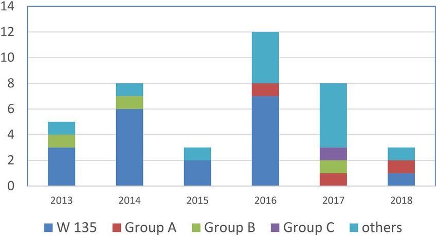
www.nature.com/scientificreports/

                                            Figure 2.  Age distribution among patients.

                                            Figure 3.  Serogroup distribution among all isolates.

                                            Figure 4.  Occurrence of Neisseria meningitidis serotypes.

                                            was 9596 (eight isolates), followed by 7979 (seven), all of which belonged to clonal complex ST-11 (Table S1,
                                            supplementary file).

                                            Discussion
                                            HMC is the main governmental hospital serving the whole state of Qatar and providing every level of care to
                                            residents of the country. Therefore, all cases of suspected meningitis are treated at HMC. We isolated a total of 39
                                            isolates of N. meningitidis that were detected by culture from 2013 to March 2018 (Table S1, supplementary file).
                                            More than half of these isolates caused invasive diseases (meningitis and/or bacteremia), and 12 cases occurred

          Scientific Reports |   (2021) 11:4812 |                https://doi.org/10.1038/s41598-021-84262-1                                                   4

Vol:.(1234567890)
www.nature.com/scientificreports/
Antimicrobial
MIC (µg/mL) breakpoints susceptibility
interpretation (%)
Antibiotics S I R S I R
Penicillin ≤ 0.006 0.12–0.25 ≥ 0.5 61 32 8
Ceftriaxone ≤ 0.12 – – 100 – –
Ciprofloxacin ≤ 0.03 0.06 ≥ 0.12 89 5 5
Meropenem ≤ 0.25 – – 100 – –
Rifampin ≤ 0.5 1 ≥2 85 12 3
Table 1. Antimicrobial susceptibility testing for Neisseria meningitidis.
in 2016 (Fig. 3), of which seven were related to serogroup W 135(58%) and three were invasive isolates (Table S1,
supplementary file). The number of cases per year varies from a minimum of 3 cases in 2015 to a maximum
of 12 in 2016 (Fig. 3). The lack of data from the Middle East, especially the Gulf Cooperation Council (GCC)
countries, provided us with an opportunity to fill that gap with this study; thus, we aimed to review the molecular
epidemiology of N. meningitis in Qatar.
The most common serogroup (19) of N. meningitidis isolated at HMC was W135 (49%), which is similar to the
N. meningitidis outbreak that occurred during pilgrimage to Saudi Arabia in 2000, with W135 (37%) being the
main serogroup, followed by group A (24%). Many countries became involved as the patients returned to their
home countries. An earlier outbreak with serogroup A occurred in Middle East and North African countries,
including Qatar, in 19885.
The predominant serogroups vary with the region. The main serogroup in sub-Saharan Africa was group A,
but since the introduction of a vaccine for group A, outbreaks caused by this serogroup have started to disap-
pear, and serogroups X and W135 have started to increase, while in the USA, serogroups B, C, and Y are the
main causes of meningitis. Serogroup B is the main serogroup causing meningitis in Europe, followed by W135
and C5,17.
Serogroup B vaccines have also been used to control outbreaks in the Saguenay Lac-Saint-Jean region in
Canada and other outbreaks at US u niversities18,19.
In 2015, national immunization programs introduced Serogroup B vaccines to infants in the UK and Ireland,
and regional programs were introduced in Italy20.
In our study, 9 serogroups were untypable, and further research is required to assess the exact serogroups
of these isolates.
In Qatar, the currently available vaccine for meningococcal infections is the quadrivalent conjugated menin-
gococcal vaccine for serogroup ACYW135, which has been widely introduced in many countries.
Our main age group consisted of persons between 20 and 40 years old. Since most of our patients were labor-
ers and expatriates, it will be highly prudent to develop labor surveillance studies for carriage and vaccination
among this group if a high carrier rate is detected among them (Fig. 2).
This is similar to what is happening in the USA and Europe, where people aged 16 through 23 and those less
than 1 year old have the highest rates of meningococcal d isease19. In our study, 2 isolates out of 39 were from
children aged < 10 years, which may suggest the possibility of underestimation of the invasive disease burden
caused by N. meningitidis in children due to early intake of antibiotics. Therefore, meningococcal disease surveil-
lance programs are strongly recommended to determine the carrier state within this group, which may provide
a clear view of the circulation of the organism within this population and may encourage the implementation of
school-based vaccination programs if high carriage rates are found. Kim et al. reported in prospective popula-
tion surveillance performed among children aged < 5 years that N. meningitidis was the most prevalent pathogen
for invasive d isease20.
Bogaert et al. studied the prevalence of N. meningitidis carriage among Dutch children aged 1–19 years and
found a 1.5% incidence, especially among children aged 1 and above 15 years o ld21.
While carrier surveillance study among children up to 14 years of age in Egypt and Turkey, presented a wide
range of carriage (1.2–18.8%), it was even higher among military staff and students, while in Israel, 29% of chil-
dren aged from 11 to 15 years old were carriers of N. meningitidis5.
Our main ST clonal complex (24 isolates) was ST-11, of which 18 isolates had serogroup W135 (75%) (Table 1,
supplementary file). This finding is similar to the findings of the 2017 report of The European Centre for Disease
Prevention and Control (ECDC), which reported an increase in serogroup W 135 with a single clone, ST-11,
in the UK as well as other countries in Europe, such as Italy and the Netherlands. In the meningitis belt within
sub-Saharan Africa, there are 2 main hypervirulent clonal complexes, ST-5 and ST-11 (mainly the W135 sero-
group), but there are fewer W135 epidemics than group A epidemics in this part of the world. A detailed guide
was drafted by the WHO to hamper the rate of incidence for outbreaks within this region, including a strategy
for chemoprophylaxis, vaccines, accurate laboratory diagnosis, and quality e valuation22,23.
Among our isolates, penicillin susceptibility was 61%, ciprofloxacin susceptibility was 89%, and meropenem
and ceftriaxone susceptibility was 100%. In a 2004 N. meningitidis surveillance study in Croatia, 17 out of 23
isolates were serogroup B, 4 were serogroup C, 1 was serogroup W135, and 1 was untypable. All serogroup C
isolates were resistant to penicillin, while all other serogroups were sensitive to all antibiotics (penicillin, ceftri-
axone, chloramphenicol, and ciprofloxacin)24.
Scientific Reports | (2021) 11:4812 | https://doi.org/10.1038/s41598-021-84262-1 5
Vol.:(0123456789)www.nature.com/scientificreports/
In a Turkish study of children (0–18 years) who had meningococcal meningitis, 8.7% of the isolates were
resistant to penicillin, 22.8% were intermediately susceptible, and all isolates were susceptible to cefotaxime25.
A Chinese study found an increase in resistance to quinolones (67.7%) among N. meningitidis isolates acquired
from horizontal transfer from commensal Neisseriae (99.3%)26. In a UK study, two people who had returned from
Hajj were found to have conjunctivitis caused by N. meningitidis, and a third person who had been in contact
with one of them had invasive disease from the same untypable N. meningitidis isolate, which was resistant to
ciprofloxacin and intermediately susceptible to p enicillin27.
Regular continuous surveillance of N. meningitidis disease and stratified carriage among different popula-
tions, especially children and laborers, and regular publication are essential to determine the magnitude of the
disease in a population and thereafter provide adequate intervention. Moreover, active early detection and study
of circulating invasive strains, either locally or internationally, will help to prevent future outbreaks and decrease
the incidence of invasive disease.
Received: 1 September 2020; Accepted: 15 February 2021
References
1. Oppenheim, B. A. Antibiotic resistance in Neisseria meningitidis. Clin. Infect. Dis. 24(Suppl 1), S98-101. https://doi.org/10.1093/
clinids/24.supplement_1.s98 (1997).
2. Romero-Gomez, M. P., Rentero, Z., Pano, J. R. & Mingorance, J. Bacteraemic pneumonia caused by Neisseria meningitidis serogroup
Y. Respir. Med. Case Rep. 5, 23–24. https://doi.org/10.1016/j.rmedc.2011.11.005 (2012).
3. Harrison, L. H. Epidemiological profile of meningococcal disease in the United States. Clin. Infect. Dis. 50(Suppl 2), S37-44. https
://doi.org/10.1086/648963 (2010).
4. (NCIRD), N. C. f. I. a. R. D. Meningeococcal disease. https://www.cdc.gov/meningococcal/index.html.
5. Ceyhan, M. et al. Meningococcal disease in the Middle East and North Africa: an important public health consideration that
requires further attention. Int. J. Infect Dis. IJID 16, e574-582. https://doi.org/10.1016/j.ijid.2012.03.011 (2012).
6. Dang, V. et al. Epidemiology of serogroup B invasive meningococcal disease in Ontario, Canada, 2000 to 2010. BMC Infect. Dis.
12, 202. https://doi.org/10.1186/1471-2334-12-202 (2012).
7. Jolley, K. A., Brehony, C. & Maiden, M. C. Molecular typing of meningococci: recommendations for target choice and nomenclature.
FEMS Microbiol. Rev. 31, 89–96. https://doi.org/10.1111/j.1574-6976.2006.00057.x (2007).
8. Maiden, M. C. et al. Multilocus sequence typing: a portable approach to the identification of clones within populations of patho-
genic microorganisms. Proc. Natl. Acad. Sci. U.S.A. 95, 3140–3145. https://doi.org/10.1073/pnas.95.6.3140 (1998).
9. Elsaid, M. F. et al. Clinical presentation of acute bacterial meningitis in Qatar. Neurosciences 7, 266–271 (2002).
10. Almog, R. et al. First recorded outbreaks of meningococcal disease in the Israel Defence Force: three clusters due to serogroup C
and the emergence of resistance to rifampicin. Infection 22, 69–71. https://doi.org/10.1007/BF01739006 (1994).
11. Blondeau, J. M. et al. Neisseria meningitidis with decreased susceptibility to penicillin in Saskatchewan, Canada. J. Clin. Microbiol.
33, 1784–1786. https://doi.org/10.1128/JCM.33.7.1784-1786.1995 (1995).
12. Van Esso, D. et al. Neisseria meningitidis strains with decreased susceptibility to penicillin. Pediatr. Infect. Dis. J. 6, 438–439. https
://doi.org/10.1097/00006454-198705000-00003 (1987).
13. Yazdankhah, S. P. et al. Distribution of serogroups and genotypes among disease-associated and carried isolates of Neisseria men-
ingitidis from the Czech Republic, Greece, and Norway. J. Clin. Microbiol. 42, 5146–5153. https: //doi.org/10.1128/JCM.42.11.5146-
5153.2004 (2004).
14. Weinstein, M. P. Methods for Dilution Antimicrobial Susceptibility Tests for Bacteria That Grow Aerobically 11th edn. (Clinical and
Laboratory Standards Institute, Wayne, 2018).
15. Frasch, C. E., Zollinger, W. D. & Poolman, J. T. Serotype antigens of Neisseria meningitidis and a proposed scheme for designation
of serotypes. Rev. Infect. Dis. 7, 504–510. https://doi.org/10.1093/clinids/7.4.504 (1985).
16. Russell, J. E., Jolley, K. A., Feavers, I. M., Maiden, M. C. & Suker, J. PorA variable regions of Neisseria meningitidis. Emerg. Infect.
Dis. 10, 674–678. https://doi.org/10.3201/eid1004.030247 (2004).
17. Watson, P. S. & Turner, D. P. Clinical experience with the meningococcal B vaccine, Bexsero((R)): prospects for reducing the
burden of meningococcal serogroup B disease. Vaccine 34, 875–880. https://doi.org/10.1016/j.vaccine.2015.11.057 (2016).
18. Gasparini, R., Amicizia, D., Lai, P. L. & Panatto, D. Meningococcal B vaccination strategies and their practical application in Italy.
J. Prev. Med. Hyg. 56, E133-139 (2015).
19. Prevention (CDC), T. C. f. D. C. a. Meningeococcal disease. https://www.cdc.gov/meningococcal/surveillance/index.html (2020).
20. Kim, S. A. et al. An expanded age range for meningococcal meningitis: molecular diagnostic evidence from population-based
surveillance in Asia. BMC Infect. Dis. 12, 310. https://doi.org/10.1186/1471-2334-12-310 (2012).
21. Bogaert, D. et al. Epidemiology of nasopharyngeal carriage of Neisseria meningitidis in healthy Dutch children. Clin. Infect. Dis.
40, 899–902. https://doi.org/10.1086/428351 (2005).
22. European Centre for Disease Prevention and Control. Invasive meningococcal disease, Annual Epidemiological Report for 2017.
https://www.ecdc.europa.eu/sites/default/files/documents/AER_for_2017-invasive-meningococcal-disease.pdf (2019).
23. Organization, W. H. Meningitis outbreak response in sub-Saharan Africa, WHO guideline. https: //apps.who.int/iris/bitstr eam/handl
e/10665/ 144727 /WHO_HSE_PED_CED_14.5_eng.pdf;jsessi onid= 2584CF AA526 32C2E
BDBE6 3391B D85FA D?sequen ce=1 (2014).
24. Boras, A. et al. Establishment of an active laboratory-based surveillance for bacterial meningitis in Croatia and molecular char-
acterization of Neisseria meningitidis isolates causing meningococcal disease that were collected in the year 2000, the first year of
activity. J. Clin. Microbiol. 42, 1803–1806. https://doi.org/10.1128/jcm.42.4.1803-1806.2004 (2004).
25. Karadag Oncel, E. et al. Surveillance of penicillin resistance of Neisseria meningitidis strains from invasive infections between 2013
and 2018 in Turkey. J. Chemother. 32, 213–216. https://doi.org/10.1080/1120009X.2020.1721176 (2020).
26. Chen, M., Zhang, C., Zhang, X. & Chen, M. Meningococcal quinolone resistance originated from several commensal Neisseria
species. Antimicrob. Agents Chemother. 64, 45. https://doi.org/10.1128/AAC.01494-19 (2020).
27. Zumla, A. & Memish, Z. A. Risk of antibiotic resistant meningococcal infections in Hajj pilgrims. BMJ 366, l5260. https://doi.
org/10.1136/bmj.l5260(2019).
Acknowledgements
The project was funded by the medical research center in Qatar.
Scientific Reports | (2021) 11:4812 | https://doi.org/10.1038/s41598-021-84262-1 6
Vol:.(1234567890)www.nature.com/scientificreports/
Author contributions
Dr. M.H. is the main author; she developed the research concept and completed the paperwork to submit the
research to the Hamad Research Center. She wrote most of the study, reviewed and analyzed the results with the
other authors, revised it and resubmitted it each time. She is the corresponding author. F.A.M.M. helped from the
start of the study and in writing up the manuscript in every step of the research and helped to overcome every
obstacle; thus, the authorship is divided with him. V.K.G. and G.S. performed the MLST study; J.A.K. revived
the isolates and sent them for molecular studies; H.E.A.-R. provided the public health information about the
vaccination and other public health data; A.S.D., E.B.I.E., S.H.D., and G.J.W. helped with the data analysis and
review of the final draft; and M.A.S.J., M.A.S.A.M., A.A.K., A.S.A.K. and F.B.A. helped write and review the
article, retrieve clinical demographic data and interpret the clinical data. S.H.D. helped correct the article in
response to the reviewer’s comments. Dr. M.H. was involved in each step, coordinated all the needed work, was
responsible for the design of the work, was involved in the data analysis and writing of the article, and drafted
the work and substantively revised it.
Competing interests
The authors declare no competing interests.
Additional information
Supplementary Information The online version contains supplementary material available at https://doi.
org/10.1038/s41598-021-84262-1.
Correspondence and requests for materials should be addressed to M.M.H.
Reprints and permissions information is available at www.nature.com/reprints.
Publisher’s note Springer Nature remains neutral with regard to jurisdictional claims in published maps and
institutional affiliations.
Open Access This article is licensed under a Creative Commons Attribution 4.0 International
License, which permits use, sharing, adaptation, distribution and reproduction in any medium or
format, as long as you give appropriate credit to the original author(s) and the source, provide a link to the
Creative Commons licence, and indicate if changes were made. The images or other third party material in this
article are included in the article’s Creative Commons licence, unless indicated otherwise in a credit line to the
material. If material is not included in the article’s Creative Commons licence and your intended use is not
permitted by statutory regulation or exceeds the permitted use, you will need to obtain permission directly from
the copyright holder. To view a copy of this licence, visit http://creativecommons.org/licenses/by/4.0/.
© The Author(s) 2021
Scientific Reports | (2021) 11:4812 | https://doi.org/10.1038/s41598-021-84262-1 7
Vol.:(0123456789)You can also read