Effect of eccentric and concentric squat exercise on quadriceps thickness and lower extremity performance in healthy young males
←
→
Page content transcription
If your browser does not render page correctly, please read the page content below
ACTA GYMNICA, 2021, Volume 51, Article e2021.015                                                                                         OPEN
https://doi.org/10.5507/ag.2021.015                                                                                                      ACCESS

ORIGINAL RESEARCH

Effect of eccentric and concentric squat exercise on quadriceps
thickness and lower extremity performance in healthy young males
Nihal Büker1*, Raziye Şavkın1, Akın Süzer2, and Nuray Akkaya3
1
 School of Physical Therapy and Rehabilitation, Pamukkale University, Denizli, Turkey; 2Therapy and Rehabilitation Department,
Burdur Vocational School of Health Services, Burdur Mehmet Akif Ersoy University, Burdur, Turkey; and 3Department of Physical
Medicine and Rehabilitation, Medical School, Pamukkale University, Denizli, Turkey

Abstract
Background: In clinical practice, resistance training, which includes concentric and eccentric dynamic muscle movements, is widely
used by physiotherapists to strengthen the quadriceps muscle. However, although eccentric training is assumed to induce greater
hypertrophy compared to concentric contractions, there are also studies reporting that similar increases in muscle thickness can be
seen in both eccentric and concentric training. Objective: This study aims to assess the effect of the eccentric and concentric squat
exercise on quadriceps thickness, and lower extremity performance during jumping and walking in healthy young sedentary males.
Methods: Participants were randomly divided into three groups: concentric exercise group (CE; n = 19), eccentric exercise group (EE;
n = 13) and control group (CG; n = 16). Both exercises were performed seven days a week, for eight weeks with a gradual strength
increase. The CG was not given any exercise. Ultrasound assessment of quadriceps muscle thickness, performance in Six-Minute Walk
Test and vertical jump was measured. Results: Thickness of dominant side of rectus femoris (p = .008) and vastus lateralis (p = .021)
differed significantly among the three groups; post hoc analysis revealed the thickness of rectus femoris in CG was significantly lower
than in the CE (p = .046) and EE (p = .006) and the thickness of vastus lateralis in the EE was significantly higher than in the CG
(p = .018). Six-Minute Walk Test score in the EE was significantly higher than in the CG (p = .025) and the vertical jump score in the
CG significantly lower than in the EE (p = .002) and CE (p < .001). Conclusions: Eccentric and concentric training both benefits muscle
hypertrophy and lower extremity functional performance. However, eccentric training also appears to offer a small advantage over
concentric training.
Keywords: muscle contraction, isotonic contraction, muscle hypertrophy, functional performances, ultrasonography

Introduction                                                                  proximal and distal body segments receive resistance train-
The quadriceps femoris is the most voluminous muscle                          ing at the same time, allowing weight-bearing for the lower
                                                                              extremity, and produce superior eccentric contraction and
group of the human body (Bordoni & Varacallo, 2021).
                                                                              co-contraction of the muscles, also reduce shear forces while
This muscle group contracts concentrically or eccentrically
                                                                              adding compressive forces to the joints. In this context,
in daily physical activities, such as walking, ascending, and
                                                                              squat exercise, which is frequently used in the clinical set-
descending stairs, standing up from a chair or sitting on a
                                                                              ting, simulate functional activities, create minimal stress on
chair (Gür et al., 2002). Furthermore, activation of pro-
                                                                              the patellofemoral joint in the functional range of motion,
prioceptive afferents of the muscle improves contralateral                    and allow the use of muscles that contribute to joint stabil-
quadriceps femoris coordination, thereby contributing to                      ity (Dionisio et al., 2013; Escamilla, 2001).
maintaining proper posture and increasing postural balance                         Resistance training, which includes concentric and
(Bordoni & Varacallo, 2021). Despite all these important                      eccentric dynamic muscle actions, is widely used as a
functions, atrophy and weakness of the quadriceps muscle                      method of gaining muscle strength to increase athletic per-
group can be observed due to various knee problems (Giles                     formance, prevent injuries and maintain a healthy lifestyle
et al., 2013; Petterson et al., 2008).                                        (Roig et al., 2009). It is assumed that eccentric training
    In clinical practice, open (seated knee extension,                        leads to greater hypertrophy compared to concentric con-
straight leg raise etc.) and closed kinetic chain exercises (leg              tractions, as it provides greater morphological and neuro-
press, squatting, sit-to-stand etc.) are widely used by phys-                 muscular adaptation, a faster increase in protein synthesis,
iotherapists to strengthen the quadriceps muscle (Kooiker                     and a greater mechanical load that occurs with active elon-
et al., 2014). However, conventional multi-joint, closed                      gation (Farthing & Chilibeck, 2003; Roig et al., 2009). On
kinetic chain exercises have several advantages because both                  the contrary, studies are reporting that similar increases can

* Corresponding author: Nihal Büker, e-mail bukernihal@gmail.com, ORCID® record https://orcid.org/0000-0001-7259-7983
Article history: Received January 4 2021, Accepted June 8 2021, Published July 28 2021
Copyright: © 2021 The Author(s). Published by Palacký University Olomouc. This is an open access article distributed under the terms of the Creative
Commons Attribution License (https://creativecommons.org/licenses/by/4.0/), which permits unrestricted use, distribution, and reproduction in any
medium, provided the original author and source are credited. This license does not cover any third-party material that may appear with permission
in the article.

                                                                         1
N. Büker et al. Acta Gymnica, 2021, 51, e2021.015
be observed regarding muscle thickness in both eccentric difficult” (Farthing & Chilibeck, 2003; Franchi et al.,
and concentric training (Blazevich et al., 2007; Cadore et 2015; Petschnig et al., 1998). According to the partici-
al., 2014; Franchi et al., 2015; Santos et al., 2018; Timmins pants’ rating, the progress of the exercise intensity was
et al., 2016). It is thought that these contradictory results achieved by adding weight to the backpacks with weights
may arise from the different evaluation methods used (Roig of 0.5 kg each. Throughout all sets, oral encouragement
et al., 2009). was given to elicit maximal effort.
The main purpose of this study was to determine the The participants were instructed to complete the exer-
effect of the eccentric and concentric squat exercises on cises with the trunk upright position. The purpose was to
quadriceps thickness, and lower extremity performance dur- minimize gluteal muscle activity and relax the calf muscles
ing jumping and walking in healthy young sedentary males. to increase the demands of the knee extensor muscles while
standing on a 25˚ decline board.
Methods Concentric exercise group
Participants The starting position was standing (trunk upright) on the
This study was conducted at the School of Physical Ther- 25˚decline board with the entire body weight on the domi-
apy and Rehabilitation at Pamukkale University and was nant leg with the knee in 70˚ flexion. From that position,
approved by the Non-invasive Clinical Research Ethics the knee was slowly straightened to full extension (Figure
Committee and an informed consent form was signed by 1). To return to the starting position, the nondominant
all participants. leg was used. Eccentric quadriceps activity was avoided as
Inclusion criteria were as follows: male gender, age much as possible.
between 20 to 25 years, not participating in any resis-
tance training, exercise, and physical activity program in Eccentric exercise group
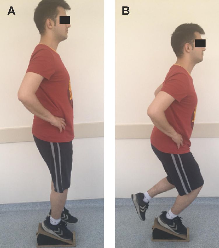
the previous 3 months and who were inactive categories The starting position was standing (trunk upright) on the
according to International Physical Activity Questionnaire- 25˚ decline board with the entire body weight on the domi-
Short Form. Exclusion criteria were as follows: history of nant leg. From that position, the knee was slowly flexed
metabolic, cardiovascular, hormonal, respiratory diseases, to 70˚ (Figure 2). To return to the starting position, the
or musculoskeletal disorders (e.g., patellofemoral pain syn- nondominant leg was used. Concentric quadriceps activity
drome, knee surgeries, and recent muscle strains). was avoided as much as possible.
A total of 60 male participants were randomly divided
into three groups: concentric exercise group (CE; n = 20), Control group
eccentric exercise group (EE; n = 20) and control group The control group was not given any exercise.
(CG; n = 20). In CE, one participant was excluded
because of incomplete data. In the EE, seven participants Participants in all groups were asked not to partici-
were excluded because of not attending exercise sessions pate in sports, recreation, and exercise activities for eight
regularly (n = 3), did not participate in the last evaluation weeks period.
(n = 2), inability to measure the thickness of the quadriceps
muscle (n = 2). In the CG, four participants were excluded Data collection
because of the inability to measure the thickness of the All participants were evaluated by the same researcher, who
quadriceps muscle (n = 2) and incomplete data (n = 2). was blinded to the groups at baseline and the end of the
The final study sample consisted of 48 participants, with 8th week.
19 in CE, 13 in EE and 16 in CG.
Ultrasound assessment
Intervention The ultrasonographic examinations were performed using
The participants in the concentric and eccentric groups a 7–13 MHz linear probe (Logiq P5, GE Medical Systems,
arrived at the clinic in the morning (08:30 to 11:30) and Wauwatosa, WI, USA) by a physician who was blinded to
cycled on an ergometer for five minutes at an intensity that the groups. Ultrasonographic muscle thickness measure-
resulted in a heart rate of around 130 beats per minute. ments were taken bilaterally (dominant/non-dominant).
Later, the participants completed eccentric and concen- All thickness measurements were repeated three times for
tric squats described by Jonsson and Alfredson (2005). each site, and the average values of the three successive
The exercise program consisted of three sets of ten repeti- measurements for each region were recorded. Measure-
tions each, performed seven days a week, for eight weeks ments were performed according to recommendations of
under supervision. The rest interval between sets was two the European Society of Musculoskeletal Radiology (Beggs
minutes. The intensity of exercise was standardized using et al., 2016).
the Rating of Perceived Exertion scale 6–20. After each
exercise set, participants were asked to rate their exertion Rectus femoris and vastus lateralis thickness
on the scale during the exercise, taking into account feel- Participants were lying in a supine position with their
ings of physical stress and fatigue (“How hard you feel knee fully extended and toes pointing to the ceiling. The
your body has worked?”). The exercises were aimed to be transducer was placed on the thigh on a longitudinal plane.
performed at a moderate intensity (12–14), “somewhat Images were taken at the level of the mid-thigh (mid-point
2N. Büker et al. Acta Gymnica, 2021, 51, e2021.015
Figure 1 Starting (A) and end (B) position for concentric quad- Figure 2 Starting (A) and end (B) position for eccentric quadri-
riceps exercise ceps exercise
of the distance from the lateral condyle of the femur to the Statistical analysis
central palpable point of the greater trochanter) at baseline Obtained data were analyzed using SPSS Statistics (Version
and after the exercise program with the application of the 21; IBM, Armonk, NY, USA). Continuous variables were
same standardized procedure. presented as mean ± standard deviation, maximum and
minimum and categorical variable values were presented
Quadriceps and patellar tendon thickness
as absolute frequency and percentages. The conformity of
Participants were lying in a supine position with their knee
flexed at ~30°. Measurements were performed with a lon- continuous variables with normal distribution was evalu-
gitudinal probe position at midpoints of the quadriceps ated using the Shapiro-Wilk test. For pairwise comparisons,
(between the proximal musculotendinous part and the if parametric test conditions were satisfied paired samples
most proximal part of the patellar insertion), and patellar t-test; and if parametric test conditions were not satisfied
tendon (between the distal pole of the patella and the most Wilcoxon signed-rank test was used (dominant and non-
proximal part of the tibial tubercle insertion of the tendon). dominant extremity rectus femoris thickness of EE group,
dominant extremity vastus lateralis thickness of CE group,
Six-Minute Walk Test Six-Minute Walk Test score of all groups and a vertical jump
Participants were sat at rest in a chair for at least 10 minutes
score of CE and CG groups were not distributed normally).
before the test. The walking course was 30 m in length. The
test was carried out following the American Thoracic Soci- The percentage of individual changes was calculated with
ety Guideline (ATS Committee on Proficiency Standards the following formula: [(Post – Pre)/Pre]*100. To compare
for Clinical Pulmonary Function Laboratories, 2002). percentage changes among groups, we used analysis of
Walking distance was recorded in meters. variance (ANOVA; post-hoc: Tukey test) when parametric
test conditions were satisfied (dominant extremity rectus
Vertical jump femoris thickness, non-dominant extremity patellar tendon
The difference in distance between participant’s standing thickness were distributed normally) and Kruskal-Wallis
reach (the point of the fingertips when the participant variance analysis (post-hoc: Mann-Whitney U test with
reaches upward with the hand closest to the wall with their
Bonferroni correction) when parametric test conditions
feet flat on the ground) and the height to which he can
were not satisfied. Effect size estimates were calculated as
jump (the participant jumps vertically as high as possible,
keeping away from the wall, using both their arms and legs follows: Cohen’s d (t/√N) for parametric test conditions, r
to help lift the body upward) and touch were determined. coefficient (Z/√N) for nonparametric test conditions and
The test was performed three times and the best of three eta-squared (η2) for ANOVA. A p-value ≤ .05 was consid-
attempts is recorded in centimeters (Petschnig et al., 1998). ered statistically significant.
3N. Büker et al. Acta Gymnica, 2021, 51, e2021.015
Results post hoc analysis revealed the rectus femoris in CG signifi-
Forty-eight males with age 23.11 ± 0.87 years in CE and cantly lower than in the CE (p = .046, d = .649) and EE
23.23 ± 1.16 years in EE and 22.81 ± 0.65 years in CG (p = .006, d = .539) and the vastus lateralis in the EE was
group participated in the study. Seventeen (89.5%) sub- significantly higher than in the CG (p = .018, r = .710).
jects in the CE group, 10 (76.9%) in the EE group, and 16 At the same time, nondominant side vastus lateralis thick-
(100%) in the CG group presented right-leg dominance. ness were significantly lower in CG than EE (p = .032,
Descriptive data of participants were shown in Table 1. r = .450; Table 2).
There was no difference between the groups at baseline, There was a statistically significant increase in the Six-
except dominant extremity quadriceps tendon thickness Minute Walk Test (CE: p = .010, r = .594; EE: p = .012,
(p = .029) and Six-Minute Walk Test (p = .035). Post hoc
analysis revealed the quadriceps tendon thickness in CG
Table 1 Descriptive data of participants at baseline
significantly higher than in the CE (p = .052) and the Six-
Minute Walk Test score of the CE significantly lower than Concentric Eccentric Controls
exercise (n = 19) exercise (n = 13) (n = 16)
in the EE (p = .044).
Age (years)
Dominant side rectus femoris (CE: p = .002, d = .919;
M ± SD 23.11 ± 0.87 23.23 ± 1.16 22.81 ± 0.65
EE: p = .006, r = 0.618), vastus lateralis (CE: p = .023,
Range 22–25 21–25 22–24
r = 0.552; EE: p < .001, d = 1.072) and quadriceps ten- Body mass index (kg/m2)
don thickness (CE: p = .044, d = .548; EE: p = .046, M ± SD 22.56 ± 3.86 22.09 ± 2.13 23.82 ± 2.29
d = .477) was increased significantly after eight weeks in Range 16.76–30.12 17.75–24.77 21.26–30.61
the CE and EE groups (Figure 3). Dominant side rectus Dominant lower extremity (n (%))
femoris (p = .008, η2 = .191) and vastus lateralis (p = .021, Right 17 (89.5) 10 (76.9) 16 (100.0)
r = .157) differed significantly among the three groups; Left 2 (10.5) 3 (23.1) 0 (0.0)
Table 2 Ultrasonographic evaluation of muscle thickness
Variable Pre Post Intra-group p Effect size Δ change (%) Inter-group p
Dominant extremity
Rectus femoris (cm) .0081–3,2–3
CE 1.569 ± 0.308 1.723 ± 0.323 .002 0.919 12.4 ± 12.7
EE 1.610 ± 0.461 1.800 ± 0.313 .006 0.618 16.0 ± 19.0
CG 1.840 ± 0.288 1.817 ± 0.274 .404 0.251 –1.1 ± 4.7
Patellar tendon (cm) .511
CE 0.325 ± 0.052 0.340 ± 0.051 .266 0.279 6.3 ± 18.45
EE 0.350 ± 0.051 0.359 ± 0.042 .452 0.172 3.9 ± 15.6
CG 0.343 ± 0.030 0.344 ± 0.026 .901 0.037 0.7 ± 8.7
Vastus lateralis (cm) .0212–3
CE 1.938 ± 0.478 2.055 ± 0.389 .023 0.552 8.9 ± 19.3
EE 1.948 ± 0.441 2.162 ± 0.400 < .001 1.072 12.2 ± 10.5
CG 2.258 ± 0.289 2.212 ± 0.287 .139 0.460 –1.9 ± 4.5
Quadriceps tendon (cm) .621
CE 0.417 ± 0.058 0.455 ± 0.060 .044 0.548 12.2 ± 22.7
EE 0.439 ± 0.055 0.472 ± 0.045 .046 0.477 9.4 ± 18.5
CG 0.450 ± 0.040 0.468 ± 0.044 .295 0.333 4.9 ± 13.1
Non-dominant extremity
Rectus femoris (cm) .191
CE 1.597 ± 0.359 1.578 ± 0.322 .774 0.071 0.4 ± 17.2
EE 1.480 ± 0.348 1.597 ± 0.342 .381 0.388 10.2 ± 20.8
CG 1.771 ± 0.354 1.752 ± 0.309 .794 0.077 0.2 ± 14.2
Patellar tendon (cm) .523
CE 0.322 ± 0.047 0.338 ± 0.044 .127 0.439 5.9 ± 12.3
EE 0.349 ± 0.065 0.344 ± 0.028 .715 0.085 0.9 ± 15.3
CG 0.338 ± 0.027 0.347 ± 0.030 .080 0.558 2.5 ± 4.6
Vastus lateralis (cm) .0322–3
CE 1.858 ± 0.480 1.923 ± 0.391 .214 0.324 5.9 ± 15.0
EE 1.896 ± 0.415 2.021 ± 0.299 .052 0.464 9.2 ± 17.1
CG 2.230 ± 0.316 2.156 ± 0.287 .087 0.543 –3.0 ± 6.1
Quadriceps tendon (cm) .887
CE 0.422 ± 0.044 0.436 ± 0.049 .359 0.254 3.980 ± 13.452
EE 0.424 ± 0.066 0.440 ± 0.035 .263 0.265 5.976 ± 17.286
CG 0.418 ± 0.042 0.439 ± 0.039 .248 0.352 6.069 ± 14.377
Note. CE = concentric exercise; EE = eccentric exercise; CG = controls. Statistically significant differences in post-hoc tests between groups: 1–3CE vs. CG; 2–3EE vs. CG.
4N. Büker et al. Acta Gymnica, 2021, 51, e2021.015
r = .699) and vertical jump (CE group: p = .001, r = .792; the EE (p = .002, r = .650) and CE (p < .001, r = .640;
EE group: p = .002, d = 1.056) after eight weeks in the CE Table 3).
and EE groups (Figure 4). Six-Minute Walk Test differed
significantly among the three groups (p = .047, r = .180); Discussion
post hoc analysis revealed the Six-Minute Walk Test score in
In this study, we aimed to compare the effect of eccen-
the EE was significantly higher than in the CG (p = .025, tric and concentric squat exercise in terms of quadriceps
r = .470). Vertical jump differed significantly among the thickness, and lower extremity performance during jump-
groups (p < .001, r = .289); post hoc analysis revealed the ing and walking in healthy sedentary young males. Both
vertical jump score in the CG significantly lower than in eccentric and concentric squats increased the thickness of
Figure 3 Comparison of ultrasonographic evaluation of rectus femoris, vastus lateralis and quadriceps tendon thickness on the domi-
nant side
RECTUS FEMORIS VASTUS LATERALIS QUADRICEPS TENDON
2.5 3 0.6
2.5 0.5
2
Thickness (cm)
Thickness (cm)
Thickness (cm)
2 0.4
1.5
1.5 0.3
1
1 0.2
0.5 0.5 0.1
0 0 0
CE EE CG CE EE CG CE EE CG
Pre Post
Note. CE = concentric exercise; EE = eccentric exercise; CG = controls.
Figure 4 Comparison of lower extremity performance measurements
SIX-MINUTE WALK TEST VERTICAL JUMP
800 50
700
40
600
Jump height (cm)
Distance (m)
500 30
400
300 20
200
10
100
0 0
CE EE CG CE EE CG
Pre Post
Note. CE = concentric exercise; EE = eccentric exercise; CG = controls.
Table 3 Lower extremity performance measurements
Variable Pre Post Intra-group p Effect size Δ change (%) Inter-group p
Six-minute walk test (m) .0472-3
CE 530.00 ± 27.57 552.53 ± 26.86 .010 0.594 5.31 ± 5.77
EE 640.23 ± 31.18 657.38 ± 41.18 .012 0.699 3.81 ± 3.10
CG 533.75 ± 25.23 535.00 ± 23.84 .359 0.229 0.71 ± 3.10
Vertical jump (cm) < .0011–3,2–3
CE 32.16 ± 2.42 33.89 ± 2.43 .001 0.792 6.06 ± 5.72
EE 34.55 ± 3.94 35.33 ± 3.85 .002 1.056 4.54 ± 3.82
CG 34. 56 ± 1.75 35.25 ± 1.77 .234 0.297 0.07 ± 2.29
Note. CE = concentric exercise; EE = eccentric exercise; CG = controls. Statistically significant differences in post-hoc tests between groups: 1–3CE vs. CG; 2–3EE vs. CG.
5N. Büker et al. Acta Gymnica, 2021, 51, e2021.015
the dominant side rectus femoris, vastus lateralis and quad- can produce regional-specific effects on muscle growth)
riceps tendon, but the eccentric squat had a greater effect and uncontrolled movement tempo are limitations of our
compared to percentages of change between groups. Simi- study. Further studies can be conducted involving differ-
lar findings were also obtained in the Six-minute Walk Test ent resistance training models in which movement tempo
and vertical jump score outcomes. is controlled and multiple sampling sites are evaluated. In
Roig et al. (2009) concluded that eccentric training addition, the cross-sectional area of the muscle can be mea-
is superior to concentric training in terms of promoting sured by imaging methods such as magnetic resonance or
strength, but this is highly specific to the contraction type computed tomography. Muscle strength can also be deter-
and velocity in healthy adults. However, most of the studies mined with isokinetic devices or a dynamometer.
included in this meta-analysis involved isokinetic resistance
training. In isotonic resistance exercise, this situation has
been examined using varying movement tempos (Pryor et Conclusions
al., 2011; Schoenfeld et al., 2015; Wilk et al., 2018). It is Eccentric and concentric training both benefits muscle
claimed that slow isotonic resistance training is more effec- hypertrophy and lower extremity performance. However,
tive at increasing strength than fast isotonic resistance train- it also appears that eccentric training probably offers a
ing (Burd et al., 2012; Farthing et al., 2003; Pereira et al., small advantage over concentric training. According to our
2016; Roig et al., 2009). In a recent meta-analysis, eccen- results, muscle thickness and functional performance can
tric muscle training resulted in a greater effect compared be increased in healthy young adults with simple, easy, and
to concentric training, but the results did not increase to well-designed exercises that do not require special equip-
statistical significance (Schoenfeld et al., 2017). Similarly, ment and space.
we observed significant thickening in the rectus femoris
and vastus lateralis muscles in both the EE and CE groups.
However, in terms of effect size, rectus femoris thickness Acknowledgments
was greater in the CE group and vastus lateralis thickness We would like to thank Assist. Prof. Hande Senol for her
in the EE group. Studies show that particular eccentric statistical support.
and concentric cadence values can cause different adaptive
responses in the muscle. Training at different movement
tempo can provide different gains in terms of strength Conflict of interest
development and muscle hypertrophy. For this reason, The authors report no conflict of interest.
our failure to control the pace of movement may affect the
results. However, we did not control the movement tempo
in our study, which may affect the results.
References
ATS Committee on Proficiency Standards for Clinical Pulmonary Function Labora-
Functional performance tests are considered an alter- tories (2002). ATS statement: Guidelines for the six-minute walk test. American
native method for evaluating muscle strength due to their Journal of Respiratory and Critical Care Medicine, 166(1), 111–117. https://doi.
org/10.1164/ajrccm.166.1.at1102
simplicity and being more time-efficient and cost-effective Beggs, I., Bianchi, S., Bueno, A., Cohen, M., Court-Payen, J., Grainger, A., Klauser,
than isometric or isokinetic instrumentation (Kollock et al., A., Kainberger, F., Martinoli, C., McNally, E., O’Connor, P. J., Peetrons, P., Reij-
2015). Traditional resistance training is known to increase nierse, M., Remplik, P., & Silvestri, E. (2016). Musculoskeletal ultrasound tech-
nical guidelines: V. knee. https://essr.org/content-essr/uploads/2016/10/knee.pdf
aerobic capacity and muscle strength (Moro et al., 2020; Blazevich, A. J., Cannavan, D., Coleman, D. R., & Horne, S. (2007). Influence of
Sentija et al., 2009). Resistance training can increase aero- concentric and eccentric resistance training on architectural adaptation in
human quadriceps muscles. Journal of Applied Physiology, 103(5), 1565–1575.
bic capacity in young individuals by providing improve- https://doi.org/10.1152/japplphysiol.00578.2007
ments in capillary-to-fiber ratio and mitochondrial enzyme Bordoni, B., & Varacallo, M. (2021). Anatomy, bony pelvis and lower limb, thigh
activity (Ozaki et al., 2013). In our study, an increase in quadriceps muscle. https://www.ncbi.nlm.nih.gov/books/NBK513334/
Burd, N. A., Andrews, R. J., West, D. W., Little, J. P., Cochran, A. J., Hector, A. J.,
aerobic capacity was achieved in both exercise groups. The Cashaback, J. G., Gibala, M. J., Potvin, J. R., Baker, S. K., & Phillips, S. M. (2012).
effect size was moderate. However, to our knowledge, there Muscle time under tension during resistance exercise stimulates differential
muscle protein sub-fractional synthetic responses in men. Journal of Physiol-
is no study comparing the effects of eccentric and con- ogy, 590(2), 351–362. https://doi.org/10.1113/jphysiol.2011.221200
centric training on aerobic capacity. However, numerous Cadore, E. L., González-Izal, M., Pallarés, J. G., Rodriguez-Falces, J., Häkkinen, K.,
studies have investigated the effects of different knee exten- Kraemer, W. J., Pinto, R. S., & Izquierdo, M. (2014). Muscle conduction velocity,
strength, neural activity, and morphological changes after eccentric and con-
sor training on knee strength, but the results are highly centric training. Scandinavian Journal of Medicine & Science in Sports, 24(5),
variable. However, although many studies have reported a e343–e352. https://doi.org/10.1111/sms.12186
Dionisio, V. C., Azevedo, B. M. S., & Siqueira, D. A. (2013). Horizontal and
greater increase in muscle strength after eccentric training declined squats in healthy individuals: A study of kinematic and muscle pat-
(Farthing & Chilibeck, 2003; Franchi et al., 2014; Reeves terns. International Scholarly Research Notices, 2013, Article 169808. https://
et al., 2009), some studies have reported that concentric doi.org/10.1155/2013/169808
Escamilla, R. F. (2001). Knee biomechanics of the dynamic squat exer-
training has the same effect (Blazevich et al., 2007; Farthing cise. Medicine & Science in Sports & Exercise, 33(1), 127–141. https://doi.
& Chilibeck, 2003; Tomberlin et al., 1991). Similarly, org/10.1097/00005768-200101000-00020
Farthing, J. P., & Chilibeck, P. D. (2003). The effects of eccentric and concentric
according to the results of our study, both concentric and training at different velocities on muscle hypertrophy. European Journal of
eccentric exercises were beneficial in terms of increasing Applied Physiology, 89(6), 578–586. https://doi.org/10.1007/s00421-003-0842-2
muscle strength and had a large effect size. Franchi, M. V., Atherton, P. J., Reeves, N. D., Flück, M., Williams, J., Mitchell, W. K.,
Selby, A., Beltran Valls, R. M., & Narici, M. V. (2014). Architectural, functional
Lack of multiple sampling sites along the length of and molecular responses to concentric and eccentric loading in human skeletal
the quadriceps muscle (eccentric and concentric actions muscle. Acta Physiologica, 210(3), 642–654. https://doi.org/10.1111/apha.12225
6N. Büker et al. Acta Gymnica, 2021, 51, e2021.015
Franchi, M. V., Wilkinson, D. J., Quinlan, J. I., Mitchell, W. K., Lund, J. N., Williams, Orthopaedic and Sports Physical Therapy, 28(1), 23–31. https://doi.org/10.2519/
J. P., Reeves, N. D., Smith, K., Atherton, P. J., & Narici, M. V. (2015). Early struc- jospt.1998.28.1.23
tural remodeling and deuterium oxide-derived protein metabolic responses Petterson, S. C., Barrance, P., Buchanan, T., Binder-Macleod, S., & Snyder-Mack-
to eccentric and concentric loading in human skeletal muscle. Physiological ler, L. (2008). Mechanisms underlying quadriceps weakness in knee osteoar-
Reports, 3(11), Article e12593. https://doi.org/10.14814/phy2.12593 thritis. Medicine & Science in Sports & Exercise, 40(3), 422–427. https://doi.
Giles, L. S., Webster, K. E., McClelland, J. A., & Cook, J. (2013). Does quadriceps org/10.1249/MSS.0b013e31815ef285
atrophy exist in individuals with patellofemoral pain? A systematic literature Pryor, R. R., Sforzo, G. A., & King, D. L. (2011). Optimizing power output by vary-
review with meta-analysis. Journal of Orthopaedic and Sports Physical Therapy, ing repetition tempo. Journal of Strength and Conditioning Research, 25(11),
43(11), 766–776. https://doi.org/10.2519/jospt.2013.4833 3029–3034. https://doi.org/10.1519/JSC.0b013e31820f50cb
Gür, H., Cakin, N., Akova, B., Okay, E., & Küçükoğlu, S. (2002). Concentric ver- Reeves, N. D., Maganaris, C. N., Longo, S., & Narici, M. V. (2009). Differential
sus combined concentric-eccentric isokinetic training: Effects on functional adaptations to eccentric versus conventional resistance training in older
capacity and symptoms in patients with osteoarthrosis of the knee. Archives humans. Experimental Physiology, 94(7), 825–833. https://doi.org/10.1113/
of Physical Medicine and Rehabilitation, 83(3), 308–316. https://doi.org/10.1053/ expphysiol.2009.046599
apmr.2002.30620 Roig, M., O’Brien, K., Kirk, G., Murray, R., McKinnon, P., Shadgan, B., & Reid, W. D.
Jonsson, P., & Alfredson, H. (2005). Superior results with eccentric compared to (2009). The effects of eccentric versus concentric resistance training on muscle
concentric quadriceps training in patients with jumper’s knee: A prospective strength and mass in healthy adults: A systematic review with meta-analysis.
randomised study. British Journal of Sports Medicine, 39(11), 847–850. https:// British Journal of Sports Medicine, 43(8), 556–568. https://doi.org/10.1136/
bjsm.2008.051417
doi.org/10.1136/bjsm.2005.018630
Santos, R., Valamatos, M. J., Mil-Homens, P., & Armada-da-Silva, P. (2018). Mus-
Kollock, R., Van Lunen, B. L., Ringleb, S. I., & Oñate, J. A. (2015). Measures of
cle thickness and echo-intensity changes of the quadriceps femoris muscle
functional performance and their association with hip and thigh strength. Jour-
during a strength training program. Radiography, 24(4), e75–e84. https://doi.
nal of Athletic Training, 50(1), 14–22. https://doi.org/10.4085/1062-6050-49.3.49
org/10.1016/j.radi.2018.03.010
Kooiker, L., Van De Port, I. G., Weir, A., & Moen, M. H. (2014). Effects of physi-
Schoenfeld, B. J., Ogborn, D. I., & Krieger, J. W. (2015). Effect of repetition dura-
cal therapist-guided quadriceps-strengthening exercises for the treatment of
tion during resistance training on muscle hypertrophy: A systematic review
patellofemoral pain syndrome: A systematic review. Journal of Orthopaedic and
and meta-analysis. Sports Medicine, 45(4), 577–585. https://doi.org/10.1007/
Sports Physical Therapy, 44(6), 391–402. https://doi.org/10.2519/jospt.2014.4127 s40279-015-0304-0
Moro, T., Marcolin, G., Bianco, A., Bolzetta, F., Berton, L., Sergi, G., & Paoli, A. Schoenfeld, B. J., Ogborn, D. I., Vigotsky, A. D., Franchi, M. V., & Krieger, J. W.
(2020). Effects of 6 weeks of traditional resistance training or high intensity (2017). Hypertrophic effects of concentric vs. eccentric muscle actions: A
interval resistance training on body composition, aerobic power and strength systematic review and meta-analysis. Journal of Strength and Conditioning
in healthy young subjects: A randomized parallel trial. International Journal Research, 31(9), 2599–2608. https://doi.org/10.1519/JSC.0000000000001983
of Environmental Research and Public Health, 17(11), Article 4093. https://doi. Sentija, D., Marsić, T., & Dizdar, D. (2009). The effects of strength training on
org/10.3390/ijerph17114093 some parameters of aerobic and anaerobic endurance. Collegium Antropologi-
Ozaki, H., Loenneke, J. P., Thiebaud, R. S., & Abe, T. (2013). Resistance train- cum, 33(1), 111–116.
ing induced increase in VO2max in young and older subjects. European Timmins, R. G., Ruddy, J. D., Presland, J., Maniar, N., Shield, A. J., Williams, M.
Review of Aging and Physical Activity, 10, 107–116. https://doi.org/10.1007/ D., & Opar, D. A. (2016). Architectural changes of the biceps femoris long head
s11556-013-0120-1 after concentric or eccentric training. Medicine & Science in Sports & Exercise,
Pereira, P. E. A., Motoyama, Y. L., Esteves, G. J., Quinelato, W. C., Botter, L., Tanaka, 48(3), 499–508. https://doi.org/10.1249/MSS.0000000000000795
K. H., & Azevedo, P. (2016). Resistance training with slow speed of movement Tomberlin, J. P., Basford, J. R., Schwen, E. E., Orte, P. A., Scott, S. C., Laughman,
is better for hypertrophy and muscle strength gains than fast speed of move- R. K., & Ilstrup, D. M. (1991). Comparative study of isokinetic eccentric and
ment. International Journal of Applied Exercise Physiology, 5(2), 37–43. https:// concentric quadriceps training. Journal of Orthopaedic and Sports Physical
doi.org/10.30472/IJAEP.V5I2.51 Therapy, 14(1), 31–36. https://doi.org/10.2519/jospt.1991.14.1.31
Petschnig, R., Baron, R., & Albrecht, M. (1998). The relationship between iso- Wilk, M., Golas, A., Stastny, P., Nawrocka, M., Krzysztofik, M., & Zajac, A. (2018).
kinetic quadriceps strength test and hop tests for distance and one-legged ver- Does tempo of resistance exercise impact training volume? Journal of Human
tical jump test following anterior cruciate ligament reconstruction. Journal of Kinetics, 62, 241–250. https://doi.org/10.2478/hukin-2018-0034
7You can also read