Comparison of Serological Tests in Cattle and Ovine Brucellosis; An Abattoir Study in Algeria - WVJ
←
→
Page content transcription
If your browser does not render page correctly, please read the page content below
2020, Scienceline Publication
World’sVeterinary Journal
World Vet J,10(3): 457-464, September 25, 2020                                                                                                     ISSN 2322-4568

                                                                                                                 DOI: https://dx.doi.org/10.36380/scil.2020.wvj57

Comparison of Serological Tests in Cattle and Ovine
Brucellosis; An Abattoir Study in Algeria
Maamar Khames1*, Amaia Zúñiga-Ripa2, Sagrario Pérez Gómez3, Mustapha Oumouna1 and Ignacio Moriyón2
1
  Department of Biology, University of Medea, Medea, Algeria
2
  Institute of Tropical Health and Department of Microbiology and Parasitology, Faculty of Medicine, University of Navarra, Pamplona, Spain
3
  Laboratorio de Calidad Agroalimentaria, Navarra, Spain
*Corresponding author`s Email: drkhames86@gmail.com;                 : 0000-0001-6820-0496

                                                                                                                                                 Accepted: 21 Sept 2020
                                                                                                                                                 Received: 03 Aug 2020
                                                                                                                                                 pii: S232245682000057-10
                                                                                                                                                                            ORGINAL ARTICLE
ABSTRACT
Despite the paucity of data, brucellosis is considered as a major problem in Algeria. The aim of present study was to
assess the presence of bovine and ovine brucellosis in the areas close to the capital city (Algiers) where its
vaccination is not implemented. A total of 402 cattle and 203 ovine sera were collected from two slaughterhouses,
and examined by the Rose Bengal Test (RBT). Positive samples were then tested by Complement Fixation Test
(CFT) and Hypertonic Double Gel Diffusion (DDG) with a smooth lipopolysaccharide, and the extract of native
hapten was also tested by Indirect Enzyme Linked Immuno Sorbent Assay (iELISAs) with smooth
lipopolysaccharide and polyclonal or protein G conjugates. Twenty-four bovine sera (5.97%) were RBT positive. Of
these, 23 were positive in CFT, DDG, and 16 samples were also positive in iELISA when the assay was adjusted to
100% specificity. Only two ovine sera were RBT positive; one was CFT and DDG positive, and the other one had a
CFT-titer of 1/4, and was DDG negative. This preliminary study confirmed that bovine brucellosis is a major
problem in Algeria, and indicated that some field studies are needed to determine the prevalence of Brucellosis in
Algeria urgently. Similarly, other studies are necessary in areas with dominance of ovine breeding system. Further
studies in the areas with a dominance of ovine breeding system are necessary. The results of this work showed that
simple tests like RBT and DDG are not outperformed by CFT or iELISA for assessing the apparent prevalence of
brucellosis in the absence of vaccination. Finally, isolation and typing of the involved Brucella species are also
necessary in order to have a complete epidemiological picture of brucellosis in Algeria.
Key words:Abattoirs, Algeria, Brucellosis, Cattle, Serology, Sheep, Prevalence

INTRODUCTION

Bacteria of the genus Brucella which causes brucellosis is a highly contagious zoonosis affecting primarily domestic
livestock and a variety of wild-life species. Brucellosis in animals is characterized by a reproductive failure, and the
human form of this disease is a grave and debilitating condition requiring prolonged and costly antibiotic treatments.
Thus, this disease presents a serious threat to the animal industry and public health (McDermott et al., 2013). Brucellosis
is considered as one of the most widespread zoonosis in the world (Hull and Schumaker, 2018) and it has a great impact
on emerging economies and resources of poor countries (Grace et al., 2012). Although it has been eradicated in
ruminants in most European countries, the USA, Canada, Australia and a few other countries, brucellosis is still a
endemic disease in many parts of the world, especially in North and East Africa, the Middle East, South and Central Asia
and Central and South America (Corbel, 2006). Moreover, intensification of breeding caused by raising food demand
increased the risk of spread of this and other zoonosis (Jones et al., 2013).
      Humans contract the disease from animals or their products, but they are not considered as a source of contagion.
Thus, under most circumstances, human brucellosis is the best indicator of the existence of the disease in animals.
Regarding, the high number of cases for human brucellosis reported in Algeria is indicative of the important impact of
this zoonosis in the country, 819 human cases were recorded in first four months of 2016 (Robert, 2016). The
intervention against brucellosis includes implementation of dairy food sanitation, specific measures to protect risk groups
(farmers, abattoir workers and veterinarians), and control and eradication of the disease in livestock. In Algeria, despite
the control program initiated in 1970 and strengthened in 1995 which was based principally on a test-and-slaughter
strategy, bovine brucellosis is a serious problem in dairy herds in all likelihood as suggested by the consistent presence
of clinical symptoms compared with the disease and occasional serological testing. About 30 years ago, Benelmouffok et
al. (1984) summarized the previous evidence indicating high (up to 25%) apparent seroprevalence as determined by the
Standard Serum Agglutination Test (SAT). More recently, apparent herd seroprevalence of 26.3% using Rose Bengal
Test (RBT) and 15.7% by complement fixation test (CFT) were reported for cattle in the Western area of Algeria (Tiaret)

                                                                             457
To cite this paper: Khames M, Zúñiga-Ripa A, Pérez Gómez S, Oumouna M and Moriyón I (2021). Comparison of Serological Tests in Cattle and Ovine Brucellosis; An
Abattoir Study in Algeria. World Vet. J., 10 (3):457-464. DOI: https://dx.doi.org/10.36380/scil.2020.wvj57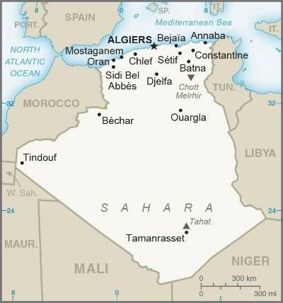
(Aggad and Boukraa, 2006). Although these reports suggest a high prevalence of bovine brucellosis, the paucity of data
and their long-time span clearly indicated the need for additional studies. Similarly, there is a paucity of works on
brucellosis in small ruminants (Hamdi-Chérif et al., 1999; Meskoud-taibi and Benzadi, 2009). However, while cattle are
usually infected by B. abortus, sheep and goats are infected by B. melitensis, the Brucella species are considered to be
more infectious for human (O’Callaghan, 2020). Accordingly, a first goal of the research described here was to obtain
additional data on the status of brucellosis in Algeria. To this end, a serological study was carried out on two
slaughterhouses that process animals coming from different areas of the country.
Because of the absence of pathognomonic symptoms, the diagnosis of brucellosis requires laboratory tests.
Bacteriological culture is cumbersome and dangerous, but serological tests are routinely used. However, there is a
bewildering list of serological tests (Ducrotoy et al., 2016), and most new developments are focused on tests suitable for
surveillance of large numbers of animals in Brucella-free areas that require conditions not met in resource-limited
settings (McGiven, 2013; Ducrotoy et al., 2015). Thus, a second aim of the present work was to compare apparent
prevalence figures obtained with simple RBT, more sophisticated and highly-sensitive tests like indirect ELISA
(iELISA) with tests of higher specificity that also differ in the degree of sophistication (CFT and double gel diffusion
(DDG) with smooth-lipopolysaccharide (S-LPS) and native hapten polysaccharide (NH).
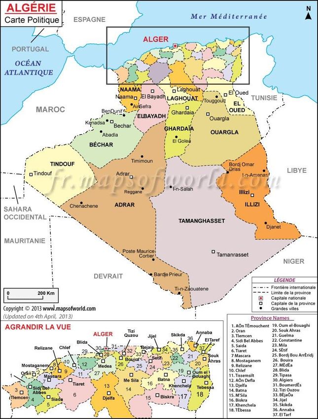
Figure 1A.General map of Algeria (http://www.state.gov/p/nea/ci/ag/)
458
To cite this paper: Khames M, Zúñiga-Ripa A, Pérez Gómez S, Oumouna M and Moriyón I (2021). Comparison of Serological Tests in Cattle and Ovine Brucellosis; An
Abattoir Study in Algeria. World Vet. J., 10 (3):457-464. DOI: https://dx.doi.org/10.36380/scil.2020.wvj57Figure 1B.The Wilayates close to Algiers (http://www.algerieprofonde.net/algerie/cartes-dalgerie/). The caption shows
the total number of cattle tested from each place and the prevalence percent:Ain-Defla, n: 74 (0.00%); Alger (Algiers), n:
86 (1.16%); Blida, n: 58 (13.80%); Bouira, n: 151 (7.28%); Boumerdes, n: 27 (7.40%); Chlef and Relizane, n: 5
(20.00%); Médéa, n: 1 (100.00%).
459
To cite this paper: Khames M, Zúñiga-Ripa A, Pérez Gómez S, Oumouna M and Moriyón I (2021). Comparison of Serological Tests in Cattle and Ovine Brucellosis; An
Abattoir Study in Algeria. World Vet. J., 10 (3):457-464. DOI: https://dx.doi.org/10.36380/scil.2020.wvj57MATERIALSAND METHODS
Ethical approval
Samples were collected as per the standard sample collection procedure without any stress or harm to the animals,
according to the rules of the Veterinary School of Algiers, Algeria.
Study site, period and sample size
The study was carried out in two slaughterhouses in Algiers, Algeria. Originally in the outskirts, the Rouiba
slaughterhouse is now in the urban area. This slaughterhouse is working each day of the week, and animals from
different areas are processed. So, it made it possible to trace the geographical origin of the cattle. El-Harrach
slaughterhouse (about 2600 m2) is located in the urban area, and it was not possible to trace the origin of the animals due
to the work’s conditions there. For the Rouiba slaughterhouse, the study included the entire cattle population (402heads:
264females and138males) of every breed (local, cross-breed and montbeliarde) and age sacrificed from 26 February to1
April 2012, and63 non-vaccinated sheep (19 females and 44 males) were sacrificed from1to 28 February 2012; for El-
Harrach slaughterhouse, 140 non-vaccinated sheep of different breeds like ouled djellal and el rembi, and different age
categories (86 females and 54 males) were sacrificedfrom1 to 31 March 2012.
Ante mortem inspection
An inspection to determine the sex, age and identification status on both ears was performed, and three types of
cattle were defined; imported (Montbeliarde cattle; foreign identification tag), local (small, uniform colored dress - black
or brown - no tag), Magpie black or red magpie crossbreed cattle (with or without tag (deleted or removed)).
Sample collection
Five milliliters of blood were collected from the jugular or the caudal vein with Vacutainer-type tubes, or directly
at the moment of scarification, and all data (breed, age, sex and pregnancy) were recorded. The blood was let to clot at
the room temperature, the sera were collected by centrifugation, 3000 tours per minute for 15 minutes, and finally stored
at -20°C until being analyzed.
Serological tests
All cattle’s serum samples were analyzed by the RBT using the official Spanish antigen (kindly provided by Dr.
J.M. Blasco, Centro de Investigación en Tecnologia Agraria, Gobierno de Aragón, Spain) according to the standard
procedure (OIE, 2016). Briefly, after equilibrating the antigen suspension at the room temperature, an equal volume
(25µl) of antigen and serum were mixed on a white tile with a toothpick, and shaked by hands for 4 minutes. No matter
the intensity, definite clumping/agglutination was considered as a positive reaction. Sheep’s serum samples were
analyzed by the modified RBT (mRBT) (Blasco et al., 1994;Diaz-Aparicio et al., 1994;OIE, 2016). A volume of 25µl of
antigen and 75µl of serum were used. In all cases, RBT-positive serum samples were then retested for CFT which was
performed by the standard micro method technique (OIE, 2016)(a titer equal to 4 contained 20 international CFT units
per ml). In addition, RBT-positive sera(or MRBT for sheep) were analyzed by the DGD immunoprecipitation test with a
hot extract of B. melitensis16M rich in native hapten (NH)and Smooth lipopolysaccharides (S-LPS) in 10% NaCl
hypertonic gels (Diaz et al., 1981). Two antigen concentrations were used: 1 and 5mg/ml. Finally, an in-house iELISA
with polyclonal and protein G conjugates and smooth B. abortus S-LPS was performed as described before (Alonso-
Urmeneta et al., 1998). Plates included both a positive and a negative control, and the results were expressed as the
optical density (%OD) of the positive control. The iELISA and DDG antigens were supplied by the Brucellosis
Laboratory, Institute of Tropical Health, University of Navarra. Complement fixation test (CFT) was performed at the
laboratory for food quality, Government of Navarra, Navarra, Spain. The Laboratorio de Calidad Agroalimentaria,
Gobierno de Navarra, Villaba, Navarra, Spain.
Statistical analysis
The results were analyzed using the Chi square test (SPSS, version 19) and P values < 0.05 were considered
significant. Confidence intervals were calculated according to Toma et al. (1996)
RESULTS AND DISCUSSION
Cattle
Twenty-four samples of bovine sera were positive by RBT (Apparent prevalence: 5.97%, 95% confidence interval,
5.95 – 5.99). Of these, 23 were positive by CFT, DDG (i.e. apparent prevalence: 5.72% CI (5.7–5.74in all cases), and 16
samples by iELISA (i.e. apparent prevalence: 3.98% CI (3.96–4)). By CFT, 18 samples had titers equal to or higher
460
To cite this paper: Khames M, Zúñiga-Ripa A, Pérez Gómez S, Oumouna M and Moriyón I (2021). Comparison of Serological Tests in Cattle and Ovine Brucellosis; An
Abattoir Study in Algeria. World Vet. J., 10 (3):457-464. DOI: https://dx.doi.org/10.36380/scil.2020.wvj57than1/256; 2, 1/128; 1, 1/64; 1, 1/16 and 1, 1/4. By DDG with the antigen at 1mg/ml, 21 serum samples developed both
the LPS and NH precipitin lines, one only with the S-LPS precipitin line, one only with the NH precipitin line, and one
was negative. The serum that developed only the S-LPS precipitin line which reacted weakly in RBT had a CFT titer
equal to 1/4, and was positive in iELISA. The serum that was negative by DDG was also negative in the iELISA, but
positive in the RBT and the CFT. For the DDG, the results were not improved by increasing the antigen concentration to
5mg/ml.
It has been consistently observed that a positive result of immunoprecipitation tests with extracts rich in S-LPS and
NH correlated with a positive CFT (i.e. ≥1:4 (20 international units) or higher, so that the former can be substituted for
the latter in the classical diagnostics scheme that started with RBT screening. However, while CFT is technically
demanding, the DDG with these extracts represented a much simpler and repeatable test (Diaz et al., 1979;Jones et al.,
1980;Díaz et al., 1984;Diaz-Aparicio et al., 1993). Moreover, it has been observed that a positive result in the DDG test
correlates with the shedding of Brucella which makes this test particularly useful in difficult epidemiological situations
when identification of shedders becomes necessary to control brucellosis (Jones et al., 1980). The results presented here
confirmed the previous investigations that demonstrated that a combination of RBT and DDG can be implemented
without a loss of performance with respect to that obtained with more sophisticated CFT and iELISA (Diaz et al., 1979;
Jones et al., 1980; Diaz et al., 1981; Diaz et al., 1983; Muñoz et al., 2005). It has been argued that the
immunoprecipitation tests with S-LPS and NH are not sensitive enough to be useful in the diagnosis of brucellosis, and
that they are outperformed by immunoenzymatic tests with diagnostic sensitivity and specificity adjusted according to
the Youden or performance indexes (Nielsen, 2002). However, because brucellosis does not spread epizootically, early
detection requiring 100% diagnostic sensitivity is not as critical as in highly transmissible epizootic diseases. Moreover,
the diagnosis of brucellosis should be interpreted on a herd/flock rather than on an individual basis, and when the
objective is the culling of positive animals in eradication contexts, it is repeated frequently over time which facilitates the
detection of infected animals. Thus, 100 percent of DSe is not strictly necessary for brucellosis tests, and low DSp
generates unnecessary culling with the ensuing conflicts with farmers, needless quarantines, and trade and policy
problems (Ducrotoy et al., 2018). Quantitative tests such as iELISA are usually adjusted according to the Youden index
which maximizes the DSe/DSp balance. Yet, as shown here, when the DSp was adjusted to 100%, the DSe decreased
with regard to the simpler RBT or DDG. As these last two tests are robust, and can be performed with relatively little
equipment, they can be recommended for use in laboratories where manpower is not the limiting factor, and access to
automatized systems is not available. In fact, these immunoprecipitation tests have been used extensively and
successfully as confirmatory tests after a RBT screening in eradication campaigns in northern Spain (Asarta, 1989;
Blasco et al., 2016). Indeed, a problem of DGD is the availability of the NH and S-LPS extract. Although there are
simple extraction protocols (Díaz et al., 1984), the antigen is not presently commercially available.
In cattle, the close parallelism among the apparent prevalence figures obtained with the different tests is
remarkable. Compared to the CFT or iELISA tests, it has been shown that the immunoprecipitation test with NH is only
not affected by false positive reactions caused by bacteria cross-reacting with Brucella at the LPS level (Muñoz et al.,
2005). Taken together, this high specificity and the absence of S19 vaccination in the origin areas of the tested animals
support the conclusion that the apparent prevalence figures obtained with this test actually represent real infections by
either B. abortus or B. melitensis. The latter species was known to be a cause of infections in cattle in areas where they
are raised and kept with the small ruminants (Verger et al., 1989), and as serological tests cannot differentiate infections
by those two species (Ducrotoy et al., 2016), bacteriological studies are necessary for a full understanding of the
epidemiology of brucellosis in Algeria. Cattle identification system allowed us to trace back their origin, thereby, all
livestock markets were represented at the Rouiba abattoir (i.e. Bouira (Center), Médéa (Center), Blida (Center), Ain El-
Defla (West), Algiers (Center), Chlef and Relizane (West), Boumerdès (Center); supplemental figure S1), only one (Ain
El-Defla) did not yield any seropositive animal. This illustrated that cattle brucellosis is widespread in the country.
Concerning other epidemiological factors, table 1 summarizes the results broken down according to sex, age, breed
and other characteristics recorded. As can be seen, the proportion of seropositive individuals was much higher in females
than in males. The literature contained contradictory reports on the influence of sex as a risk factor in cattle brucellosis.
Whereas some authors reported that males are at a higher risk (Boukary et al., 2013), others found opposite results
(Kebede et al., 2008; Mohammed et al., 2011; Rahman et al., 2011; Olabode et al., 2012). However, it is known that the
management determined the rates of infection in males. Artificial insemination is extended in Algeria, and in the present
study, all138 males were slaughtered for meat when they were less than two years old, instead of using them as breeding
males which prevented them from getting infected from the females. Among the females, only one was less than 2 years
old. This bias in the sample reflected the management of the animals in the dairies of Algeria where old females are
removed from the farms when the productivity decreases. Similarly, most of the tested animals belonged to the
crossbreed category, and this also reflected the preference of the farmers for these animals because of the better milk
yields and comparative lower market prices. However, the data showed that there were no statistically significant
differences in apparent seroprevalence among this and the other two groups identified in the study.
461
To cite this paper: Khames M, Zúñiga-Ripa A, Pérez Gómez S, Oumouna M and Moriyón I (2021). Comparison of Serological Tests in Cattle and Ovine Brucellosis; An
Abattoir Study in Algeria. World Vet. J., 10 (3):457-464. DOI: https://dx.doi.org/10.36380/scil.2020.wvj57Out of 46 identified cattle samples, 10 (21.73%) were positive. Ten out of the 305 (3.27%) non-identified cattle and
4out of the 51(7.84%) cattle samples that presented are moved ear-tag were found positive (Table 1). Statistically, there
was a significant association (p5years 263 23 (8.74%) 22(8.36%) 22 (8.36%) 16(6.08%)
Local 33 1 (3.03%) 1(3.03%) 1(3.03%) 1(3.03%)
Breed Cross-breed 344 22 (6.39%) 21(6.10%) 21 (6.10%) 14 (4.07%)
Montbeliarde 25 1 (4.00%) 1(4.00%) 1 (4.00%) 1(4.00%)
Yes 46 10(21.73%) 10(21.73%) 10 (21.73%) 8(17.39%)
Identification
No 305 10 (3.27%) 10(3.27%) 10 (3.27%) 6(1.96%)
status
Ear tag removed 51 4 (7.84%) 3(5.88%) 3 (5.88%) 2(3.92%)
Pregnancy Pregnant 21 3 (14.28%) 2(9.52%) 2(9.52%) 2(9.52%)
(females) Non-pregnant 235 20 (8.51%) 20(8.51%) 20(8.51%) 14(5.95%)
N°: number; RBT: Rose Bengal Test; CF: complement fixation; DDG-NH: double gel diffusion-native hapten; iELISA: indirect Enzyme Linked
ImmunoSorbent Assay
Sheep
Concerning the ovine sera, two samples (0.98%) were positive by mRBT. By CFT, one sample had a titer of 1/4,
and the other one had a titer of 1/16. This last serum was found positive by DDG-NH, but only with antigen at 1mg/ml.
This was consistent with previous observations that the NH-S-LPS antigen concentration needed to be comparatively
reduced for optimal sensitivity in immunoprecipitation with sera from small ruminants (Diaz-Aparicio et al.,
1993).These two animals belonged to the larger sheep sample (El-Harrach abattoir).
The apparent seroprevalence found in sheep was lower than 3.08 % reported by Hamdi-Chérif et al.(1999) in the
east of Algeria (Setif and M’sila, 1999), or the 7.2% found by the National Institute of Veterinary Medicine of Algiers in
2007 (Meskoud-taibi and Benzadi, 2009), and several circumstances could explain the differences. First, these two
surveys which were conducted before the benefits of Rev 1 vaccination campaigns (started in the spring of 2006) could
be detectable. It was worth noting that those few positive results cannot be the result of post-vaccinal antibodies. None of
the sheep carried the green tag was used to label vaccinated animals in Algeria. An alternative explanation is that the
tested sheep came from a few intensive farms surrounding Algiers where the animals which were kept for fattening, and
then sold to the slaughterhouse which favored control. The two seropositive sexually mature rams introduced from an
infected breeding area, but this practice did not seem a significant risk. In cattle, as in sheep, males play no relevant role
in spreading the disease by direct sexual contact (Alton, 1990; Nielsen and Duncan, 1990), while artificial insemination
can be a risk, which is not practiced in Algeria. However, the survey in sheep revealed deficiencies in the
implementation of those measures (permanent tagging, movements of animals of unknown status) that are essential to
carry out brucellosis control and eradication programs. Finally, since the present study was carried on ovine brucellosis
462
To cite this paper: Khames M, Zúñiga-Ripa A, Pérez Gómez S, Oumouna M and Moriyón I (2021). Comparison of Serological Tests in Cattle and Ovine Brucellosis; An
Abattoir Study in Algeria. World Vet. J., 10 (3):457-464. DOI: https://dx.doi.org/10.36380/scil.2020.wvj57in a slaughterhouse in the capital city Algiers, the results did not reflect the epidemiological situation in Algeria, and
more studies in areas with dominance of ovine/caprine breeding are necessary to determine the origin of the human
disease.
CONCLUSIONS
The results of this survey confirmed the importance of cattle brucellosis in Algeria, and that a combination of Rose
Bengal test (RBT) and double gel diffusion in gel (DDG)RBT and DDG can be implemented instead of the more
sophisticated like complement fixation test (CFT) and indirect Enzyme Linked Immuno Sorbent Assay (iELISA) to
determine the apparent prevalence figures. As RBT and DDG are robust and technically simple, they should be useful in
laboratories with basic equipment and where manpower is not limiting. Further studies including bacteriological
methods, are urgent to clarify the epidemiology of brucellosis in Algeria. Finally, the study highlights a significant
absence of proper implementation of general measures (identification and tagging, control of animal movements) that are
necessary for successful brucellosis control.
Acknowledgements
We thank Alberto Delgado López for skillful technical assistance and the veterinarians of the slaughterhouses of
Rouiba and El-Harrach for their help. The authors of this study would like to thank the Institute of Tropical health for
hosting Maamar Khames to perform the serological tests and the School of Sciences and Technology of the University of
Medea (Algeria) for funding MK stay in Spain.
Competing interests
The authors declare that they have no conflict of interest with respect to the research, authorship, and/or
publications of this article. The authors declare that they have no competing interests.
Author’s contribution
Maamar Khames, Amaia Zúñiga-Ripa and Ignacio Moriyon designed the experiment and laboratory analyses.
Ignacio Moriyon and Mustapha Oumouna designed article writing, revision and approval. Maamar Khames, Ignacio
Moriyon and Mustapha Oumouna designed tabulation of experimental data, manuscript writing, commenting and
approval. Sagrario Pérez Gómez helped in field study, collected data, laboratory analyses, statistical analysis, manuscript
writing. All authors have read and approved the final manuscript.
REFERENCES
Aggad H and Boukraa L (2006). Prevalence of bovine and human brucellosis in western Algeria: Comparison of screening tests. Eastern
Mediterranean Health Journal, 12: 119-128. Available at: http://www.emro.who.int/emhj-volume-12-2006/volume-12-issue-1-2/prevalence-of-
bovine-and-human-brucellosis-in-western-algeria-comparison-of-screening-tests.html
Alonso-Urmeneta B, Marín C, Aragón V, Blasco J M, Díaz R and Moriyón I (1998). Evaluation of lipopolysaccharides and polysaccharides of
different epitopic structures in the indirect enzyme-linked immunosorbent assay for diagnosis of brucellosis in small ruminants and cattle.
Clinical and Diagnostic Laboratory Immunology, 5: 749-754. Available at: https://www.ncbi.nlm.nih.gov/pmc/articles/PMC96196/
Alton GG (1990). Brucella melitensis, 1887 TO 1987. In: Animal Brucellosis, Available at: https://www.routledge.com/Animal-
Brucellosis/Nielsen/p/book/9781315890586
Asarta A (1989). Erradicación de la brucelosis en Navarra. In: Microbiología, S.E. de (Ed.), XII Congreso Nacional de Microbiología, Sociedad
Española de Microbiología. Gráficas San JUan, HUarte-Pamplona, Navarra, Spain, Pamplona, pp. 371–375. Available at:
http://crinoidea.semicrobiologia.org/info/XIICongreso/
Benelmouffok A, Cherif A and Taril A (1984). Bovine brucellosis in Algeria. Results of serological detection from 1969 to 1976. Archive de l'Institut
Pasteur d'Alger, 53: 120–126. Available at: https://europepmc.org/article/med/6436120
Blasco JM, Garin-Bastuji B, Marin CM, Gerbier G, Fanlo J, Jiménez de Bagués MP and Cau C (1994). Efficacy of different Rose Bengal and
complement fixation antigens for the diagnosis of Brucella melitensis infection in sheep and goats. Veterinary Research, 134: 415–420.
DOI: http://www.doi.org/10.1136/vr.134.16.415
Blasco JM and Molina-Flores B (2011). Control and Eradication of Brucella melitensis Infection in Sheep and Goats. Veterinary Clinics of North
America: Food Animal Practice, 27: 95–104. DOI: http://www.doi.org/10.1016/j.cvfa.2010.10.003
Blasco JM, Moreno E and Moriyón I (2016). Brucellosis vaccines and vaccine candidates. In: Metwally, S., Viljoen, G.J., El Idrissi, A. (Eds.),
Veterinary Vaccines for Developing Countries. FAO (Rome), Rome. DOI: http://www.doi.org/10.18869/modares.iem.2.4.32
Boukary AR, Saegerman C, Abatih E, Fretin D, Bada R A, De Deken R, Harouna H A, Yenikoye A and Thys E (2013). Seroprevalence and potential
risk factors for Brucella Spp. infection in traditional cattle, sheep and goats reared in urban, periurban and rural areas of Niger. PLoS One 8: 1–
12. DOI: https://www.doi.org/10.1371/journal.pone.0083175
Corbel MJ (2006). Brucellosis in Humans and Animals, WHO/FAO/WOAH, Available at:
https://www.who.int/csr/resources/publications/Brucellosis.pdf?ua=1
Diaz R, Garatea P, Jones LM and Moriyon I (1979). Radial immunodiffusion test with a Brucella polysaccharide antigen for differentiating infected
from vaccinated cattle. Journal of Clinical Microbiology, 10: 37–41. Available at: https://www.ncbi.nlm.nih.gov/pmc/articles/PMC273089/
Diaz R, Toyos J, Salvo MD, Fernandez-Lago L, Alonso B, Moriyon I and Dorronsoro I (1983). 3rd International Symposium on Brucellosis, Algiers,
463
To cite this paper: Khames M, Zúñiga-Ripa A, Pérez Gómez S, Oumouna M and Moriyón I (2021). Comparison of Serological Tests in Cattle and Ovine Brucellosis; An
Abattoir Study in Algeria. World Vet. J., 10 (3):457-464. DOI: https://dx.doi.org/10.36380/scil.2020.wvj57Algeria, 1983. In: Studies on the Polysaccharide B and Native Haptene of Brucella and Yersinia Enterocolitica Serotype 9, Available at:
https://pubmed.ncbi.nlm.nih.gov/6436095/
Díaz R, Toyos J, Salvo M D, Fernández-Lago L, Alonso-Urmeneta B, Moriyón I and Dorronsoro I (1984). Studies on the polysaccharide B and native
haptene of Brucella and Yersinia enterocolitica serotype 9. Developments in biological standardization, 56: 213–220. Available at:
https://pubmed.ncbi.nlm.nih.gov/6436095/
Diaz R, Toyos J, Salvó MD and Pardo ML (1981). A simple method for the extraction of polysaccharide B from Brucella cells for use in the radial
immunodiffusion test diagnosis of bovine brucellosis. Annals Research Veterinary, 12: 35–39. Available at:
https://pubmed.ncbi.nlm.nih.gov/6805406/
Diaz-Aparicio E, Aragon V, Marin C, Alonso B, Font M, Moreno E, Perez- Ortiz S, Blasco J M, Diaz R and Moriyon I (1993). Comparative analysis
of Brucella serotype A and M and Yersinia enterocolitica O:9 polysaccharides for serological diagnosis of brucellosis in cattle, sheep, and goats.
Journal of Clinical Microbiology, 31: 3136–3141. Available at: https://www.ncbi.nlm.nih.gov/pmc/articles/PMC266364/
Diaz-Aparicio E, Marin C, Alonso-Urmeneta B, Aragon V, Perez-Ortiz S, Pardo M, Blasco J M, Diaz R and Moriyon I (1994). Evaluation of
serological tests for diagnosis of Brucella melitensis infection of goats. Journal of Clinical Microbiology, 32: 1159–1165. Available at:
https://jcm.asm.org/content/32/5/1159
Ducrotoy M, Bertu WJ, Matope G, Cadmus S, Conde-Álvarez R, Gusi M, Welburn S, Ocholi R, Blasco JM and Moriyón (2015). Brucellosis in Sub-
Saharan Africa: Current challenges for management, diagnosis and control. Acta Tropica, 1–15. DOI:
https://www.doi.org/10.1016/j.actatropica.2015.10.023
Ducrotoy MJ, Conde-Álvarez R, Blasco JM and Moriyón I (2016). A review of the basis of the immunological diagnosis of ruminant. Veterinary
Immunology and Immunopathology, 171: 81–102. DOI: https://www.doi.org/10.1016/j.vetimm.2016.02.002
Ducrotoy MJ, Pilar M Muñoz, Raquel Conde-Álvarez,José M Blasco and Ignacio Moriyón (2018). A systematic review of current immunological tests
for the diagnosis of cattle brucellosis. Preventive Veterinary Medicine, 151: 57-72. DOI: https://www.doi.org/10.1016/j.prevetmed.2018.01.005
Grace D, Mutua F, Ochungo P, Kruska R, Jones K, Brierley L, Lapar L, Said M, Herrero M, Pham DP et al. (2012). Mapping of poverty and likely
zoonoses hotspots 1–119. Available at: http://hdl.handle.net/10568/21161
Hamdi-Chérif M, Hamouda RA, Touabti A, Sedjal R, Kharchi R, Zellagui A, Kaouane R, Laala A, Lecheheb A, Mahnane A and Laouamri S (1999).
La brucellose dans les hauts plateaux setifiens et les hautes steppes de M’sila, Relevé épidemiologique mensuel, Available at:
http://www.insp.dz/index.php/Non-categorise/rapports-periodiques.html
Hull NC and Schumaker BA (2018). Comparisons of brucellosis between human and veterinary medicine. Infection Ecology and Epidemiology, 8:
1500846. DOI: https://www.doi.org/10.1080/20008686.2018.1500846
Robert H (2016). Brucellosis outbreak in Algeria exceeds 800 people year to date, Available at: http://outbreaknewstoday.com/
Jones BA, Grace D, Kock R, Alonso S, Rushton J, Said MY, McKeever D, Mutua F, Young J, McDermott J and Pfeiffer DU (2013). Zoonosis
emergence linked to agricultural intensification and environmental change. Proceedings of the National Academy of Sciences, U.S.A., 110:
8399–8404. DOI: http://www.doi.org/10.1073/pnas.1208059110
Jones LM, Berman DT, Moreno E, Deyoe B L, Gilsdorf MJ, Huber JD and Nicoletti P (1980). Evaluation of a radial immunodiffusion test with
polysaccharide B antigen for diagnosis of bovine brucellosis. Journal of Clinical Microbiology, 12: 753–760. Available at:
https://jcm.asm.org/content/12/6/753
Kebede T, Ejeta G and Ameni G (2008). Seroprevalence of bovine brucellosis in smallholder farms in central Ethiopia (Wuchale-Jida district). Revue
de Médecine Vétérinaire. (Toulouse), 159: 3–9. Available at: https://www.revmedvet.com/artdes-us.php?id=1584
McDermott J, Grace D and Zinsstag J (2013). Economics of brucellosis impact and control in low-income countries. Revue scientifique et technique.
Office international des épizooties, 32: 249–261. DOI: http://www.doi.org/10.20506/rst.32.1.2197
McGiven JA (2013). New developments in the immunodiagnosis of brucellosis in livestock and wildlife. Revue scientifique et technique. Office
international des épizooties, 32: 163–176. DOI: http://www.doi.org/10.20506/rst.32.1.2205
Meskoud-taibi M and Benzadi O (2009). Méthodologie de prélèvement Bilan du programme de dépistage des maladies contagieuses, Available at:
https://www.univ-blida.dz/
Mohammed FU, Ibrahim S, Ajogi I and Olaniyi BJO (2011). Prevalence of Bovine Brucellosis and Risk Factors Assessment in Cattle Herds in Jigawa
State. ISRN Veterinary Science, 2011: 1–4. DOI: https://www.doi.org/10.5402/2011/132897
Muñoz PM, Marín CM, Monreal D, González D, Garin-Bastuji B, Díaz R, Mainar-Jaime RC, Moriyón I and Blasco JM (2005). Efficacy of several
serological tests and antigens for diagnosis of bovine brucellosis in the presence of false-positive serological results due to Yersinia enterocolitica
O:9. Clin. Diagn. Lab. Immunol, 12: 141-151. DOI: http://www.doi.org/10.1128/CDLI.12.1.141-151.2005
Nielsen K and Duncan JR (1990). Animal Brucellosis. Nepean, Ontario. Available at: https://www.routledge.com/Animal-
Brucellosis/Nielsen/p/book/9781315890586
Nielsen KH (2002). Diagnosis of brucellosis by serology. Vet. Microbiol, 90: 447–459. DOI: http://www.doi.org/10.1016/s0378-1135(02)00229-8
O'Callaghan D (2020). Human brucellosis: recent advances and future challenge. In: Infectious Diseases of Poverty, 9: 101. DOI:
https://www.doi.org/10.1186/s40249-020-00715-1
OIE (2016). Brucellosis (Brucella abortus, B. Melitensis and B. Suis). In: Manual of Diagnostic Tests and Vaccines for Terrestrial Animals, Available
at: https://www.oie.int/standard-setting/terrestrial-manual/
Olabode HOK, Adah B, Nafarnda WD, Aworh MKF and Bello RH(2012). Sero-prevalence of brucella abortus antibodies in slaughtered cattle and
meat by-product handlers in Ilorin abattior , Kwara state – Nigeria. Prime. Journal of Microbiology Research, 2: 109–113. Available at:
http://www.primejournal.org/PJMR/
ONS (2008). Recensement général de la population et de l’habitat. Alger, Availbale at: http://www.ons.dz/spip.php?rubrique33
Rahman MS, Faruk MO, Her M, Kim JY, Kang SI and Jung SC (2011). Prevalence of brucellosis in ruminants in Bangladesh. Vet. Med. (Praha), 56:
379–385. DOI: http://www.doi.org/10.17221/1555-VETMED
Toma B, Dufour B, Sanaa M, Bénet JJ, Ellis P, Moutou F and Louzä A (1996). Epidémiologie appliquée à la lutte collective contre les maladies
animales transmissibles majeures, Médecine et Maladies Infectieuses, Available at: https://www.oie.int/fr/
Verger JM, Garin-Bastuji B, Grayon M and Mahé AM (1989). La brucellose bovine à Brucella melitensis en France.Annales de Recherches
Vétérinaires, INRA Editions, 20 (1): 93-102. hal-00901847. Available at: https://hal.archives-ouvertes.fr/ARINRA-ARV
464
To cite this paper: Khames M, Zúñiga-Ripa A, Pérez Gómez S, Oumouna M and Moriyón I (2021). Comparison of Serological Tests in Cattle and Ovine Brucellosis; An
Abattoir Study in Algeria. World Vet. J., 10 (3):457-464. DOI: https://dx.doi.org/10.36380/scil.2020.wvj57You can also read