Adapting Off-the-Shelf Source Segmenter for Target Medical Image Segmentation
←
→
Page content transcription
If your browser does not render page correctly, please read the page content below
Adapting Off-the-Shelf Source Segmenter for
                                                Target Medical Image Segmentation

                                                 Xiaofeng Liu1 , Fangxu Xing1 , Chao Yang2 , Georges El Fakhri1 , and
                                                                           Jonghye Woo1
                                             1
                                                 Gordon Center for Medical Imaging, Department of Radiology, Massachusetts
arXiv:2106.12497v1 [cs.CV] 23 Jun 2021

                                                     General Hospital and Harvard Medical School, Boston, MA, 02114
                                                          2
                                                             Facebook Artificial Intelligence, Boston, MA, 02142

                                                  Abstract. Unsupervised domain adaptation (UDA) aims to transfer
                                                  knowledge learned from a labeled source domain to an unlabeled and
                                                  unseen target domain, which is usually trained on data from both do-
                                                  mains. Access to the source domain data at the adaptation stage, how-
                                                  ever, is often limited, due to data storage or privacy issues. To alleviate
                                                  this, in this work, we target source free UDA for segmentation, and
                                                  propose to adapt an “off-the-shelf” segmentation model pre-trained in
                                                  the source domain to the target domain, with an adaptive batch-wise
                                                  normalization statistics adaptation framework. Specifically, the domain-
                                                  specific low-order batch statistics, i.e., mean and variance, are gradually
                                                  adapted with an exponential momentum decay scheme, while the con-
                                                  sistency of domain shareable high-order batch statistics, i.e., scaling and
                                                  shifting parameters, is explicitly enforced by our optimization objective.
                                                  The transferability of each channel is adaptively measured first from
                                                  which to balance the contribution of each channel. Moreover, the pro-
                                                  posed source free UDA framework is orthogonal to unsupervised learn-
                                                  ing methods, e.g., self-entropy minimization, which can thus be simply
                                                  added on top of our framework. Extensive experiments on the BraTS
                                                  2018 database show that our source free UDA framework outperformed
                                                  existing source-relaxed UDA methods for the cross-subtype UDA seg-
                                                  mentation task and yielded comparable results for the cross-modality
                                                  UDA segmentation task, compared with a supervised UDA methods with
                                                  the source data.

                                         1       Introduction
                                         Accurate tumor segmentation is a critical step for early tumor detection and
                                         intervention, and has been significantly improved with advanced deep neural
                                         networks (DNN) [25,18,10,9,17]. A segmentation model trained in a source do-
                                         main, however, usually cannot generalize well in a target domain, e.g., data
                                         acquired from a new scanner or different clinical center, in implementation. Be-
                                         sides, annotating data in the new target domain is costly and even infeasible
                                         [11]. To address this, unsupervised domain adaptation (UDA) was proposed to
                                         transfer knowledge from a labeled source domain to unlabeled target domains
                                         [13].
2 X. Liu et al.
The typical UDA solutions can be classified into three categories: statistic
moment matching, feature/pixel-level adversarial learning [15,14,12], and self-
training [33,16]. These UDA methods assume that the source domain data are
available and usually trained together with target data. The source data, how-
ever, are often inaccessible, due to data storage or privacy issues, for cross-clinical
center implementation [1]. Therefore, it is of great importance to apply an “off-
the-shelf” source domain model, without access to the source data. For source-
free classification UDA, Liang et al. [8] proposed to enforce the diverse predic-
tions, while the diversity of neighboring pixels is not suited for the segmentation
purpose. In addition, the class prototype [13] and variational inference meth-
ods [11] are not scalable for pixel-wise classification based segmentation. More
importantly, without distribution alignment, these methods relied on unreliable
noisy pseudo labeling.
Recently, the source relaxed UDA [1] was presented to pre-train an additional
class ratio predictor in the source domain, by assuming that the class ratio, i.e.,
pixel proportion in segmentation, is invariant between source and target do-
mains. At the adaptation stage, the class ratio was used as the only transferable
knowledge. However, that work [1] has two limitations. First, the class ratio can
be different between the two domains, due to label shift [11,13]. For example, a
disease incident rate could vary between different countries, and tumor size could
vary between different subtypes and populations. Second, the pre-trained class
ratio predictor used in [1] is not typical for medical image segmentation, thereby
requiring an additional training step using the data in the source domain.
In this work, to address the aforementioned limitations, we propose a practi-
cal UDA framework aimed at the source-free UDA for segmentation, without an
additional network trained in the source domain or the unrealistic assumption
of class ratio consistency between source and target domains. More specifically,
our framework hinges on the batch-wise normalization statistics, which are easy
to access and compute. Batch Normalization (BN) [6] has been a default setting
in the most of modern DNNs, e.g., ResNet [5] and U-Net [30], for faster and
more stable training. Notably, the BN statistics of the source domain are stored
in the model itself. The low-order batch statistics, e.g., mean and variance, are
domain-specific, due to the discrepancy of input data. To gradually adapt the
low-order batch statistics from the source domain to the target domain, we de-
velop a momentum-based progression scheme, where the momentum follows an
exponential decay w.r.t. the adaptation iteration. For the domain shareable high-
order batch statistics, e.g., scaling and shifting parameters, a high-order batch
statistics consistent loss is applied to explicitly enforce the discrepancy mini-
mization. The transferability of each channel is adaptively measured first, from
which to balance the contribution of each channel. Moreover, the proposed unsu-
pervised self-entropy minimization can be simply added on top of our framework
to boost the performance further.
Our contributions are summarized as follows:
• To our knowledge, this is the first source relaxed or source free UDA framework
for segmentation. We do not need an additional source domain network, or theBatch statistics for Source-Relaxed Segmentation UDA 3 Fig. 1: Comparison of (a) conventional UDA [28] and (b) our source-relaxed OS- UDA segmentation framework based on the pre-trained “off-the-shelf” model with BN. We minimize the domain discrepancy based on the adaptively com- puted batch-wise statistics in each channel. The model consists of a feature encoder (Enc) and a segmentor (Seg) akin to [3,32]. unrealistic assumption of the class ratio consistency [1]. Our method only relies on an “off-the-shelf” pre-trained segmentation model with BN in the source domain. • The domain-specific and shareable batch-wise statistics are explored via the low-order statistics progression with an exponential momentum decay scheme and transferability adaptive high-order statistics consistency loss, respectively. • Comprehensive evaluations on both cross-subtype (i.e., HGG to LGG) and cross-modality (i.e., T2 to T1/T1ce/FLAIR) UDA tasks using the BraTS 2018 database demonstrate the validity of our proposed framework and its superiority to conventional source-relaxed/source-based UDA methods. 2 Methodology We assume that a segmentation model with BN is pre-trained with source do- main data, and the batch statistics are inherently stored in the model itself. At the adaptation stage, we fine-tune the model based on the batch-wise statistics and the self-entropy (SE) of target data prediction. The overview of the differ- ent setups of conventional UDA and our “off-the-shelf (OS)” UDA is shown in Fig. 1. Below, we briefly revisit the BN in Subsec. 2.1 first and then introduce our OSUDA in Subsec. 2.2. The added unsupervised SE minimization and the overall training protocol are detailed in Subsec. 2.3. 2.1 Preliminaries on Batch Normalization As a default setting in the most of modern DNNs, e.g., ResNet [5] and U- Net [30], Batch Normalization (BN) [6] normalizes the input feature in the l- th layer fl ∈ RB×Hl ×Wl ×Cl within a batch in a channel-wise manner to have
4           X. Liu et al.

zero mean and unit variance. B denotes the number of images in a batch, and
Hl , Wl , and Cl are the height, width, and channels of layer l. We have sam-
ples in a batch, with index b ∈ {1, · · · , B}, spatial index n ∈ {1, · · · , Hl × Wl },
and channel index c ∈ {1, · · · , Cl }. BN calculates the mean of each channel
                  PB PHl ×Wl
µl,c = B×H1l ×Wl b         n       fl,b,n,c , where fl,b,n,c ∈ R is the feature value. The
             2             1
                                   PB PHl ×Wl
variance {σ }l,c = B×Hl ×Wl b               n      (fl,b,n,c − µl,c )2 . Then, the input fea-
ture is normalized as
                                                     q
                 f˜l,b,n,c = γl,c (fl,b,n,c − µl,c )/ {σ 2 }l,c +  + βl,c ,              (1)

where  ∈ R+ is a small scalar for numerical stability. γl,c and βl,c are learnable
scaling and shifting parameters, respectively.
    In testing, the input is usually a single sample rather than a batch with B
samples. Therefore, BN stores the exponentially weighted average of the batch
statistics at the training stage and used it in testing. Specifically, the mean and
variance over the training are tracked progressively, i.e.,
    µkl,c = (1 − η) · µk−1        k
                       l,c + η · µl,c ;         {σ 2 }kl,c = (1 − η) · {σ 2 }k−1         2 k
                                                                             l,c + η · {σ }l,c , (2)

where η ∈ [0, 1] is a momentum parameter. After K training iterations, µK        l,c ,
{σ 2 }K
      l,c , γ K
              l,c , and β K
                          l,c are stored and used for testing normalization [6].

2.2      Adaptive source-relaxed batch-wise statistics adaptation
Early attempts of BN for UDA simply added BN in the target domain, with-
out the interaction with the source domain [7]. Recent studies [2,20,26,19] in-
dicated that the low-order batch statistics, i.e., mean µl,c and variance {σ 2 }l,c ,
are domain-specific, because of the divergence of cross-domain representation
distributions. Therefore, brute-forcing the same mean and variance across do-
mains can lead to a loss of expressiveness [29]. In contrast, after the low-order
batch statistics discrepancy is partially reduced, with domain-specific mean and
variance normalization, the high-order batch statistics, i.e., scaling and shifting
parameters γl,c and βl,c , are shareable across domains [20,26].
    However, all of the aforementioned methods [2,20,29,26,19] require the source
data at the adaptation stage. To address this, in this work, we propose to mitigate
the domain shift via the adaptive low-order batch statistics progression with
momentum, and explicitly enforce the consistency of the high-order statistics in
a source-relaxed manner.
Low-order statistics progression with an exponential momentum de-
cay scheme. In order to gradually learn the target domain-specific mean and
variance, we propose an exponential low-order batch statistics decay scheme. We
initialize the mean and variance in the target domain with the tracked µK   l,c and
   2 K
{σ }l,c in the source domain, which is similar to applying a model with BN in
testing [6]. Then, we progressively update the mean and variance in the t-th
adaptation iteration in the target domain as
    µtl,c = (1 − η t ) · µtl,c + η t · µK
                                        l,c ;   {σ 2 }tl,c = (1 − η t ) · {σ 2 }tl,c + η t · {σ 2 }K
                                                                                                   l,c , (3)
Batch statistics for Source-Relaxed Segmentation UDA 5
where η t = η 0 exp(−t) is a target adaptation momentum parameter with an ex-
ponential decay w.r.t. the iteration t. µtl,c and {σ 2 }tl,c are the mean and variance
of the current target batch. Therefore, the weight of µK 2 K
l,c and {σ }l,c are smoothly
decreased along with the target domain adaptation, while µtl,c and {σ 2 }tl,c grad-
ually represent the batch-wise low-order statistics of the target data.
Transferability adaptive high-order statistics consistency. For the high-
order batch statistics, i.e., the learned scaling and shifting parameters, we ex-
plicitly encourage its consistency between the two domains with the following
high-order batch statistics (HBS) loss:
Cl
L X
X
K t K t
LHBS = (1 + αl,c ){|γl,c − γl,c | + |βl,c − βl,c |}, (4)
l c
K K
where γl,c and βl,c are the learned scaling and shifting parameters in the last
t t
iteration of pre-training in the source domain. γl,c and βl,c are the learned scaling
and shifting parameters in the t-th adaptation iteration. αl,c is an adaptive
parameter to balance between the channels.
We note that the domain divergence can be different among different layers
and channels, and the channels with smaller divergence can be more transferable
[22]. Accordingly, we would expect that the channels with higher transferability
contribute more to the adaptation. In order to quantify the domain discrepancy
in each channel, a possible solution is to measure the difference between batch
statistics. In the source-relaxed UDA setting, we define the channel-wise source-
target distance in the t-th adaptation iteration as
µK
l,c µtl,c
dl,c = | q −q |. (5)
{σ 2 }K
l,c + {σ 2 }tl,c +
L×C×(1+d )−1
Then, the transferability of each channel can be measured by αl,c = P P (1+dl,c
l,c )
−1 .
l c
Therefore, the more transferable channels will be assigned with higher impor-
tance, i.e., with larger weight (1 + αl,c ) in Ll,c .
2.3 Self-entropy minimization and overall training protocol
The training in the unlabeled target domain can also be guided by an unsu-
pervised learning framework. The SE minimization is a widely used objective
in modern DNNs to encourage the confident prediction, i.e., the maximum soft-
max value can be high [4,8,24,1]. SE for pixel segmentation is calculated by the
averaged entropy of the classifier’s softmax prediction given by
×W0
B H0X
1 X
LSE = {δb,n logδb,n }, (6)
B × H0 × W 0 n b
where H0 and W0 are the height and width of the input, and δb,n is the histogram
distribution of the softmax output of the n-th pixel of the b-th image in a batch.
Minimizing LSE leads to the output close to a one-hot distribution.6 X. Liu et al.
Table 1: Comparison of HGG to LGG UDA with the four-channel input for
our four-class segmentation, i.e., whole tumor, enhanced tumor, core tumor, and
background. ± indicates standard deviation. SEAT [23] with the source data for
UDA training is regarded as an “upper bound.”
Source Dice Score [%] ↑ Hausdorff Distance [mm] ↓
Method
data WholeT EnhT CoreT Overall WholeT EnhT CoreT Overall
Source only no UDA 79.29 30.09 44.11 58.44±43.5 38.7 46.1 40.2 41.7±0.14
CRUDA [1] Partial3 79.85 31.05 43.92 58.51±0.12 31.7 29.5 30.2 30.6±0.15
OSUDA no 83.62 32.15 46.88 61.94±0.11 27.2 23.4 26.3 25.6±0.14
OSUDA-AC no 82.74 32.04 46.62 60.75±0.14 27.8 25.5 27.3 26.5±0.16
OSUDA-SE no 82.45 31.95 46.59 60.78±0.12 27.8 25.3 27.1 26.4±0.14
SEAT [23] Yes 84.11 32.67 47.11 62.17±0.15 26.4 21.7 23.5 23.8±0.16
At the source-domain pre-training stage, we follow the standard segmentation
network training protocol. At the target domain adaptation stage, the overall
training objective can be formulated as L = LHBS + λLSE , where λ is used to
balance between the BN statistics matching and SE minimization. We note that
a trivial solution of SE minimization is that all unlabeled target data could have
the same one-hot encoding [4]. Thus, to stabilize the training, we linearly change
the hyper-parameter λ from 10 to 0 in training.
3 Experiments and Results
The BraTS2018 database is composed of a total of 285 subjects [21], includ-
ing 210 high-grade gliomas (HGG, i.e., glioblastoma) subjects, and 75 low-grade
gliomas (LGG) subjects. Each subject has T1-weighted (T1), T1-contrast en-
hanced (T1ce), T2-weighted (T2), and T2 Fluid Attenuated Inversion Recovery
(FLAIR) Magnetic Resonance Imaging (MRI) volumes with voxel-wise labels
for the enhancing tumor (EnhT), the peritumoral edema (ED), and the necrotic
and non-enhancing tumor core (CoreT). Usually, we denote the sum of EnhT,
ED, and CoreT as the whole tumor. In order to demonstrate the effectiveness
and generality of our OSUDA, we follow two UDA evaluation protocols using
the BraTS2018 database, including HGG to LGG UDA [23] and cross-modality
(i.e., T2 to T1/T1ce/FLAIR) UDA [32].
For evaluation, we adopted the widely used Dice similarity coefficient and
Hausdorff distance metrics as in [32]. The Dice similarity coefficient (the higher,
the better) measures the overlapping part between our prediction results and the
ground truth. The Hausdorff distance (the lower, the better) is defined between
two sets of points in the metric space.
3.1 Cross-subtype HGG to LGG UDA
HGG and LGG have different size and position distributions for tumor regions
[23]. Following the standard protocol, we used the HGG training set (source
3
An additional class ratio predictor was required to be trained with the source data.Batch statistics for Source-Relaxed Segmentation UDA 7
Fig. 2: The comparison with the other UDA methods, and an ablation study of
adaptive channel-wise weighting and SE minimization for HGG to LGG UDA.
domain) to pre-train the segmentation model and adapted it with the LGG
training set (target domain) [23]. The evaluation was implemented in the LGG
testing set. We adopted the same 2D U-Net backbone in [23], sliced 3D volumes
into 2D axial slices with the size of 128×128, and concatenated all four MRI
modalities to get a 4-channel input.
The quantitative evaluation results are shown in Table 1. Since the pixel
proportion of each class is different between HGG and LGG domains, the class
ratio-based CRUDA [1] only achieved marginal improvements with its unsuper-
vised learning objective. We note that the Dice score of the core tumor was worse
than the pre-trained source-only model, which can be the case of negative trans-
fer [27]. Our proposed OSUDA achieved the state-of-the-art performance for
source-relaxed UDA segmentation, approaching the performance of SEAT [23]
with the source data, which can be seen as an “upper-bound.”
We used OSUDA-AC and OSUDA-SE to indicate the OSUDA without the
adaptive channel-wise weighting and self-entropy minimization, respectively. The
better performance of OSUDA over OSUDA-AC and OSUDA-SE demonstrates
the effectiveness of adaptive channel-wise weighting and self-entropy minimiza-
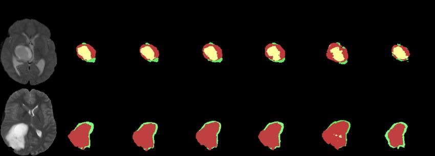
tion. The illustration of the segmentation results is given in Fig. 2. We can see
that the predictions of our proposed OSUDA are better than the no adaptation
model. In addition, CRUDA [1] had a tendency to predict a larger area for the
tumor; and the tumor core is often predicted for the slices without the core.
3.2 Cross-modality T2 to T1/T1ce/FLAIR UDA
Because of large appearance discrepancies between different MRI modalities, we
further applied our framework to the cross-modality UDA task. Since clinical
annotation of the whole tumor is typically performed on T2-weighted MRI, the
typical cross-modality UDA setting is to use T2-weighted MRI as the labeled
source domain, and T1/T1ce/FLAIR MRI as the unlabeled target domains [32].
We followed the UDA training (80% subjects) and testing (20% subjects) split8 X. Liu et al.
Table 2: Comparison of whole tumor segmentation for the cross-modality UDA.
We used T2-weighted MRI as our source domain, and T1-weighted, FLAIR, and
T1ce MRI as the unlabeled target domains.
Source Dice Score [%] ↑ Hausdorff Distance [mm] ↓
Method data T1 FLAIR T1CE Average T1 FLAIR T1CE Average
Source only no UDA 6.8 54.4 6.7 22.6±0.17 58.7 21.5 60.2 46.8±0.15
CRUDA [1] Partial4 47.2 65.6 49.4 54.1±0.16 22.1 17.5 24.4 21.3±0.10
OSUDA no 52.7 67.6 53.2 57.8±0.15 20.4 16.6 22.8 19.9±0.08
OSUDA-AC no 51.6 66.5 52.0 56.7±0.16 21.5 17.8 23.6 21.0±0.12
OSUDA-SE no 51.1 65.8 52.8 56.6±0.14 21.6 17.3 23.3 20.7±0.10
CycleGAN [31] Yes 38.1 63.3 42.1 47.8 25.4 17.2 23.2 21.9
SIFA [3] Yes 51.7 68 58.2 59.3 19.6 16.9 15.01 17.1
DSFN [32] Yes 57.3 78.9 62.2 66.1 17.5 13.8 15.5 15.6
Fig. 3: Comparison with the other UDA methods and an ablation study for the
cross-modality whole tumor segmentation UDA task. From top to bottom, we
show a target test slice of T1, T1ce, and FLAIR MRI.
as in [32], and adopted the same single-channel input backbone. We note that
the data were used in an unpaired manner [32].
The quantitative evaluation results are provided in Table 2. Our proposed
OSUDA outperformed CRUDA [1] consistently. In addition, in CRUDA, the ad-
ditional class ratio prediction model was required to be trained with the source
data, which is prohibitive in many real-world cases. Furthermore, our OSUDA
outperformed several UDA methods trained with the source data, e.g., Cycle-
GAN [31] and SIFA [3], for the two metrics. The visual segmentation results of
three target modalities are shown in Fig. 3, showing the superior performance
of our framework, compared with the comparison methods.Batch statistics for Source-Relaxed Segmentation UDA 9
4 Discussion and Conclusion
This work presented a practical UDA framework for the tumor segmentation
task in the absence of the source domain data, only relying on the “off-the-shelf”
pre-trained segmentation model with BN in the source domain. We proposed a
low-order statistics progression with an exponential momentum decay scheme
to gradually learn the target domain-specific mean and variance. The domain
shareable high-order statistics consistency is enforced with our HBS loss, which is
adaptively weighted based on the channel-wise transferability. The performance
was further boosted with the unsupervised learning objective via self-entropy
minimization. Our experimental results on the cross-subtype and cross-modality
UDA tasks demonstrated that the proposed framework outperformed the com-
parison methods, and was robust to the class ratio shift.
Acknowledgements
This work is partially supported by NIH R01DC018511, R01DE027989, and
P41EB022544.
References
1. Bateson, M., Kervadec, H., Dolz, J., Lombaert, H., Ayed, I.B.: Source-relaxed
domain adaptation for image segmentation. In: International Conference on Medi-
cal Image Computing and Computer-Assisted Intervention. pp. 490–499. Springer
(2020)
2. Chang, W.G., You, T., Seo, S., Kwak, S., Han, B.: Domain-specific batch normal-
ization for unsupervised domain adaptation. In: Proceedings of the IEEE/CVF
Conference on Computer Vision and Pattern Recognition. pp. 7354–7362 (2019)
3. Chen, C., Dou, Q., Chen, H., Qin, J., Heng, P.A.: Synergistic image and feature
adaptation: Towards cross-modality domain adaptation for medical image segmen-
tation. In: Proceedings of the AAAI Conference on Artificial Intelligence. vol. 33,
pp. 865–872 (2019)
4. Grandvalet, Y., Bengio, Y.: Semi-supervised learning by entropy minimization. In:
NIPS (2005)
5. He, K., Zhang, X., Ren, S., Sun, J.: Deep residual learning for image recognition. In:
Proceedings of the IEEE Conference on Computer Vision and Pattern Recognition
(CVPR) (June 2016)
6. Ioffe, S., Szegedy, C.: Batch normalization: Accelerating deep network training by
reducing internal covariate shift. In: International conference on machine learning.
pp. 448–456. PMLR (2015)
7. Li, Y., Wang, N., Shi, J., Hou, X., Liu, J.: Adaptive batch normalization for prac-
tical domain adaptation. Pattern Recognition 80, 109–117 (2018)
8. Liang, J., Hu, D., Feng, J.: Do we really need to access the source data? source hy-
pothesis transfer for unsupervised domain adaptation. In: International Conference
on Machine Learning. pp. 6028–6039. PMLR (2020)
9. Liu, X., Fan, F., Kong, L., Diao, Z., Xie, W., Lu, J., You, J.: Unimodal regularized
neuron stick-breaking for ordinal classification. Neurocomputing 388, 34–44 (2020)10 X. Liu et al.
10. Liu, X., Han, X., Qiao, Y., Ge, Y., Li, S., Lu, J.: Unimodal-uniform constrained
wasserstein training for medical diagnosis. In: Proceedings of the IEEE/CVF In-
ternational Conference on Computer Vision Workshops. pp. 0–0 (2019)
11. Liu, X., Hu, B., Jin, L., Han, X., Xing, F., Ouyang, J., Lu, J., El Fakhri, G.,
Woo, J.: Domain generalization under conditional and label shifts via variational
bayesian inference. In: IJCAI (2021)
12. Liu, X., Hu, B., Liu, X., Lu, J., You, J., Kong, L.: Energy-constrained self-training
for unsupervised domain adaptation. ICPR (2020)
13. Liu, X., Liu, X., Hu, B., Ji, W., Xing, F., Lu, J., You, J., Kuo, C.C.J., Fakhri, G.E.,
Woo, J.: Subtype-aware unsupervised domain adaptation for medical diagnosis.
AAAI (2021)
14. Liu, X., Xing, F., El Fakhri, G., Woo, J.: A unified conditional disentanglement
framework for multimodal brain mr image translation. In: ISBI. pp. 10–14. IEEE
(2021)
15. Liu, X., Xing, F., Prince, J.L., Carass, A., Stone, M., El Fakhri, G., Woo, J.: Dual-
cycle constrained bijective vae-gan for tagged-to-cine magnetic resonance image
synthesis. In: ISBI. pp. 1448–1452. IEEE (2021)
16. Liu, X., Xing, F., Stone, M., Zhuo, J., Timothy, R., Prince, J.L., El Fakhri, G.,
Woo, J.: Generative self-training for cross-domain unsupervised tagged-to-cine mri
synthesis. In: MICCAI (2021)
17. Liu, X., Xing, F., Yang, C., Kuo, C.C.J., ElFakhri, G., Woo, J.: Symmetric-
constrained irregular structure inpainting for brain mri registration with tumor
pathology. MICCAI BrainLes (2020)
18. Liu, X., Zou, Y., Song, Y., Yang, C., You, J., K Vijaya Kumar, B.: Ordinal re-
gression with neuron stick-breaking for medical diagnosis. In: Proceedings of the
European Conference on Computer Vision (ECCV) Workshops. pp. 0–0 (2018)
19. Mancini, M., Porzi, L., Bulo, S.R., Caputo, B., Ricci, E.: Boosting domain adap-
tation by discovering latent domains. In: Proceedings of the IEEE Conference on
Computer Vision and Pattern Recognition. pp. 3771–3780 (2018)
20. Maria Carlucci, F., Porzi, L., Caputo, B., Ricci, E., Rota Bulo, S.: Autodial: Au-
tomatic domain alignment layers. In: Proceedings of the IEEE International Con-
ference on Computer Vision. pp. 5067–5075 (2017)
21. Menze, B.H., Jakab, A., Bauer, S., Kalpathy-Cramer, J., Farahani, K., Kirby, J.,
Burren, Y., Porz, N., Slotboom, J., Wiest, R., et al.: The multimodal brain tumor
image segmentation benchmark (BRATS). IEEE transactions on medical imaging
34(10), 1993–2024 (2014)
22. Pan, X., Luo, P., Shi, J., Tang, X.: Two at once: Enhancing learning and gener-
alization capacities via ibn-net. In: Proceedings of the European Conference on
Computer Vision (ECCV). pp. 464–479 (2018)
23. Shanis, Z., Gerber, S., Gao, M., Enquobahrie, A.: Intramodality domain adaptation
using self ensembling and adversarial training. In: Domain Adaptation and Rep-
resentation Transfer and Medical Image Learning with Less Labels and Imperfect
Data, pp. 28–36. Springer (2019)
24. Wang, D., Shelhamer, E., Liu, S., Olshausen, B., Darrell, T.: Fully test-time adap-
tation by entropy minimization. arXiv preprint arXiv:2006.10726 (2020)
25. Wang, J., Liu, X., Wang, F., Zheng, L., Gao, F., Zhang, H., Zhang, X., Xie, W.,
Wang, B.: Automated interpretation of congenital heart disease from multi-view
echocardiograms. Medical Image Analysis 69, 101942 (2021)
26. Wang, X., Jin, Y., Long, M., Wang, J., Jordan, M.: Transferable normalization: To-
wards improving transferability of deep neural networks. arXiv preprint arXiv:2019
(2019)Batch statistics for Source-Relaxed Segmentation UDA 11
27. Wang, Z., Dai, Z., Póczos, B., Carbonell, J.: Characterizing and avoiding negative
transfer. In: Proceedings of the IEEE/CVF Conference on Computer Vision and
Pattern Recognition. pp. 11293–11302 (2019)
28. Wilson, G., Cook, D.J.: A survey of unsupervised deep domain adaptation. ACM
Transactions on Intelligent Systems and Technology (TIST) 11(5), 1–46 (2020)
29. Zhang, J., Qi, L., Shi, Y., Gao, Y.: Generalizable semantic segmentation
via model-agnostic learning and target-specific normalization. arXiv preprint
arXiv:2003.12296 (2020)
30. Zhou, X.Y., Yang, G.Z.: Normalization in training u-net for 2-D biomedical seman-
tic segmentation. IEEE Robotics and Automation Letters 4(2), 1792–1799 (2019)
31. Zhu, J.Y., Park, T., Isola, P., Efros, A.A.: Unpaired image-to-image translation
using cycle-consistent adversarial networks. In: ICCV (2017)
32. Zou, D., Zhu, Q., Yan, P.: Unsupervised domain adaptation with dualscheme fusion
network for medical image segmentation. In: Proceedings of the Twenty-Ninth
International Joint Conference on Artificial Intelligence, IJCAI-20, International
Joint Conferences on Artificial Intelligence Organization. pp. 3291–3298 (2020)
33. Zou, Y., Yu, Z., Liu, X., Kumar, B., Wang, J.: Confidence regularized self-training.
In: Proceedings of the IEEE/CVF International Conference on Computer Vision.
pp. 5982–5991 (2019)You can also read