Androgen Receptor in Prostate Cancer
←
→
Page content transcription
If your browser does not render page correctly, please read the page content below
0163-769X/04/$20.00/0                                                                                               Endocrine Reviews 25(2):276 –308
Printed in U.S.A.                                                                                          Copyright © 2004 by The Endocrine Society
                                                                                                                            doi: 10.1210/er.2002-0032

Androgen Receptor in Prostate Cancer
CYNTHIA A. HEINLEIN                AND   CHAWNSHANG CHANG
George Whipple Laboratory for Cancer Research, Departments of Pathology, Urology, and Radiation Oncology, and the
Cancer Center, University of Rochester, Rochester, New York 14642

The normal development and maintenance of the prostate is                       lators have been found to occur with prostate cancer progres-
dependent on androgen acting through the androgen recep-                        sion and may contribute to differences in AR ligand specificity
tor (AR). AR remains important in the development and pro-                      or transcriptional activity. Prostate cancer progression is also
gression of prostate cancer. AR expression is maintained                        associated with increased growth factor production and an
throughout prostate cancer progression, and the majority of                     altered response to growth factors by prostate cancer cells.
androgen-independent or hormone refractory prostate can-                        The kinase signal transduction cascades initiated by mito-
cers express AR. Mutation of AR, especially mutations that                      genic growth factors modulate the transcriptional activity of
result in a relaxation of AR ligand specificity, may contribute                 AR and the interaction between AR and AR coactivators. The
to the progression of prostate cancer and the failure of endo-                  inhibition of AR activity through mechanisms in addition to
crine therapy by allowing AR transcriptional activation in                      androgen ablation, such as modulation of signal transduction
response to antiandrogens or other endogenous hormones.                         pathways, may delay prostate cancer progression. (Endocrine
Similarly, alterations in the relative expression of AR coregu-                 Reviews 25: 276 –308, 2004)

 I. Introduction                                                                 V. Prostate Cancer Progression Associated with Relaxation of
                                                                                     AR Ligand Specificity
 II. AR in the Normal Prostate
                                                                                     A. AR mutations
     A. Androgens and AR in normal prostate development
                                                                                     B. Role of coactivators in ligand activation
     B. Androgens and AR in the maintenance of prostate
                                                                                     C. Antiandrogen withdrawal syndrome
        epithelia                                                               VI. Nongenomic Androgen Action
III. AR Expression and Prostate Carcinogenesis                                  VII. Summary and Future Directions
     A. AR expression in prostate cancer
     B. Androgen availability in the prostate after androgen
        ablation                                                                                       I. Introduction
     C. Androgen deprivation and prostate cancer prolifera-
        tion and apoptosis
     D. Androgen regulation of prostate-specific antigen (PSA)
                                                                                A      NDROGENS, ACTING THROUGH the androgen re-
                                                                                        ceptor (AR), are required for prostate development
                                                                                and normal prostate function (1). Androgen action can be
IV. Prostate Cancer Progression and the Modulation of AR
                                                                                considered to function through an axis involving the testic-
     Transcriptional Activity
                                                                                ular synthesis of testosterone, its transport to target tissues,
     A. AR trinucleotide CAG and GGN repeats: effect on pros-
                                                                                and the conversion by 5␣-reductase to the more active me-
        tate cancer development and progression
     B. AR amplification
                                                                                tabolite 5␣-dihydrotestosterone (DHT). Testosterone and
     C. AR coregulator overexpression                                           DHT exert their biological effects through binding to AR and
     D. AR and tumor suppressor genes                                           inducing AR transcriptional activity (Fig. 1). The androgen-
     E. Growth factor modulation of AR activity                                 induced transcriptional activation of AR is modulated by the
                                                                                interaction of AR with coregulators and by phosphorylation
                                                                                of AR and AR coregulators in response to growth factors
   Abbreviations: AIB1, Amplified in breast cancer-1; AR, androgen              (1– 4). AR and the modulators of AR activity remain impor-
receptor; ARE, androgen response element; bFGF, basic FGF; DES, di-
                                                                                tant in prostate cancer. Approximately 80 –90% of prostate
ethylstilbestrol; DHEA, dehydroepiandrosterone; DHT, 5␣-dihydro-
testosterone; DIM, 3,3⬘-diindolymethane; EGF, epidermal growth fac-             cancers are dependent on androgen at initial diagnosis, and
tor; EGFR, EGF receptor; ESE2, epithelium-specific Ets factor 2; Ets, E         endocrine therapy of prostate cancer is directed toward the
twenty-six; FGF, fibroblast growth factor; HF, hydroxyflutamide; IGF-           reduction of serum androgens and inhibition of AR (5). How-
IR, IGF-I receptor; IGFBP, IGF binding protein; LBD, ligand binding             ever, androgen ablation therapy ultimately fails, and pros-
domain; nmt55, nuclear matrix protein, 55 kDa; PDEF, prostate-derived
Ets factor; PI3K, phosphatidylinositol 3-kinase; PKA, protein kinase A;         tate cancer progresses to a hormone refractory state. AR is
PSA, prostate-specific antigen; PTEN, phosphatase and tensin homolog;           expressed throughout prostate cancer progression and per-
PYK2, proline-rich tyrosine kinase 2; Rb, retinoblastoma susceptibility         sists in the majority of patients with hormone refractory
gene; SH2, Src homology 2; SMRT, silencing mediator of retinoid and             disease (6 –10). Also, most identified AR mutations from
thyroid hormone receptor; SRC, steroid receptor coactivator; STAT,
                                                                                hormone refractory prostate cancer are capable of transcrip-
signal transducer and activator of transcription; Tfm, testicular femi-
nized; TIF, transcriptional intermediary factor.                                tional activity (Table 1). These observations suggest that loss
Endocrine Reviews is published bimonthly by The Endocrine Society               of AR function is not a major cause of androgen ablation
(http://www.endo-society.org), the foremost professional society serv-          failure and that AR-negative prostate cancer cells do not have
ing the endocrine community.                                                    a significant growth or survival advantage. Instead, the avail-

                                                                          276
AR in Prostate Cancer • Heinlein and Chang Endocrine Reviews, April 2004, 25(2):276 –308 277
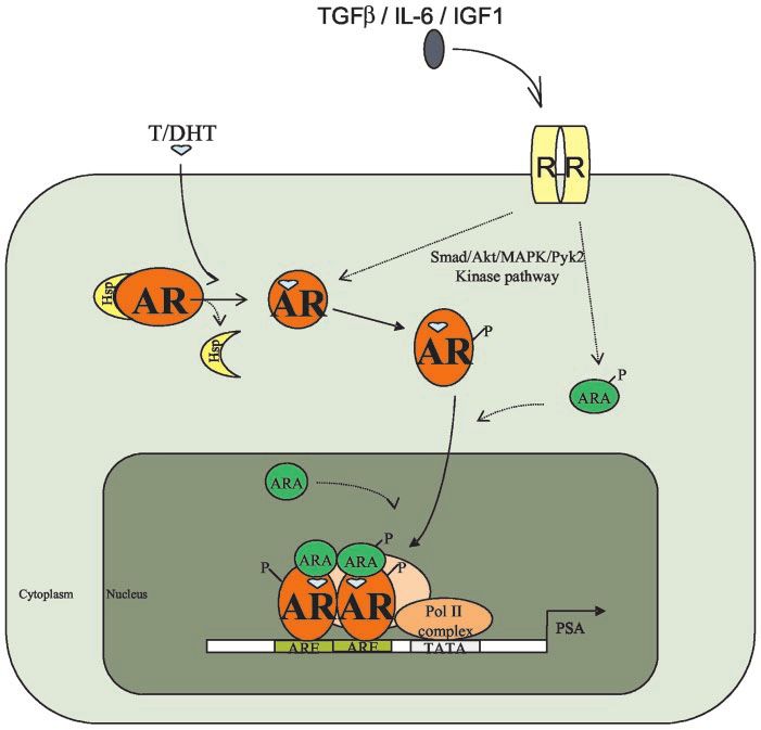
FIG. 1. Androgen-AR action in the prostate.
Testosterone (T) and DHT bind to AR and
promote the association of AR coregulators
(ARAs). AR then translocates to the nucleus
and binds to AREs in the promoter regions of
target genes to induce cell proliferation and
apoptosis. Other signal transduction path-
ways, such as those involving TGF, IL-6, and
IGF-I, can also enhance AR activity via phos-
phorylation of AR and/or ARAs. Hsp, Heat
shock protein; R, membrane receptor; P, pro-
tein phosphorylation.
able clinical and experimental evidence suggests that pros- complete prostate morphogenesis (14). The initiation of prostate
tate cancer progression occurs through alteration of the nor- development is dependent on a functional AR. The prostate is
mal androgen axis by dysregulation of AR activity through absent in AR knockout mice (15), testicular feminized (Tfm)
signal transduction cascades, alteration in the expression of mice, and individuals with complete androgen insensitivity
AR coregulators, and mutations of AR that enable it to be- due to an inactivating mutation of AR (4, 16, 17). The formation
come transcriptionally active in response to ligands in ad- of prostatic buds results from epithelia-stroma interaction that
dition to testosterone and DHT. requires the presence of a functional AR in the urogenital mes-
enchyme but not in the epithelia, suggesting that DHT-regu-
lated growth factors are secreted by the mesenchyme and act
II. AR in the Normal Prostate upon the developing prostate epithelium (4, 11). In tissue re-
combinants, the urogenital epithelia of fetal Tfm mice can form
A. Androgens and AR in normal prostate development
prostatic buds when placed in contact with wild-type fetal
The prenatal development of the prostate is dependent on stroma and grafted into intact male mice. However, wild-type
androgen, particularly on DHT. Although the fetal testis pro- urogenital epithelia is unable to form prostatic buds when com-
duces testosterone, the intracellular reduction of testosterone by bined with Tfm urogenital stroma (18, 19). Consistent with
5␣-reductase is necessary for complete prostate morphogenesis. these experiments, AR is expressed in the fetal mouse urogen-
At about 10 wk gestation in the human, the ductal structure of ital mesenchyme but not in the prostate epithelial buds. Prostate
the prostate arises from epithelial outgrowths of the urogenital epithelium expresses AR in late fetal or early neonatal devel-
sinus and moves into the surrounding mesenchyme immedi- opment when AR function has been implicated in the final
ately below the developing bladder. The 5␣-reductase enzyme morphogenesis of the prostate and the initiation of prostate
is present in the urogenital sinus before and during prostate secretory protein expression (20 –22).
development (11, 12). In individuals lacking a functional 5␣- The hypothesis that a receptor might be needed to mediate
reductase gene, the prostate is small or undetectable. In rats, the biological effects of androgens developed in the 1960s. At
inhibition of 5␣-reductase during fetal development results in first, numerous efforts were made to isolate/purify the AR
partial prostate development (13). The partial prostate forma- without success. Autoimmune anti-AR antibodies from hu-
tion that occurs with 5␣-reductase deficiency suggests that the man serum were found to be able to precipitate an [3H]-
initiation of prostate development can occur in response to R1881-AR complex (22a), yet attempts to use those autoim-
extremely low levels of DHT or in response to testosterone mune anti-AR antibodies to purify AR or isolate AR cDNA
alone, but also that a threshold level of DHT is necessary for were still unsuccessful, due to their lack of specificity. Even-278 Endocrine Reviews, April 2004, 25(2):276 –308 AR in Prostate Cancer • Heinlein and Chang
TABLE 1. Mutations associated with prostate cancer
Change codon/
Exon Position Characteristics Ref.
amino acid
TIS ⫹2 bp CAG3 CAT Germline mutation from a prostate cancer patient with no known family 404
history of prostate cancer. Mutation abolishes the initiation CAG of
the transcription initiation site II (TISII).
5⬘ UTR ⫹214 bp GCC3 GAC Germline mutation from a patient with a familial history of prostate 404
cancer.
1 Contraction of the polyglutamine repeat from 20 to 18 151
1 Contraction of the polyglutamine repeat from 24 to 18. Detected in a 405
radical prostatectomy sample prior to hormonal therapy.
1 57 CTG3 CAG Isolated from a TURP sample prior to hormonal therapy. 330
L3 Q
1 111 CAG3 CAT Isolated from a TURP sample prior to hormonal therapy. 330
Q3 H
1 167 GGC3 AGC Identified in a TURP sample from a hormone refractory tumor after 406
G3 S combined treatment of orchiectomy and bicalutamide.
1 179 AAA3 AGA Isolated from a TURP sample prior to hormonal therapy. 330
K3 R
1 198 GAA3 GGA Isolated from a bone marrow metastasis from a patient with hormone 407
E3 G refractory cancer after treatment with bicalutamide.
1 269 CCA3 TCA Isolated from a TURP sample prior to hormonal therapy. 330
P3 S
1 330 TCC3 CCC Isolated from a bone marrow metastasis from a patient with hormone 407
S3 P refractory cancer after treatment with flutamide
1 527 GAT3 GGT Isolated from a TURP sample prior to hormonal therapy. 330
D3 G
2 546 TTG3 TTC Deletion resulting in a frameshift mutation expected to result in 12 327
L3 F missense amino acids prior to a stop codon. Detected in an archival
latent prostatic tumor sample from a Japanese man.
2 553 CCA3 CCC Deletion causing a frameshift expected to result in 5 missense amino 327
P3 P acids prior to a stop codon. Detected in two archival latent prostatic
tumor samples from Japanese men.
2 574 ACA3 GCA Isolated from a pelvic lymph node metastasis. This mutant can be 152, 328
T3 A weakly activated by DHEA in vitro.
2 579 AAG3 AGG Isolated from a pelvic lymph node metastasis. This mutant exhibits 152, 328
K3 R weak constitutive activity and can be transcriptionally activated by
DHT, DHEA, flutamide, bicalutamide, hydrocortisone, estradiol, and
progesterone.
2 585 GCC3 GTC Isolated from a pelvic lymph node metastasis sample. This mutant is 152, 328
A3 V transcriptionally inactive in vitro.
2 586 GTC3 TCT Isolated from a pelvic lymph node metastasis. This mutant can be 152, 328
A3 S activated by DHEA in vitro.
3 618 TGT3 TAT Identified in a pelvic lymph node metastasis. This mutation prevents AR 328, 408
C3 Y DNA binding, resulting in loss of transcriptional activity.
4 670 ATC3 ACC Isolated from a TURP sample prior to hormonal therapy. This mutant 152, 330
I3 T can be weakly activated by DHEA in vitro.
4 683 GGT3 GCT Isolated from two separate individuals with hormone refractory tumors 150 –152
G3 A carrying an amplification of AR. Conflicting results regarding the in
vitro activity of this mutation have been reported, with one group
finding that the transcriptional activity does not differ from the wild-
type receptor and another group observing that the response of the
mutant receptor to DHT is significantly reduced compared to wild
type.
4 700 CTC3 CAC Isolated from a prostatic autopsy sample from a patient treated with 152, 329
L3 H castration and chlormadinone acetate whose cancer had become
hormone resistant. Cancerous tissues from metastatic sites from the
same patient showed a separate AR mutation (T876A). This mutant
can be transcriptionally activated by DHT, DHEA, estradiol,
hydrocortisone, progesterone, bicalutamide, and flutamide.
4 714 GTG3 ATG Isolated from a fine-needle biopsy sample from a patient with metastatic 152, 340,
V3 M prostate cancer. Prior to biopsy, the patient had been treated by 341, 409
castration followed by flutamide and estracyte. At the time of biopsy,
the cancer was hormone refractory. There is no significant difference
in the relative binding affinity between this mutant and the wild-type
AR. However, this mutant demonstrates an enhanced transcriptional
activation in response to HF, progesterone, DHEA, estradiol,
androstone, androstanediol and androstenedione.
4 719 AAG3 GAG Isolated from a bone metastasis. Transcriptional activation of the 152, 410
K3 E mutant receptor in vitro does not differ significantly from the wild-
type receptor in response to R1881 or DHT.AR in Prostate Cancer • Heinlein and Chang Endocrine Reviews, April 2004, 25(2):276 –308 279
TABLE 1. Continued
Change codon/
Exon Position Characteristics Ref.
amino acid
4 720 GCC3 ACC Isolated from a hormone refractory bone marrow metastasis from a 152, 336,
A3 T patient treated with flutamide and an LHRH agonist. Does not become 341
transcriptionally active in response to estradiol or progesterone. Shows
moderate transcriptional activation in response to 1 M nilutamide,
but not HF or bicalutamide. Demonstrates a reduced response to DHT
in vitro compared to the wild-type receptor.
5 725 CGC3 CTC Germline mutation observed in 2% of Finnish sporadic prostate cancer 152, 342,
R3 L patients. The AR CAG repeat length is 26 in 85% of mutation carriers. 411
This mutation does not alter the ability of the mutant AR to bind
mibolerone, DHT, estradiol, or progesterone. However, this mutation
enhances AR transcription in response to estradiol compared to the
wild-type receptor in CV-1 cells. This mutation also can be activated
by DHEA in vitro.
5 729 GTG3 ATG Isolated from a patient with organ-confined prostate cancer who had not 409, 412
V3 M received hormonal therapy. This mutant shows enhanced
transactivation in response to HF, androsterone, and androstanediol.
However, the relative binding affinity of the mutant receptor for
R1881, androstenediol, androstanediol, and androsterone is the same
as the wild-type receptor.
5 740 TGG3 TAG Detected in an archival latent prostatic tumor sample from a Japanese 327
W3 Stop man.
5 740 TGG3 TGT Identified from a TURP sample from a hormone refractory tumor treated 406, 407
W3 C with a combination of orchiectomy and bicalutamide. The same amino
acid substitution has also been isolated from a bone marrow
metastasis from a patient with hormone refractory cancer after
treatment with bicalutamide.
5 742 GGG3 GGC Deletion causing a frameshift mutation resulting in a stop codon after 1 327
G3 G Stop amino acid. Detected in an archival latent prostatic tumor sample
from a Japanese man. A separate latent tumor foci in the same
individual contained the L743F mutation.
5 743 CTC3 TTC Detected in an archival latent prostatic tumor sample from a Japanese 327
L3 F man. A separate latent tumor foci from the same individual contained
the ⌬742 mutation.
5 747 GGC3 GTC Detected in an archival latent prostatic tumor sample from a Japanese 152, 327
A3 V man. This mutation results in a decreased transcriptional response to
DHT compared to the wild-type receptor in vitro. Can also be
transcriptionally activated by DHEA in vitro.
5 748 ATG3 ATA Detected in an archival latent prostatic tumor sample from a Japanese 152, 327
M3 I man. This mutant does not become transcriptionally active in vitro in
response to DHT, DHEA, progesterone, estradiol, hydrocortisone,
flutamide, or bicalutamide.
5 748 ATG3 ATA Isolated from a TURP sample from a hormone refractory tumor after 406
M3 I treatment by orchiectomy and bicalutamide.
5 749 GGC3 AGC Detected in an archival latent prostatic tumor sample from a Japanese 152, 327
G3 S man. This mutant can be transcriptionally activated in vitro by DHEA.
5 750 TGG3 TAG Detected in two archival latent prostatic tumor samples from Japanese 327
W3 Stop men.
5 754 ACC3 GCC Detected in an archival latent prostatic tumor sample from a Japanese 327
T3 A man.
5 756 GTC3 GCC Isolated from a pelvic lymph node metastasis. The mutant receptor can 152, 328
V3 A be transcriptionally activated by DHEA in vitro.
5 758 TCC3 CCC Detected in an archival latent prostatic tumor sample from a Japanese 152, 327
S3 P man. This mutant shows a reduced transcriptional activity in response
to DHT in vitro compared to the wild-type receptor.
5 762 TAC3 TGC Detected in an archival latent prostatic tumor sample from a Japanese 152, 327
Y3 C man. This mutant become transcriptionally active in vitro in response
to DHT but not estradiol, DHEA, progesterone, hydrocortisone,
flutamide, or bicalutamide.
6 781 AGC3 AAC Isolated from a TURP sample prior to hormonal therapy. The mutant 152, 330
S3 N can be transcriptionally activated by DHT and DHEA in vitro.
6 795 TGG3 TGA Isolated from a TURP sample prior to hormonal therapy. 330
W3 STOP
7 845 AGA3 GGA Isolated from a pelvic lymph node metastasis. This mutation can show 152, 328
R3 G transcriptional activation in vitro in response to DHT and DHEA.
8 865 GTG3 ATG Detected in an archival latent prostatic tumor sample from a Japanese 152, 327
V3 M man. This mutation fails to be activated by DHT, DHEA, estradiol,
progesterone, hydrocortisone, flutamide, or bicalutamide in vitro.
Continued on next page280 Endocrine Reviews, April 2004, 25(2):276 –308 AR in Prostate Cancer • Heinlein and Chang
TABLE 1. Continued
Change codon/
Exon Position Characteristics Ref.
amino acid
8 873 CAT3 TAT Isolated from a hormone refractory bone marrow metastasis from a 152, 336,
H3 Y patient treated with flutamide and an LHRH agonist. The tumor was 341
initially treated with local radiotherapy. Shows enhanced in vitro
transcriptional activity in response to DHEA, estradiol, and
progesterone compared to the wild-type receptor. Transcription can
also be induced by the antiandrogens nilutamide and HF, but not
bicalutamide.
8 876 ACT3 AGT Isolated from a hormone refractory bone marrow metastasis from a 335, 336,
T3 S patient treated with flutamide and an LHRH agonist. The tumor was 341, 343
initially treated with local radiotherapy. Also identified in a patient
whose tumor contained both the T876S and T876A mutations. In
addition to testosterone and DHT, this mutant can be induced by HF,
casodex, and cyptoterone acetate.
8 876 ACT3 GCT In one study, this mutation was found in 31% of patients examined with 329, 334,
T3 A hormone refractory disease after surgical or chemical castration plus 335, 337
flutamide treatment. A separate study found this mutation in three
metastatic cancerous loci in 1 of 8 (12.5%) of patients with hormone-
resistant prostate cancer that had been treated with chlormadinone
acetate. This patient had a different AR mutation (L700H) in
cancerous foci in the prostate. A separate study found this mutation in
archival TURP sections from 6 of 24 (25%) prostate cancer patients.
This mutation is present in the AR expressed in LNCaP cells.
8 878 GAC3 GCC Isolated from a bone marrow metastasis from a patient with hormone 407
D3 G refractory cancer after treatment with bicalutamide. Shows enhanced
in vitro transcriptional activity in response to estradiol and
progesterone compared to the wild-type receptor. In addition, the
antagonists nilutamide and HF, but not bicalutamide, activate
transcription of this mutant.
8 889 GAC3 AAC Isolated from a bone marrow metastasis from a patient treated with 152, 335
D3 N LHRH agonist therapy with hormone refractory disease. Mutation is
associated with an increase in transcription in response to DHEA in
vitro.
8 901 CAA3 CGA Isolated from a hormone refractory bone marrow metastasis from a 336, 341
Q3 R patient treated with flutamide and an LHRH agonist. The tumor was
initially treated by radical prostatectomy. In transfection assays,
shows approximately 37% of wild-type AR activity at 1 nM DHT.
Demonstrates marginal transcriptional activity in response to 0.01–10
nM androstenedione and no activity in response to up to 1 M estradiol
or progesterone.
8 908 GGG3 GAG Detected in an archival latent prostatic tumor sample from a Japanese 327
G3 E man.
Because AR contains two trinucleotide repeat regions that are polymorphic in length, amino acid positions may vary between publications.
In this table, amino acid positions are as numbered in Ref. 99. Silent mutations have not been included. TURP, Transurethral resection of the
prostate; UTR, untranslated region.
tually, use of a DNA oligonucleotide probe that was homol- thelia, the primary cell type thought to be transformed in
ogous to other steroid receptors allowed Chang et al. (22b) prostate adenocarcinoma (23). In the normal prostate, the
and Lubahn et al. (22c) to isolate full-length human AR rate of cell death is 1–2% per day, which is balanced by a
cDNAs, from which in vitro transcribed/translated protein 1–2% rate of proliferation (24, 25). The reduction of serum
was generated, and found to bind to [3H]-R1881 with a Kd of and prostatic DHT levels by castration results in a loss of 70%
0.3 nm (22b). Structural analysis of AR revealed that it con- of the prostate secretory epithelial cells due to apoptosis in
tains four functional domains, similar to other members of adult male rats, but the basal epithelia and stromal cell pop-
the steroid receptor superfamily: a conserved DNA binding ulations are relatively unaffected (26). In the intact rat pros-
domain (DBD), a hinge region, a ligand-binding domain tate, the secretory epithelial cells show strong AR immuno-
(LBD), and a less conserved amino-terminal domain (22b, reactivity, whereas the majority of basal epithelial cells are
22c). Further analysis of AR structure revealed two tran- AR negative (27), suggesting an explanation for their differ-
scriptional activation function domains, including the ent sensitivity to androgen. However, AR is also expressed
N-terminal ligand-independent AF-1 domain and the C- in the prostatic stroma, although castration results in the loss
terminal ligand-dependent AF-2 domain. of stromal AR expression (27, 28). The prostatic stroma there-
fore has the capacity to respond to androgen, but androgen
B. Androgens and AR in the maintenance of
is not required for its survival. Physiological testosterone
prostate epithelia
levels prevent secretory rat prostate epithelial apoptosis.
After the development of the prostate, androgens continue However, normal epithelial function is dependent on pros-
to function in promoting the survival of the secretory epi- tatic DHT levels (29). Superphysiological levels of serumAR in Prostate Cancer • Heinlein and Chang Endocrine Reviews, April 2004, 25(2):276 –308 281
androgen in dogs and in human habitual anabolic steroid gens, either alone or in combination with surgical or chemical
users result in an increase in cellular proliferation in the castration (referred to as combined androgen blockade).
prostate (30, 31). In humans, the proliferation occurs pre- Over 80% of patients show a positive response to androgen
dominantly in the transitional zone of the prostate, the region ablation. However, patients with metastatic prostate cancer
that is primarily affected in benign prostatic hypertrophy but eventually experience disease progression in a median of 12
is seldom the initial site of prostate carcinoma formation (30, to 18 months after androgen deprivation therapy. The tu-
32). Although individual cases of prostate cancer have been mors of these patients are considered to be hormone refrac-
reported in anabolic steroid users (33), epidemiological stud- tory. Although these tumors are refractory in the sense that
ies have failed to establish a link between elevated serum they have progressed despite a reduction in serum androgen
testosterone, DHT, or adrenal androgens and prostate cancer and/or treatment with antiandrogens, the majority of these
risk (reviewed in Ref. 34), suggesting that elevated testicular tumors are unlikely to be completely resistant to androgen.
and adrenal androgens alone do not significantly promote In 97% of patients with hormone refractory metastatic pros-
prostate carcinogenesis. tate cancer, exogenous androgen treatment results in disease
In addition to apoptosis of secretory epithelial cells, flare and unfavorable response (reviewed in Ref. 44). Sec-
castration also results in apoptosis and degeneration of ondary therapy for patients with hormone refractory pros-
prostatic capillaries and constriction of larger blood ves- tate cancer is also predominantly targeted at androgen pro-
sels, which precedes the appearance of epithelial apoptosis duction and AR function and includes administration of a
(35, 36). These observations suggest that the reduction of secondary antiandrogen, inhibition of adrenal androgen pro-
blood flow to the prostate may contribute to epithelial
duction, and further LH inhibition with progesterone or es-
apoptosis. However, castration does not induce necrosis or
trogenic agents (45). Although secondary hormonal therapy
apoptosis in all prostatic cell types, suggesting that if
also eventually fails, the ability of therapies directed toward
secretory epithelial cell loss is influenced by the alteration
AR to provide positive therapeutic benefit suggests that AR
in blood flow, these cells are more sensitive to this change
activity is an important mediator of prostate cancer growth
than other prostate cell types. Administration of testos-
and survival.
terone to castrated rats results in vascular regrowth fol-
lowed by reconstitution of the secretory epithelia (37).
However, the vascular endothelial cells of the rat prostate
do not express AR (27). In the normal prostate, cellular A. AR expression in prostate cancer
homeostasis is modulated in part by paracrine growth AR expression is observed in primary prostate cancer and
factor regulation between epithelial and stromal cells (38). can be detected throughout progression in both hormone-
A subset of these growth factors, including basic fibroblast sensitive and hormone refractory cancers (8, 9, 46). Immu-
growth factor (bFGF) and vascular endothelial growth nohistochemical studies have shown that AR expression is
factor, can be regulated by androgens and can influence heterogeneous in prostate cancer and that the degree of het-
vascular survival (38 – 41). It is possible that castration
erogeneity does not generally correlate with response to an-
initially alters prostatic growth factor production in the
drogen deprivation therapy (8, 46). However, we and others
stroma, which contributes to a decrease in vascular func-
have observed that a higher degree of AR positivity corre-
tion. The resulting reduction in blood flow, combined with
lates with a greater degree of differentiation or lower Gleason
an altered growth factor environment and decreased ex-
score (9, 47, 48), although this is not a universal observation
pression of other androgen regulated proteins, may con-
tribute to apoptosis of the secretory epithelia. (10, 46). Although animal models of prostate cancer have
suggested that elevation of AR expression can initiate pros-
tate cancer development (49) or is associated with recurrent
growth in the presence of low androgen (50), the persistent
III. AR Expression and Prostate Carcinogenesis heterogeneity of human prostate cancer suggests that in-
Although serum androgens alone may not promote pros- creased AR expression is not generally associated with pros-
tate carcinogenesis, androgen action and the functional sta- tate cancer initiation, and that hormone refractory prostate
tus of AR are important mediators of prostate cancer pro- cancers are not clonally selected from AR-negative foci. The
gression. Low serum testosterone levels in men with newly cause of the loss of AR expression in some cells of tumor foci
diagnosed and untreated prostate cancer have been found to is unclear. X chromosome losses, including loss of the AR
correlate with higher AR expression, increased capillary ves- gene, are extremely rare in prostate cancer (51–53). Epige-
sel density within the tumor, and higher Gleason score (42). netic silencing of AR expression by methylation may occur
Recent analysis of clinical prostate cancer specimens also and has been observed in 8% of primary prostate cancers (54).
collected from patients without preoperative treatment dem- Another possibility for the loss of AR expression in some
onstrated that high AR expression correlated with lower tumor cells is a decrease in AR protein stability that reduces
recurrence-free survival and disease progression (43). The the AR protein level to one difficult to detect immunohis-
endocrinological treatment of prostate cancer primarily in- tologically. AR is degraded by ubiquitin targeting to the
volves the modulation of AR activity through the depriva- proteasome (55). Ubiquitination of AR is promoted by Akt
tion of circulating testicular androgens by surgical castration kinase-mediated phosphorylation of the receptor, suggesting
or chemical castration with LHRH agonists. The activity of that cells with increased Akt activation may have a reduced
AR may also be blocked by the administration of antiandro- AR protein level (55).282 Endocrine Reviews, April 2004, 25(2):276 –308 AR in Prostate Cancer • Heinlein and Chang
B. Androgen availability in the prostate after ence tumor cell number up to 2 wk after castration (66, 67).
androgen ablation In CWR22 xenograft tumors, castration initially induced
growth arrest in tumor cells. However, foci of Ki-67 immu-
Androgen ablation by surgical castration or treatment
nopositive cells were detected by 120 d after castration (50).
with LHRH agonists results in a 90 –95% decrease in serum
The clinical and animal prostate cancer data suggest that a
testosterone levels. However, intraprostatic DHT levels only
significant proportion of prostate tumors are resistant to
decline by approximately 50% (5, 56, 57). Although this re-
androgen ablation-induced apoptosis at the time of treat-
duction is able to cause the death of over 70% of normal
ment and that the observed therapeutic benefit may be the
prostate secretory epithelial cells as discussed above, pros-
result of a decrease in the proliferation rate of the tumor cells.
tate cancer cells surviving this treatment would be exposed
The mechanism of the resistance to apoptosis in prostate
to a relative abundance of DHT. In contrast, GnRH agonist
tumors remains to be determined, however apoptosis resis-
treatment has been reported to reduce intraprostatic DHT by
tance may be at least partially due to an increase in the
90% (58). Combined treatment of castration and flutamide
expression of apoptosis suppressor genes. Elevation of the
has been found to reduce prostate DHT levels to approxi-
antiapoptosis proteins bcl-2, bcl-x, and mcl-1 has been found
mately 20% of pretreatment levels (56, 59). Flutamide is a
in prostatic intraepithelial neoplasia (68, 69), suggesting that
nonsteroidal antiandrogen, and the mechanism of its effect
resistance to apoptosis may be an early event in prostate
on prostatic DHT levels has not yet been determined. In
cancer. The expression of these antiapoptotic proteins is
contrast to castration alone, the presence of flutamide in
found to further increase with prostate cancer progression
combined androgen ablation would be expected to substan-
(68, 69). Overexpression of bcl-2 in the androgen-depen-
tially block the ability of the residual prostatic DHT to ac-
dent prostate cancer cell line LNCaP enables cell growth in
tivate AR transcription (57). Although the differences in re-
androgen-depleted media and enhances tumor formation
maining DHT and accessibility of the DHT to AR might be
in castrated male mice (70). The suppression of bcl-2 expres-
expected to correlate with a difference in prostate cancer
sion through AR-mediated androgen action has been pro-
prognosis after different androgen ablation regimes, a recent
posed as a mechanism for enhanced bcl-2 expression upon
meta-analysis of studies comparing patient survival with
androgen deprivation (71). These observations suggest that
combined androgen ablation or with either castration or
elevated expression of antiapoptotic proteins, particularly
long-term LHRH agonist treatment alone found no statisti-
bcl-2, in prostate tumors may contribute to the resistance of
cally significant difference between the two groups (60). Al-
some tumors to androgen deprivation-induced apoptosis.
though monotherapy and combined androgen blockade may
The stimulation of antiapoptotic genes may represent a sec-
not differ in overall prostate cancer survival, it is possible that
ondary event in response to modulation by other factors,
these treatment regimes may differ in the molecular mech-
such as growth factors. For example, fibroblast growth factor
anism used by the tumors to become androgen insensitive.
2 (FGF2) has been found to promote growth and survival of
prostate cancer cells through induction of bcl-2 expression
C. Androgen deprivation and prostate cancer proliferation
(72).
and apoptosis
On the basis of cell line models, it has been suggested that
prostate cancer cells surviving androgen deprivation therapy
Although androgen deprivation results in a dramatic re- may be sensitive to apoptosis induced by androgen (73). The
duction of a population of prostate secretory cells through growth of LNCaP cells is normally dependent on androgen.
apoptosis, there is some evidence to suggest that prostate However, LNCaP cell growth is inhibited at high concen-
cancer cells acquire a relative resistance to androgen abla- trations (300 nm) of DHT (74). The growth of several LNCaP
tion-induced apoptosis early in transformation and that an- sublines selected for growth in reduced androgen-containing
drogen primarily regulates the proliferation of prostate can- media can be inhibited by physiological levels of DHT (1–10
cer cells in vivo. In analysis of castration-induced involution nm) (75–77). Additionally, DHT, acting through AR, has been
of the normal rat prostate, the majority of epithelial cell loss shown to potentiate the apoptotic effect of 12-O-tetrade-
occurs within 7 d (25, 26). Several studies of human prostate canoylphorbolacetate, a protein kinase C activator, via in-
cancer samples obtained several months after the initiation terruption of nuclear factor B signaling and activation of the
of androgen deprivation have detected the presence of pros- c-Jun NH2-terminal kinase pathway in LNCaP cells (S. Al-
tate cancer foci that appear to be morphologically altered by tuwaijri, and C. Chang, unpublished observations). Simi-
androgen deprivation but have not undergone necrosis or larly, stable transfection of AR into the AR-negative prostate
apoptosis (61– 63). Histochemical analysis of human prostate cancer cell line PC-3 has been reported to generate sublines
tumors 7 d after orchiectomy showed that 88% of tumors that undergo growth arrest or apoptosis in the presence of
demonstrated a decrease in proliferation as determined by physiological levels of androgen (78, 79). However, it is un-
Ki-67 immunopositivity (64). However, in 60% of tumors, clear to what extent these cell lines represent prostate cancer
castration either had no effect or reduced the rate of apo- in vivo. Treatment of prostate cancer patients with hormone
ptosis (64). In a separate study, androgen deprivation was refractory metastases with either physiological or super-
found to result in only a 3.4% apoptotic index (65). Similar physiological doses of androgen results in a negative re-
results have been obtained in the androgen-sensitive Dun- sponse in 97% of patients (44), suggesting that androgen-
ning R3327 rat prostate tumor model. Castration induced induced growth arrest in tissue culture cell lines does not
involution of the normal prostate epithelia and reduced the represent a common physiological response in vivo.
mitotic index of tumor cells, but did not significantly influ- Although continuous androgenic therapy may not be ther-AR in Prostate Cancer • Heinlein and Chang Endocrine Reviews, April 2004, 25(2):276 –308 283
apeutically beneficial for the majority of prostate cancer pa- tion factor, GAGATA binding protein, has been identified
tients, intermittent androgen ablation has been proposed as and found to affect androgen-mediated expression of PSA
a therapy to delay the development of tumors that cannot through binding to an alternative enhancer site (GAGATA)
respond to androgen deprivation (80, 81). It has been pro- in the PSA promoter (95). Two E twenty-six (Ets) family
posed that androgen-dependent cells surviving androgen transcription factors, epithelium-specific Ets factor 2 (ESE2)
ablation adapt to a low androgen environment and eventu- and prostate-derived Ets factor (PDEF), have also been found
ally become androgen insensitive (81, 81a). According to this to induce transcription of a PSA reporter gene in the AR-
model, periodic exposure to androgen would prevent cells negative cell line CV-1 (96, 97). PDEF is highly expressed in
that can grow independent of androgen from becoming pre- the prostate and weakly expressed in the ovary (97). Al-
dominant. Consistent with this model, the development of though PDEF is capable of inducing PSA expression in the
androgen-independent LNCaP xenografts in castrated nude absence of AR, PDEF can heterodimerize with AR to enhance
mice is delayed by intermittent treatment with testosterone AR-induced transcription (97). ESE2 is weakly expressed in
(82). Phase II clinical trials have suggested that intermittent the normal prostate, and it is not yet known whether this
androgen deprivation may improve quality of life and sexual transcription factor directly interacts with AR (96). The abil-
function compared with continuous androgen deprivation, ity of Ets transcription factors to regulate PSA expression in
although it remains unclear whether intermittent therapy prostate cancer remains to be determined. A small percent-
provides a survival benefit (reviewed in Refs. 80 and 81). age of cells in local prostate tumors have been found to
express PSA but lack detectable AR expression by immuno-
histochemistry (46). It is possible that PSA expression in these
D. Androgen regulation of prostate-specific antigen (PSA)
cells is regulated by an Ets transcription factor. As discussed
PSA is generally considered to be the most sensitive bio- above, the majority of prostate tumors express AR, and there-
chemical marker available for monitoring the presence of fore the significance of AR-independent PSA expression is
prostatic disease, particularly prostate cancer, and response unclear.
to therapy. PSA is a glycoprotein and a member of the kal-
likrein family of serine proteases (83). In the normal prostate,
PSA is secreted into the glandular ducts where it functions IV. Prostate Cancer Progression and the Modulation
to degrade high molecular weight proteins produced in of AR Transcriptional Activity
the seminal vesicles to prevent coagulation of the semen A. AR trinucleotide CAG and GGN repeats: effect on
(84). PSA levels in the normal prostate are approximately 1 prostate cancer development and progression
million-fold higher than in the serum. PSA normally enters
the serum only through leakage into the prostatic extracel- The NH2-terminal transactivation domain of AR contains
lular fluid. During prostate cancer progression, serum PSA two trinucleotide repeat regions, both of which are poly-
levels become progressively elevated due to aberration of the morphic in length. The CAG repeat, encoding a polyglu-
normal prostate ductal structure by the neoplastic epithelial tamine region, is located within a region of the NH2-terminal
cells. The increasingly abnormal ductal structure allows PSA that is required for full ligand-inducible transcription (98 –
to be actively secreted into the extracellular space and enter 101). Charged, glutamine-rich regions have been identified
the circulation (85, 86). In addition to the prostate, a low level in other transcription factors, including cAMP response
of PSA expression is found in amniotic fluid, in the lactating element-binding protein (CREB), amplified in breast cancer-
breast, and in a subset of breast and ovarian tumors (87). 1 (AIB1), and specificity protein 1 (Sp1), where they mediate
The primary regulator of PSA expression is AR, which protein-protein interactions with coregulators or members of
induces PSA expression through three androgen response the basal transcriptional machinery (102–104). The second
element-containing enhancer elements located in the prox- trinucleotide repeat is the GGN or polyglycine repeat that lies
imal 6 kb of the PSA promoter (88, 89). The androgen-inde- 3⬘ of the CAG repeat. The two repeat regions are separated
pendent prostate cancer cell line PC-3, which does not ex- by 248 amino acids of nonpolymorphic sequence. Polymor-
press either AR or PSA, was induced to produce PSA after phic variation in the trinucleotide repeat lengths of the NH2-
transfection of AR and treatment with androgen. This result terminal of AR is associated with altered AR transcriptional
highlights the importance of AR activity for PSA expression activity in vitro (105–107), as well as variations in prostate
in this cell line (90). In addition to androgens, PSA expression growth upon testosterone substitution in hypogonadal men
has been reported to be induced by glucocorticoids in T47D (108), and may therefore contribute to prostate cancer risk or
breast cancer cells (91) and LNCaP cells transfected with the progression (109, 110). The occurrence of prostate cancer
glucocorticoid receptor (92). Progestins are also able to stim- demonstrates familial aggregation, with a 2- to 4-fold in-
ulate PSA expression at low concentrations (10⫺11 to 10⫺10 m) creased risk among men reporting prostate cancer in a father
in breast cancer cell lines (91, 93), and oral contraceptives or brother after adjustment for age and dietary factors (111–
containing progestin can induce PSA expression in breast 113). Recently, several hereditary prostate cancer loci have
tissue (94). However, these cell lines and tissues are all been identified. The cancer-associated alleles of these loci are
known to express a functional AR, and the cell culture assays rare, autosomal dominant or X-linked, and show high pen-
were done in media that is expected to contain residual etrance (114 –117). In contrast, epidemiological studies sug-
androgen. Therefore, it remains possible that the glucocor- gest that AR trinucleotide repeat polymorphisms associated
ticoid receptor or progesterone receptor may cooperate with with prostate cancer are common alleles of relatively low
AR to promote PSA expression. Recently, a novel transcrip- penetrance (118).284 Endocrine Reviews, April 2004, 25(2):276 –308 AR in Prostate Cancer • Heinlein and Chang The AR CAG repeat normally varies between eight and 30 central zone of the prostate (136), considered to be the most contiguous repeats in length (119). However, the modal CAG hormonally sensitive prostatic region (30). The reason for the repeat number varies between ethnic groups, with 18 repeats inconsistent association between the AR CAG repeat number being the most abundant allele in African-Americans and 21 and prostate cancer or in vivo parameters of androgen action and 22 repeat alleles most abundant in non-Hispanic whites is unclear. Differences in study design and reference CAG and Asians, respectively (102, 120, 121). Expansion of the lengths may contribute to the divergent results in the epi- CAG repeat to over 40 causes the rare neuromuscular dis- demiological studies. It has been proposed that the poly- order spinal and bulbar muscular atrophy, which is also morphic CAG repeats function as low penetrance prostate often associated with reduced virilization (122). Ethnic dif- cancer alleles that may require additional genetic or envi- ferences in prostate cancer incidence are inversely correlated ronmental factors to result in increased cancer risk (118, 137). to the predominant AR CAG repeat length in each group, The CAG repeat region is located in an AR domain that is with Asians having the lowest prostate cancer incidence and known to interact with some AR coregulators (2). It is pos- the longest AR CAG repeats, whereas African-Americans sible that variation in the prostatic coregulator milieu con- have the highest incidence and shortest CAG repeat length. tributes to the association between CAG repeat length and Longer CAG repeat lengths have been correlated with de- prostatic disease. Transfection assays have demonstrated creased AR transcriptional activity in vitro. AR molecules that the interaction between AR and the coactivator ARA24 carrying more than 40 CAG repeats show reduced transcrip- decreases with increasing AR CAG repeat length, resulting tional activity compared with AR molecules with 25, 20, or in decreased AR transactivation (138). Similarly, longer AR no CAG repeats (106, 107). However, analysis of the tran- CAG repeat lengths result in a decrease in the ability of AR scriptional effect of CAG repeat lengths within the normal to be coactivated by members of the steroid receptor coac- repeat range suggests that the correlation between short tivator (SRC) family of coregulators [SRC-1, transcriptional CAG repeat lengths and increased transactivation is cell-type intermediary factor 2 (TIF-2), and SRC-3] (124). The expres- dependent. In the fibroblastic COS-1 cell line, a 25% reduc- sion of SRC-1 and TIF-2 has been found to be elevated in tion in AR transcription is seen between receptors having 12 some prostate tumor specimens (139). It is possible that in- CAG repeats and those having 20 (107). Similarly, a 40% dividuals who normally have an increased expression of an progressive decrease in the level of AR transcription occurs SRC coregulator in the prostate and carry an AR allele with between a CAG repeat length of 15 and that of 31 in COS-1 a short CAG repeat length may have a greater risk of prostate cells (123). In contrast, one study found no significant dif- cancer. Alternatively, polymorphisms in the promoters of ference in AR transcription, in the epithelial prostate cancer AR target genes in combination with short CAG AR alleles cell line PC-3, between AR molecules with 15, 24, or 31 CAG may contribute to prostate cancer susceptibility. The PSA repeats (123). In a separate study using PC-3 cells, a 7% gene promoter contains a polymorphic androgen response decrease in AR transcription was observed between recep- element (ARE), referred to as the A and G alleles. Individuals tors with nine and 21 CAG repeats, and a 13% decrease was carrying an AR allele with less than 20 CAG repeats and shown between receptors with nine and 29 CAG repeats homozygous for the PSA G allele have been reported to have (124). a 5-fold increased risk of prostate cancer (125). The protease Although differences in AR transcription with CAG repeat activity of PSA has been hypothesized to contribute to pros- lengths in the normal range may be difficult to observe in tate carcinogenesis through cleavage of extracellular matrix vitro, it is possible that small differences in AR transactivation proteins or through modulation of the availability of IGF-I by may cumulatively contribute to lifetime prostate cancer risk cleavage of IGF binding protein-3 (IGFBP-3) (140 –142). It is or age of diagnosis. In healthy men without prostate cancer, possible that AR binds to the PSA G allele with greater a short AR CAG repeat length correlates to a modestly affinity and that in combination with the increased tran- higher, but statistically significant, serum PSA level (125), scriptional activity of short CAG repeat alleles of AR, con- suggesting that the CAG repeat number influences AR trans- tributes to prostate carcinogenesis (143, 143a). activation in vivo. Short CAG repeat lengths (CAG repeat The second polymorphic AR trinucleotide repeat, the length ⱕ17 to ⱕ23, depending on the study) have been found GGN or polyglycine repeat, is less well studied than the CAG to correlate with an increased prostate cancer risk (120, 126, repeat. Deletion of the GGN repeat results in a 30% reduction 127). This association has been shown in both American in AR transcriptional activation in transfection experiments non-Hispanic white men (126, 127) and in a population- (105), but it has not yet been determined whether this trinu- based study in China (120). The association of short AR CAG cleotide repeat functions as a protein interaction domain. repeat length with prostate cancer risk in both a moderate Because a comparison of AR transcriptional activation with risk non-Hispanic white population and a low-risk Chinese varying GGN repeat lengths has not been performed, it re- population suggests that this may represent a genuine pros- mains to be determined whether the reduction in AR activity tate cancer predictor. Several studies have also reported an with the deletion of the GGN region reflects a reduction in association between a short AR CAG repeat length and an AR transactivation with decreasing repeat length. Although earlier age of diagnosis (128 –130) or more advanced cancer the AR GGN repeat shows a lesser degree of polymorphism grade and stage at diagnosis (127, 131). than the CAG repeat (120, 121, 132), several studies have However, a number of studies have failed to link AR CAG examined the GGN repeat length and prostate cancer sus- repeat number to sporadic or familial prostate cancer (132– ceptibility. Short GGN repeat lengths (GGN ⱕ 14 or GGN ⱕ 135). In men without known prostate disease, CAG repeat 16, depending on the study) have been found to be associated length was not found to be related to the volume of the with increased prostate cancer risk (126, 132). If there is a
AR in Prostate Cancer • Heinlein and Chang                                      Endocrine Reviews, April 2004, 25(2):276 –308   285

direct relationship between GGN repeat length and AR              although generalized microsatellite instability may represent
transactivation, then this result is unexpected. However, one     an early event in prostate carcinogenesis (159, 160). Similarly,
study found that long GGN repeat lengths (GGN ⱖ 16) were          chromosomal aberrations are found to increase with prostate
associated with an increased risk of prostate cancer recur-       cancer progression, both in patients initially treated with
rence and increased risk of death (135). Two separate studies     hormonal therapy and in patients treated by radical prosta-
failed to find a link between GGN repeat number and pros-         tectomy without hormonal intervention (51, 53). Therefore,
tate cancer risk (133, 135). Additional molecular and epide-      genomic instability in general does not appear to be related
miological studies will be required to more firmly establish      to primary therapy. Because AR amplification has not been
the role of the AR GGN repeat in AR transcriptional activity      found to consistently result in an increase in the expression
and prostate disease.                                             of AR target genes, the amplification event may reflect an
                                                                  increased level of genome instability with prostate cancer
                                                                  progression. However, it is possible that the primary therapy
B. AR amplification
                                                                  may influence the prevalence of particular genetic changes.
   The amplification of the AR gene has been suggested as a       In addition to amplification of AR, hormone refractory tu-
mechanism that enables prostate cancer cells to become            mors of patients have a higher frequency of loss of chromo-
sensitive to the reduced level of androgens present after         somal markers on chromosomes 15, 19, and 22, compared
androgen ablation therapy. AR amplification occurs rarely in      with recurrent tumors of patients treated by radical prosta-
untreated primary prostate cancers, with an observed fre-         tectomy without hormonal therapy (51, 53). The cause and
quency between 0 and 5% (144 –147). However, amplification        functional consequences of these differences remain to be
of AR is found in 20 –30% of hormone refractory prostate          determined.
cancers (145–149). In the prostate cancers analyzed, the AR
amplification predominantly involves the wild-type se-            C. AR coregulator overexpression
quence (150, 151). In two separate cases, the amplified AR
gene contained a point mutation at codon 683 resulting in a          Because coactivators enhance the transcriptional activity
glycine to alanine substitution (150, 151). This mutation,        of steroid receptors and enable steroid receptors to become
however, does not alter the functional properties of AR in        transcriptionally active at lower ligand concentrations, it has
transfection assays (150) and does not allow AR to become         been suggested that overexpression of select coactivators
activated by other steroids or antiandrogens (152). The as-       may contribute to carcinogenesis in steroid-responsive can-
sociation between AR amplification and hormone refractory         cers such as those of the breast and prostate. Support for this
prostate cancer has led some authors to suggest that selection    model originally came from the observation that SRC-3
for increased AR gene copy number may occur under con-            is overexpressed in 64% of primary breast cancers (161).
ditions of androgen deprivation because an elevated level of      Subsequently, the SRCs peroxisome proliferator-activated
AR gene expression could contribute to the ability of cancer      receptor-␥ binding protein (PBP)/thyroid hormone receptor-
cells to proliferate in a reduced androgen environment (150,      associated protein 220 (TRAP220)/vitamin D receptor-
153, 154).                                                        interacting protein 205 (DRIP205) and TRAP100 have been
   It remains unclear whether amplification of the AR gene        found to be amplified and overexpressed in breast cancers
in hormone refractory tumors results in an increase in AR         (162, 163). In prostate cancer, several SRCs that are capable
protein levels. Using in situ hybridization, one study found      of enhancing AR transcription have also been found to be
that hormone refractory prostate tumors carrying an ampli-        overexpressed. The expression of the three members of the
fied AR expressed a higher level of AR mRNA compared              SRC, or p160, family of coactivators, SRC-1, TIF-2, and SRC-3
with untreated primary tumors with a single copy of AR per        (2, 164), is elevated in prostate cancer. SRC-1, but not TIF-2,
cell (150). However, using the more quantitative technique of     is overexpressed in 50% of androgen-dependent prostate
real time RT-PCR, hormone refractory tumors carrying an           cancers, compared with normal prostate tissue and benign
amplification of AR were not found to express a higher level      prostatic hyperplasia specimens (139). In hormone refractory
of AR mRNA than hormone refractory tumors with a normal           prostate cancers, both SRC-1 and TIF-2 are overexpressed in
AR copy number (148). Divergent results have been obtained        63% of samples (139). In a separate study, an increase in
for the influence of AR amplification on PSA expression.          SRC-3 expression was found to correlate with increased pros-
Although one study found AR amplification positively cor-         tate cancer grade and stage and decreased disease-free sur-
related with an increase in tumor PSA (149), two subsequent       vival (165). A recent study also demonstrated that enhanced
studies failed to correlate the presence of AR amplification in   expression of ACTR/AIB1/SRC-3 resulted in higher PSA
hormone refractory carcinomas with either tumor or serum          levels, with or without androgen stimulation. More specif-
PSA levels (148, 155). Therefore, the significance of AR am-      ically, it was determined that ACTR/AIB1/SRC-3 facilitates
plification in prostate cancer is currently unclear.              RNA polymerase II recruitment to a distant enhancer ele-
   Genome instability, including microsatellite instability,      ment of the PSA gene, thereby producing the observed en-
amplification of cellular oncogenes, and gain or loss of chro-    hancement of PSA expression (166). In addition to the SRC
mosomal regions, is associated with the progression of mul-       family of coactivators, the AR coactivator ARA70 (167) is also
tiple tumor types, including prostate cancer (156, 157). In-      found to be overexpressed in prostate cancer specimens (S.
stability of microsatellites, particularly those located on       Yeh, and C. Chang, unpublished observations) and in the
chromosomes 8 and 16, has been found to be associated with        CWR22 xenograft tumors that have become hormone refrac-
higher Gleason scores in prostate cancer patients (52, 158),      tory after castration (168). The cdk-activating phosphatase,
You can also read