Application of low-level laser therapy for analgesia of odinophagy caused by head and neck radiotherapy: Clinical case series report - Redalyc
←
→
Page content transcription
If your browser does not render page correctly, please read the page content below
Revista Estomatológica Herediana
                                                               ISSN: 1019-4355
                                                               ISSN: 2225-7616
                                                               faest.revista@oficinas-upch.pe
                                                               Universidad Peruana Cayetano Heredia
                                                               Perú

Application of low-level laser therapy for
analgesia of odinophagy caused by head
and neck radiotherapy: Clinical case series
report
Grando, Liliane; Mituuti, Cláudia; Santos, Aira; Ghidini, Gabriela; Smiderle, Fabiane; Simões, Alyne;
Lisboa, Mariáh
Application of low-level laser therapy for analgesia of odinophagy caused by head and neck radiotherapy: Clinical
case series report
Revista Estomatológica Herediana, vol. 31, núm. 3, 2021
Universidad Peruana Cayetano Heredia, Perú
Disponible en: https://www.redalyc.org/articulo.oa?id=421569005006
DOI: https://doi.org/10.20453/reh.v31i3.4046

Esta obra está bajo una Licencia Creative Commons Atribución 4.0 Internacional.

                                    PDF generado a partir de XML-JATS4R por Redalyc
                     Proyecto académico sin fines de lucro, desarrollado bajo la iniciativa de acceso abierto
Artículos originales
Application of low-level laser therapy
for analgesia of odinophagy caused by
head and neck radiotherapy: Clinical case
series report
Aplicación del láser de baja potencia para analgesia de la
odinofagia causada para radioterapia de cabeza y coello:
Reporte de series de casos clínicos
a *
Liliane Grando liliane.j.grando@ufsc.br
Universidade Federal de Santa Catarina, Brasil
aa
Cláudia Mituuti
Universidade Federal de Santa Catarina, Brasil
aaa
Aira Santos
Universidade Federal de Santa Catarina, Brasil
b
Revista Estomatológica Herediana, vol. Gabriela Ghidini
31, núm. 3, 2021 Universidade Federal de Santa Catarina, Brasil
c
Universidad Peruana Cayetano Heredia,
Perú
Fabiane Smiderle
Universidade Federal de Santa Catarina, Brasil
Recepción: 08 Marzo 2021 aaaa
Aprobación: 14 Junio 2021 Alyne Simões
DOI: https://doi.org/10.20453/ Universidade de São Paulo, Brasil
reh.v31i3.4046 d
Mariáh Lisboa
Redalyc: https://www.redalyc.org/ Universidade Federal de Santa Catarina, Brasil
articulo.oa?id=421569005006
Financiamiento
Fuente: None
Abstract: Objective: To propose the use of extraoral laser therapy to treat odynophagia
associated with radioinduced hypopharyngeal mucositis. Odynophagia is characterized
by swallowing pain, oen requiring diet changes, which directly impacts the patient’s
quality of life. Material and Methods: e investigation was an observational
descriptive study with case series report. Was performed application of extraoral
photobiomodulation for three oncologic patients with odynophagia associated with oral
and hypopharyngeal mucositis radioinduced, with low-level laser therapy (erapy XT
DMC®) with λ808nm, 100mW power and 4J energy, 40 seconds per point. Reports
of three patients who underwent treatment were collected, transcribed and reviewed
qualitatively. e laser application points were proposed based on a previous anatomical
study in cadavers, which sought to identify the best area of application of the extraoral
laser in the cervical region to reach the hypopharynx. Results: Patients reported reduced
odynophagia and greater comfort with the use of this extraoral laser therapy to the
hypopharynx in the carotid trigone. Conclusion: e carotid trigone was the best
extraoral region for laser application to reach the hypopharyngeal region. e laser
therapy protocol applied in this region brought comfort to the patients assessed. Further
studies are needed to assess the analgesic capacity of this treatment.
Keywords: Squamous cell carcinoma, radiotherapy, adverse effects, mucositis, laser
therapy, case report.
Resumen: Objetivo: Proponer el uso del láser de baja potencia extraoral para el
tratamiento de la odinofagia asociada a mucositis hipofaríngea radio inducida. La
PDF generado a partir de XML-JATS4R por Redalyc
Proyecto académico sin fines de lucro, desarrollado bajo la iniciativa de acceso abierto 171Revista Estomatológica Herediana, 2021, vol. 31, núm. 3, Julio-Septiembre, ISSN: 1019-4355 / 2225-7616
odinofagia se caracteriza por dolor al tragar, que a menudo requiere cambios en la dieta,
lo que impacta directamente en la calidad de vida del paciente. Material y métodos:
La investigación fue un estudio observacional descriptivo con reporte de series de casos
clínicos. Se realizó aplicación de fotobiomodulación extraoral para tres pacientes con
odinofagia asociada a mucositis oral e hipofaríngea. Se utilizó la terapia con láser de
baja potencia (erapy XT DMC®), con λ808nm, 100 mW de potencia y 4J de energía,
40 segundos por punto. Se recopilaron, transcribieron y revisaron cualitativamente los
informes de tres pacientes que se sometieron a tratamiento. Los puntos de aplicación
del láser se propusieron a partir de un estudio anatómico previo en cadáveres, que
buscó identificar la mejor zona de aplicación del láser extraoral en la región cervical
para llegar a la hipofaringe. Resultados: Los pacientes informaron una reducción de
la odinofagia y una mayor comodidad con el uso de este tratamiento en el trígono
carotídeo. Conclusión: El trígono carotídeo resultó ser la mejor región extraoral para la
aplicación de láser para llegar a la región hipofaríngea. El uso de láser de baja potencia
aplicado en esta región redujo la odinofagia de los pacientes. Se necesitan más estudios
para evaluar la capacidad analgésica de este tratamiento.
Palabras clave: Carcinoma de células escamosas, radioterapia, efectos adversos,
mucositis, láser de baja potencia, casos clínicos.
INTRODUCTION
Mouth cancer is considered a public health problem. An estimated 15 190
new cases are expected to occur in the year 2020 affecting 11 180 men
and 4 010 women, this being one of the most frequent malignancies in
the current population (1,2).
Oncological surgery of the lesion is considered the first treatment
option, followed by adjuvant radiotherapy or radiochemotherapy,
depending on the stage of the disease. e most common side effects
of radiotherapy are: oral, hypopharyngeal and esophageal mucositis,
difficulty in chewing and swallowing, hyposalivation, dry mouth, loss
of taste, trismus, radiation decay, osteoradionecrosis, among others
(3,4). Patients present painful ulcers, dysphagia, odynophagia and
difficulties in speaking, which can lead to malnutrition and treatment
discontinuation. Odynophagia is characterized by swallowing pain, oen
requiring diet changes, which directly impacts the patient’s quality of life.
Currently, photobiomodulation therapy, using low-level laser therapy is
the treatment of choice for oral mucositis (5).
e low-level laser therapy modulates the inflammatory, repairing and
analgesic process. is non-invasive and non-drug therapy is able to delay
the development of oral mucositis, as well as to attenuate both the severity
of the lesions and their duration (6).
Clinical protocols for intraoral photobiomodulation for oral mucositis
are consolidated in the literature, but very few reports are available
regarding hypopharyngeal mucositis control (7,8).
However, the pharyngeal region is difficult to access intraorally and
this is why the light beam is unable to come into direct contact with the
injured mucosa of that region, a fact that may reduce the laser therapeutic
efficacy. In view of the above, the objective of this study was to propose the
use of low- level laser therapy extraoral to treat odynophagia associated
with radioinduced hypopharyngeal mucositis.
PDF generado a partir de XML-JATS4R por Redalyc
Proyecto académico sin fines de lucro, desarrollado bajo la iniciativa de acceso abierto 172Liliane Grando, et al. Application of low-level laser therapy for analgesia of odinophagy caused by head and neck radiotherapy: Clinical c...
MATERIAL AND METHODS
is investigation was approved by the ResearchEthics Committee under
number CAAE 72989417 0 0000 0121, dated April 9, 2018. e
investigation was observational descriptive case series report.
ree patients older than 18 years undergoing oncologic head and
neck radiation therapy or radiotherapy associated with chemotherapy
(radiation doses equal to or greater than 5000cGy), who exhibited
oral and hypopharyngeal mucositis associated with odynophagia, were
assessed and voluntarily accepted to undergo this photobiomodulation
technique on the bilateral carotid artery trigone and reporting about their
perception of pain and odynophagia.
Hypopharyngeal mucositis was determined from the report of pain
in the region, associated with odynophagia. In addition to using
this technique to treat hypopharyngeal mucositis, patients received
conventional photobiomodulation therapy to prevent and treat oral
mucositis. e extraoral laser therapy clinical treatment proposed for
hypopharyngeal mucositis used a diode laser (erapy XT DMC®),
λ808nm (infra-red), 100mW power, continuous mode, 4J energy and 40
seconds per point, 0.028cm2 spot size.
Information was collected such as: gender, tumor site, irradiated
site, type of treatment performed, total radiation dose, number of
radiotherapy sessions, at what point in time the radiotherapy laser therapy
was started, pain complaints and odynophagia and use of nasoenteral
tube. At the end of the treatment, all patients issued a verbal and
spontaneous statement about the perception of their symptoms during
the photobiomodulation sessions. e statements were recorded and later
transcribed in text format.
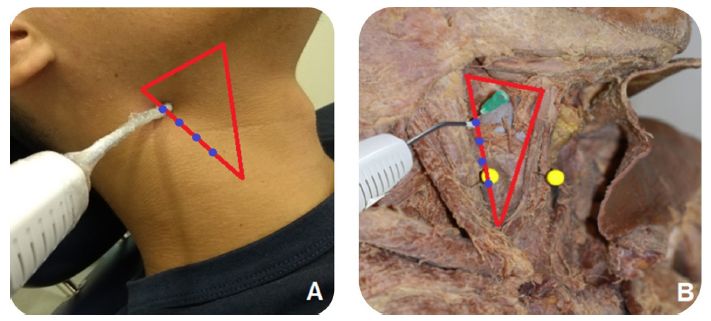
e extraoral application points selected were based on authors’ prior
observation in cadavers to determine the region where it is possible
to visualize the propagation of the visible red laser beam through the
different tissues of the cervical structures (figure 1). e study of the
anatomy of the human cervical region was performed on dissected parts
of cadavers (Human Anatomical eater, Department of Morphology).
All the cadavers used in this study were previously prepared using a
10% formaldehyde and glycerol medium. A diode laser (erapy XT
DMC®) was used, at a wavelength of λ660nm (visible red), continuous
mode, 100 mW power, 10 seconds per point, 0.028cm2 spot size over
all regions of the cervical trigones, time considered sufficient for visual
observation of the red light beam passing through the tissues. rough
direct observation, the area in which the visible red beam propagated and
reached the hypopharynx region to the point of being seen with the naked
eye was assessed.
Based on it, the carotid trigone was considered the best extraoral
region for laser application allowing to reach the hypopharyngeal region.
e anterior margin of the sternocleidomastoid muscle (SCM), one of
the boundaries of the carotid trigone, was used as the most favorable
application site, for being more easily clinically identified, through
PDF generado a partir de XML-JATS4R por Redalyc
Proyecto académico sin fines de lucro, desarrollado bajo la iniciativa de acceso abierto 173Revista Estomatológica Herediana, 2021, vol. 31, núm. 3, Julio-Septiembre, ISSN: 1019-4355 / 2225-7616
palpation. us, the low-level laser therapy was applied, in the clinical
phase of the study, in 4 points (4J per point) perpendicularly and in
contact with the skin, along the anterior margin of SCM muscle.
RESULTS
e three patients participating in this study were diagnosed with
squamous cell carcinoma on the border of the tongue and were
treated surgically and with head and neck radiotherapy. As a sequel to
treatment, all patients developed oral and hypopharyngeal mucositis,
and complained of odynophagia. Table 1 presents information related
to patients. Patients 1 and 2, who started oral and hypopharyngeal
photobiomodulation on the same day they started radiotherapy,
developed oral and hypopharyngeal mucositis, reporting mouth pain
and odynophagia; both patients maintained exclusively oral feeding
throughout the cancer treatment. roughout the treatment, both
patients reported symptoms improvement in the oral cavity, but only
patient 1 reported odynophagia improvement. e maximum score of
mucositis they developed was grade 3, according to the classification of
Sonis et al., 2004 (9).
Figure 1. Anatomy of the human cervical region (A and B). Propagation
of the visible red laser until reaching the hypopharyngeal mucosa (C).
PDF generado a partir de XML-JATS4R por Redalyc
Proyecto académico sin fines de lucro, desarrollado bajo la iniciativa de acceso abierto 174Liliane Grando, et al. Application of low-level laser therapy for analgesia of odinophagy caused by head and neck radiotherapy: Clinical c...
Table 1. Information of patients undergoing radiotherapy treatment in the head and neck region.
Figure 2. Definition of 4 points for laser therapy application along the anterior
margin of the sternocleidomastoid muscle in vivo (A) and in cadaver (B).
Patient 3 was referred to the Hospital Dentistry Center, only
when he was already in the sixteenth (16th) radiotherapy session,
exhibiting grade 4 oral mucositis, according to the Sonis, et al., 2004
(9) classification and using a nasoenteral tube. Aer performing oral and
hypopharyngeal photobiomodulation, the patient reported unmeasured
pain improvement both in the oral cavity and in swallowing.
e three patients voluntarily and spontaneously orally reported
about their experiences and sensations when being attended by the
Hospital Dentistry Center team of dentists. e transcribed reports are
presented below: Patient 1: “(...) Mouth sores appeared aer one week of
radiotherapy, it hurt a lot, I felt a lot of pain when swallowing, I chocked
a lot, I always had to drink some water aer food intake to be able to
swallow. As soon as the laser was applied, I already felt better, it didn’t stop
PDF generado a partir de XML-JATS4R por Redalyc
Proyecto académico sin fines de lucro, desarrollado bajo la iniciativa de acceso abierto 175Revista Estomatológica Herediana, 2021, vol. 31, núm. 3, Julio-Septiembre, ISSN: 1019-4355 / 2225-7616
hurting totally, but I managed to eat soon aer. e laser applied on my
neck helped me a lot with the pain to swallow (...)”. Patient 2: “(...) In the
first days of radiotherapy I lost my sense of taste, I already had a sore throat
and mouth injuries. In the mouth I didn’t feel much pain, but I felt a lot
of pain to swallow, I couldn’t eat properly. I wish there was something
penetrating in my throat to relieve the pain, just like the laser has worked
into the mouth (...). Patient 3: “(...) I felt pain all over my mouth and pain
when swallowing. I couldn’t even rinse my mouth with water, or brush my
teeth; I had to use a probe for eating. I started to recover very well aer the
laser application; with each session I felt better, mainly swallowing (...)”.
e region that allowed the best propagation of the laser beam was the
carotid trigone, allowing good access of the red light to the hypopharynx.
In this region, during application in cadavers, the visible red laser beam
was observed spreading through all muscle and cartilage layers, thus being
considered the best extraoral access site for laser application. e laser tip
was positioned perpendicularly and in contact with the skin, along the
anterior margin of the SCM muscle. e cadaveric anatomical study was
important to establish the extraoral location of laser applications. A total
of four points was then established, with 1 cm distance between them,
following the direction of the SCM muscle along the carotid trigone, for
an improved laser beam penetration and better analgesic effect (figure 2).
DISCUSSION
Mucositis is the most undesirable consequence of radiotherapy or
radiochemotherapy treatments (10,11). e low-level laser therapy has
important therapeutic properties in the treatment of mucositis. For
best results, the laser tip should be positioned as close as possible
and perpendicular to the injured tissue (12). In oral mucositis, this is
possible due to easier access to the tissues (4,7,8,11,13). In contrast, in
hypopharyngeal mucositis, this direct contact between the tip of the laser
device and the mucosa is unfeasible.
e carotid trigone has the anterior border of the median portion of
the SCM muscle as a posterolateral boundary, the upper belly of the
omohyoid muscle as an anteroinferior boundary and the posterior belly
of the digastric muscle and stylo-hyoid muscle as an upper limit. In this
study, the laser at the visible red wavelength was applied externally along
the cervical structures, seeking to observe from which anatomical region
the red light would reach the hypopharynx region. Although there is
a vast literature on the application of intra-oral laser in oral mucositis,
no clinical studies are available on the effects of photobiomodulation
therapy in the hypopharyngeal region (3,7,8,13,15,16). However, in
animal studies, significant effects have already been observed in the use of
extraoral laser in the treatment of oral mucositis (16).
Likewise, no studies were found that evaluate swallowing in a clinical or
instrumental way to verify the effect of applying the laser therapy protocol
in dysphagia. In the study by Gautam et al., comparing a group submitted
to oral laser therapy with a placebo group, the authors found that the
PDF generado a partir de XML-JATS4R por Redalyc
Proyecto académico sin fines de lucro, desarrollado bajo la iniciativa de acceso abierto 176Liliane Grando, et al. Application of low-level laser therapy for analgesia of odinophagy caused by head and neck radiotherapy: Clinical c...
need and duration of parenteral nutrition was significantly lower in the
group treated with laser to control radiotherapy- induced oral mucositis
(17). e authors considered the practice feasible to prevent and treat
oral mucositis and minimize pain and dysphagia; however, similar studies
that used laser in the hypopharynx region have not been described in
the literature. No equipment is available on the market that will allow
intraoral laser access to the hypopharynx region.
e laser photons, when crossing a material, can be absorbed or
scattered, which can cause attenuation in the light intensity. is is why,
a λ range between 600nm and 1200nm is widely used for the treatment of
deep tissues (up to ~2cm) (18). e λ808nm diode laser is used in clinical
practice for the therapeutic effects on deeper tissues (19, 20, 21).
Other authors, whose studies have evaluated the effect of low-level laser
therapy applied extraorally, highlight the lack of investigation reports on
the subject covered by our protocol. In addition, dosimetry revealed to
be quite complex for these cases (16). In this article, we are suggesting
the use of diode laser (erapy XT DMC), λ808nm, 4 points of 4J each,
1 cm apart, applied perpendicularly along the anterior belly of the SCM
muscle, bilaterally. In this way, a total of 32J is applied to the neck skin, in
the region of the carotid trigone, and so far it is impossible to know how
much of this energy actually reaches the hypopharyngeal mucosa.
e lack of measurable criteria when attending patients, lead us to opt
for qualitative analysis through three observational case studies, seeking
to understand how each patient understood empirically his individual
reality (22), without extrapolating these impressions to a whole group.
Despite being a clinical case series report, this report allows an initial
analysis of how promising the carotid trigone technique is and can further
be studied and improved (23), in an attempt to solve one of the major
problems that cancer patients experience during treatment, which is the
radio-induced hypopharyngeal mucositis.
Based on the patients’ reports, it is believed that laser therapy targeting
the carotid trigone region caused analgesia in the hypopharynx: Patient
1: "As soon as the laser was applied, I already felt improvement; it never
stopped hurting totally, but I was able to eat soon aer" and patient 3:
"I started to recover very well aer the laser applications; in each session
I felt better, especially to swallow". Although patient 2 did not report
improvement in odynophagia, it can be inferred that the laser application
contributed to patients 1 and 2 continuing to feed exclusively orally and
patient 3 progressing to oral feeding, considering that it is known that,
during radiotherapy in the head and neck region, the need to use an
alternative feeding route can reach 61% of cases (23), due to pain, weight
loss and dysphagia conditions.
e importance of developing a laser application protocol for
hypopharyngeal mucositis was also perceived by patient 1: "e laser
applied on my neck helped me a lot with the swallowing pain”. Patient
2 reveals that “... I wish there was something that entered my throat to
relieve the pain, just as laser has worked out in my mouth”.
PDF generado a partir de XML-JATS4R por Redalyc
Proyecto académico sin fines de lucro, desarrollado bajo la iniciativa de acceso abierto 177Revista Estomatológica Herediana, 2021, vol. 31, núm. 3, Julio-Septiembre, ISSN: 1019-4355 / 2225-7616
One of the limitations of this study was the difficulty in measuring the
energy that can reach the hypopharyngeal mucosa through the extraoral
application of laser therapy. In addition, clinical studies are required to
be able to classify the degree of hypopharyngeal mucositis and that better
assess the analgesia achieved by this suggested laser therapy protocol
in an extraoral region of the carotid trigone. It was concluded that
within the limitations of this study, the carotid trigone was defined as
the best extraoral region for laser application, in order to extend the
benefits of photobiomodulation to the treatment of hypopharyngeal
mucositis. Although most patients have reported improvement with the
laser application in this region, further studies are needed to gather
evidence of laser therapy effects, as well as the best protocols to be used.
Acknowledgments
Acknowledgments: Hospital Dentistry Center of the University Hospital
of the Federal University of Santa Catarina.
Bibliographic references
1. Ministério da Saúde (BR), Instituto Nacional de Câncer. Estimativa 2020:
incidência de câncer no Brasil. Brasilia: Instituto Nacional de Câncer;
2020. (Access date: February 23, 2021). Available in: https://www.inca.g
ov.br/publicacoes/livros/estimativa-2020-incidencia-de-cancer-no-brasil
2. World Health Organization. Oral Cancer. Geneva: World Health
Organization; 2020. (Access date: May 02, 2021). Available
in: https://www.who.int/cancer/ prevention/diagnosis-screening/oral-
cancer/en/ (access date: May 02, 2020).
3. Mallick S, Benson R, Rath GK. Radiation induced oral mucositis: a review of
current literature on prevention and management. European Archives of
Oto-Rhino-Laryngology. 2016; 273(9): 2285-2293.
4. Tolentino EDS, Centurion BS, Ferreira LHC, Souza APD, Damante J H,
Rubira-Bullen I R. Oral adverse effects of head and neck radiotherapy:
literature review and suggestion of a clinical oral care guideline for
irradiated patients. Journal of Applied Oral Science.
5. Fekrazad R, Chiniforush N. Oral mucositis prevention and management by
therapeutic laser in head and neck cancers. Journal of Lasers in Medical
Sciences. 2014;
6. Oton-Leite AF, Elias LSA, Morais MO, et al. Effect of low level laser therapy
in the reduction of oral complications in patients with cancer of the head
and neck submitted to radiotherapy. Special Care in Dentistry. 2013;
33(6): 294-300.
7. Zecha JA, Raber-Durlacher JE, Nair RG, et al. Low- level laser therapy/
photobiomodulation in the management of side effects of chemoradiation
therapy in head and neck cancer: part 2: proposed applications and
treatment protocols. Supportive Care in Cancer. 2016; 24(6): 2793-2805.
8. Zecha JA, Raber-Durlacher JE, Nair RG, et al. Low level laser therapy/
photobiomodulation in the management of side effects of chemoradiation
therapy in head and neck cancer: Part 1: Mechanisms of action,
PDF generado a partir de XML-JATS4R por Redalyc
Proyecto académico sin fines de lucro, desarrollado bajo la iniciativa de acceso abierto 178Liliane Grando, et al. Application of low-level laser therapy for analgesia of odinophagy caused by head and neck radiotherapy: Clinical c...
dosimetric, and safety considerations. Supportive Care in Cancer. 2016;
24(6): 2781-2792.
9. Sonis ST, Elting LS, Keefe D, et al. Perspectives on cancer therapy-
induced mucosal injury: pathogenesis, measurement, epidemiology, and
consequences for patients. Cancer: Interdisciplinary International Journal
of the American Cancer Society. 2004; 100(S9): 1995- 2025.
10. Bossi P, Alfieri S. e benefit of a multidisciplinary approach to the
patient treated with (chemo) radiation for head and neck cancer. Current
Treatment Options in Oncology. 2016; 17(10): 1-10.
11. Alterio D, Gerardi MA, Cella L, et al. Radiation-induced acute dysphagia.
Strahlentherapie und Onkologie. 2017; 193(11): 971-981.
12. Maciel CM, Piva MR, Ribeiro MAG, de Santana- Santos T, Ribeiro
C F, Martins-Filho PRS. Methylene Blue-mediated photodynamic
inactivation followed by low-laser therapy versus Miconazole gel in the
treatment of denture stomatitis. Journal of Prosthodontics. 2016; 25(1):
28-32.
13. Simões A, Eduardo FP, Luiz AC, et al. Laser phototherapy as topical
prophylaxis against head and neck cancer radiotherapy-induced oral
mucositis: comparison between low and high/low power lasers. Lasers in
Surgery and Medicine: e Official Journal of the American Society for
Laser Medicine and Surgery.2009; 41(4): 264-270.
14. Williams III DW. (1997, June). An imager’s guide to normal neck anatomy.
Seminars in Ultrasound, CT and MRI. 1997; 18(3): 157-181.
15. Peralta-Mamani M, da Silva BM, da Silva-Pinto AC, et al. Low-level laser
therapy dosimetry most used for oral mucositis due to radiotherapy for
head and neck cancer: a systematic review and meta-analysis. Critical
Reviews in Oncology/Hematology. 2019; 138: 14-23.
16. ieme S, Ribeiro JT, Dos Santos BG, et al. Comparison of
photobiomodulation using either an intraoral or an extraoral laser on oral
mucositis induced by chemotherapy in rats. Supportive Care in Cancer.
2020; 28(2): 867-876.
17. Gautam AP, Fernandes DJ, Vidyasagar MS, Maiya AG, Vadhiraja BM.
Low level laser therapy for concurrent chemoradiotherapy induced oral
mucositis in head and neck cancer patients–a triple blinded randomized
controlled trial. Radiotherapy and Oncology. 2012; 104(3): 349-354.
18. Sousa MV, Prates R, Kato IT, et al. Laser scattering by transcranial rat brain
illumination. Biophotonics: Photonic Solutions for Better Health Care
III. 2012; 8427: 842728.
19. Brignardello-Petersen R. Low-level laser therapy may reduce the time
of recovery from paresthesia aer orthognathic surgery. Journal of the
American Dental Association 2018; 149(2): e44-e44.
20. Hamid MA. Low-level laser therapy on postoperative pain aer mandibular
third molar surgery. Annals of Maxillofacial Surgery. 2017; 7(2): 207.
21. Noba C, Mello-Moura ACV, Gimenez T, Tedesco T K, Moura-Netto
C. Laser for bone healing aer oral surgery: systematic review. Lasers in
Medical Science. 2018; 33(3): 667-674.
22. Lara AMD B, Molina AA. (2015). Pesquisa Qualitativa: apontamentos,
conceitos e tipologias. Em: Arnaut C, Gonzaga M. Metodologia e
técnicas de pesquisa nas áreas de ciências humanas. Maringá, PR: EdUem.
p.121-72.
PDF generado a partir de XML-JATS4R por Redalyc
Proyecto académico sin fines de lucro, desarrollado bajo la iniciativa de acceso abierto 179Revista Estomatológica Herediana, 2021, vol. 31, núm. 3, Julio-Septiembre, ISSN: 1019-4355 / 2225-7616
23. Wopken K, Bijl HP, Langendijk JA. Prognostic factors for tube feeding
dependence aer curative (chemo-) radiation in head and neck cancer:
a systematic review of literature. Radiotherapy and Oncology. 2018;
126(1):56-67.
Notas de autor
a PhD degree, professor
aa PhD degree, professor
aaa PhD degree, professor
b Specialist
c Master Student
aaaa PhD degree, professor
d Master
Mailing address: Profa. Liliane Grando - Centro de Ciências
da Saúde; Departamento de Patologia Campus Universitário
– Bairro Trindade Universidade Federal de Santa Catarina
88040-900 Florianópolis, Santa Catarina, Brasil. Phone: 55 48
99971-6864. E-mail: liliane.j.grando@ufsc.br
Declaración de intereses
* e authors affirm that we have no financial affiliation or
involvement with any commercial organization with direct
financial interest in the subject or materials discussed in this
manuscript. Any other potential conflict of interest is disclosed.
Información adicional
Ethics approval: Research Ethics Committee at Federal University of
Santa Catarina under number CAAE 72989417 0 0000 0121, dated
April 9, 2018.
Authors' contributions: All authors contributed to this manuscript.
Enlace alternativo
https://revistas.upch.edu.pe/index.php/REH/article/view/4046/4599
(pdf)
PDF generado a partir de XML-JATS4R por Redalyc
Proyecto académico sin fines de lucro, desarrollado bajo la iniciativa de acceso abierto 180You can also read