Atypical benign partial epilepsy and a new variant of SLC35A3 gene plus 2p25.1 duplication. Phenotypic-Genotypic correlation?
←
→
Page content transcription
If your browser does not render page correctly, please read the page content below
Atypical benign partial epilepsy and a new variant of
SLC35A3 gene plus 2p25.1 duplication. Phenotypic-
Genotypic correlation?
Enrico Parano ( enrico.parano@irib.cnr.it )
Azienda Ospedaliero Universitaria Policlinico Vittorio Emanuele Catania
Piero Pavone
Azienda Ospedaliero Universitaria Policlinico Vittorio Emanuele Catania
Xena Giada Pappalardo
Consiglio Nazionale delle Ricerche
Roberto Caraballo
Paediatric Hospital Dr Juan Garrahan: Hospital de Pediatria Prof Dr Juan P Garrahan
Andrea Domenico Praticò
University Hospital Vittorio Emanuele Catania Polyclinic: Azienda Ospedaliero Universitaria Policlinico
Vittorio Emanuele Catania
Raffaele Falsaperla
Azienda Ospedaliero Universitaria Policlinico Vittorio Emanuele Catania
Research article
Keywords: Atonic seizures, pseudo-Lennox syndrome, Continuous Spike-Wave during Sleep (CSWS),
Electrical Status Epilepticus during Sleep (ESES), phenotypic-genotypic correlation
Posted Date: March 15th, 2021
DOI: https://doi.org/10.21203/rs.3.rs-315555/v1
License: This work is licensed under a Creative Commons Attribution 4.0 International License.
Read Full License
Page 1/5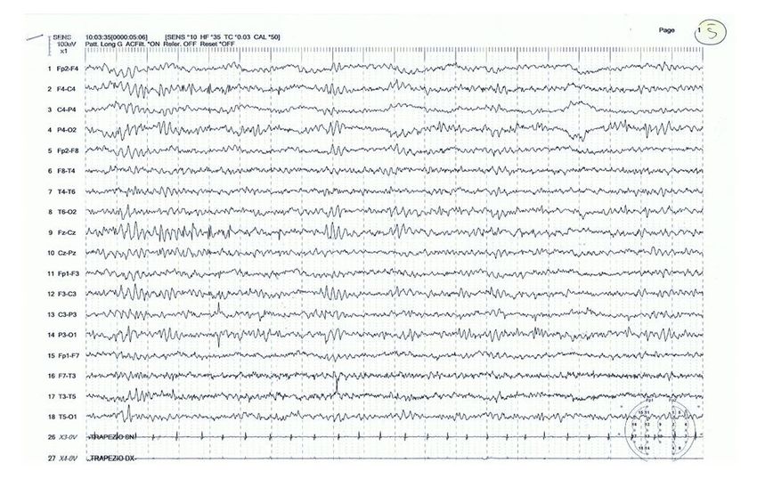
Abstract
Background Atypical Benign Partial Epilepsy (ABPE), recognized also as pseudo-Lennox syndrome, is an
uncommon form of epilepsy characterized by generalized minor seizures such as atonic, absences, or
myoclonic seizures, and electroencephalographic pattern of focal or multifocal sharp waves with
activation of epileptiform discharges during sleep. ABPE is indicated as a variant of ESES (ILAE
classification 2017). ESES is a clinical entity that is characterized by encephalopathy with cognitive/
behavioral regression and EEG pattern of electrical status epilepticus during slow sleep. Fine and gross
motor, language and social/behavioral impairment are associated symptoms, which may have reversible
or persistent course. ABPE has been ascribed to the group of the “epilepsy aphasia spectrum” disorders,
which includes also Rolandic Epilepsy, Landau-Kleffner syndrome, and electrical status epilepticus during
sleep/continuous spike-wave during sleep. We report a young boy with a previous mild motor and
language delay, who at 2-year-old presented with recurrent atonic seizures and an EEG pattern consisting
of continuous spike and waves during sleep. Methods Next Generation Sequencing and microarray
technology were used to investigate the molecular background of the proband, and the findings were then
analyzed and integrated with clinical data. Results The child has been followed up to 7 years of age
showing a progressive, complete EEG resolution and a normalization of the previous motor and cognitive
impairment in association to the levetiracetam treatment. Genetic diagnosis displayed a novel
heterozygous mutation c.310G>A of the SLC35A3 gene and a partial duplication of the short arm of
chromosome 2, which might have pathogenic correlation with the neurological signs presented by the
child. Conclusions Data generated by a genomic approach disclose a more comprehensive view of the
genotype-phenotype correlation analysis for the novel pathogenic variant and ABPE. The relationship
between the phenotypic manifestations of the child and genetic data is discussed.

Full Text
Due to technical limitations, full-text HTML conversion of this manuscript could not be completed.
However, the manuscript can be downloaded and accessed as a PDF.

Figures

                                                 Page 2/5
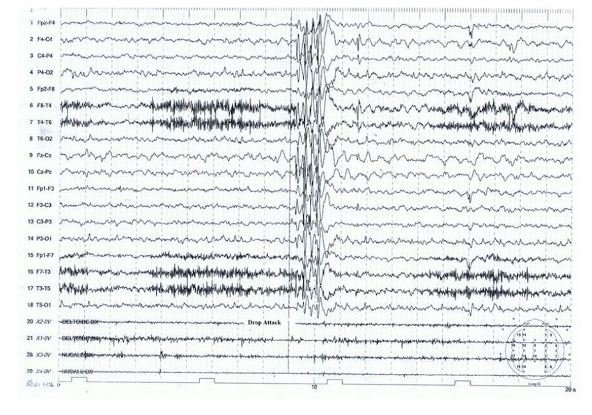
Figure 1

EEG at 28 months. To note, the continuous spike and waves during the slow sleep CSWS type.

                                               Page 3/5
Figure 2
Atonic crisis was registered during EEG record. Spikes and wave prevalent in the fronto-centro-temporal
areas during the atonic crisis.
Page 4/5Figure 3
EEG after ethosuximide and levetiracetam with noticeable improvement of the EEG. Isolated ft-onto-
central spikes remain.
Page 5/5You can also read