Atypical hemolytic uremic syndrome after childbirth: a case report - Annals of ...
←
→
Page content transcription
If your browser does not render page correctly, please read the page content below
Case Report
                                                                                                                                    Page 1 of 5

Atypical hemolytic uremic syndrome after childbirth: a case report
Hong Sang Choi1, Jae Won Yun2,3, Hee-Jin Kim2, Doyeun Oh4, Nah Ihm Kim5, Chang Seong Kim1,
Seong Kwon Ma1, Soo Wan Kim1, Eun Hui Bae1^
1
    Department of Internal Medicine, Chonnam National University Medical School, Gwangju, Korea; 2Department of Laboratory Medicine and
Genetics, Samsung Medical Center, Sungkyunkwan University School of Medicine, Seoul, Korea; 3Veterans Medical Research Institute, Veterans
Health Service Medical Center, Seoul, Korea; 4Department of Internal Medicine, School of Medicine, CHA University, School of Medicine,
Seongnam, Korea; 5Department of Pathology, Chonnam National University Medical School, Gwangju, Korea
Correspondence to: Eun Hui Bae, MD, PhD. Department of Internal Medicine, Chonnam National University Medical School, 42 Jebongro, Gwangju
61469, Korea. Email: baedak76@gmail.com.

                  Abstract: We report a case of atypical hemolytic uremic syndrome (HUS) that occurred after childbirth. A
                  33-year-old female was admitted to the emergency room, complaining of abdominal pain six days after giving
                  birth to twins. The patient was diagnosed with hemoperitoneum due to hepatic hemangioma rupture and
                  a left lateral hepatectomy was performed. Angioembolization was performed for the accompanying uterine
                  artery bleeding. After that, her kidney function worsened after the 12th day postpartum. Microangiopathic
                  anemia, thrombocytopenia and renal dysfunction were observed. Shiga toxin-producing Escherichia coli
                  was negative in the stool. Plasma ADMATS 13 activity was normal. After transfer to the nephrology
                  department with suspected atypical HUS, the patient underwent fresh frozen plasma (FFP) transfusion
                  with three hemodialysis sessions. The patient improved without additional dialysis, but a renal biopsy was
                  performed because of persistent proteinuria. Renal pathologic findings were compatible with thrombotic
                  microangiopathy. A genetic test for atypical HUS revealed variants of uncertain significance in the
                  complement factor H related (CFHR) 4 gene and the presence of CFHR3-CFHR1 copy number gain. The
                  CFHR3-CFHR1 copy number gain found in this case is a rare causative mutation of atypical HUS. This case
                  suggests that genetic testing of atypical HUS should include analysis of CFH-CFHR rearrangements as well
                  as general screening for complement-associated genes.

                  Keywords: Atypical hemolytic uremic syndrome (HUS); CFHR3-CFHR1; copy number gain; pregnancy; case
                  report

                  Submitted May 08, 2020. Accepted for publication Sep 30, 2020.
                  doi: 10.21037/atm-20-3789
                  View this article at: http://dx.doi.org/10.21037/atm-20-3789

Introduction                                                                 infection, pregnancy, organ transplantation, malignancy,
                                                                             autoimmune disease, and drugs (1). Cases of pregnancy-
Hemolytic uremic syndrome (HUS) is a comparatively
                                                                             associated atypical HUS are known to occur during the
rare hematological abnormality that accompanies
microangiopathic hemolytic anemia (MAHA),                                    postpartum period (2). More than half of thrombotic
thrombocytopenia, and acute kidney injury (AKI). Atypical                    microangiopathies associated with pregnancy have genetic
HUS is caused by inborn or acquired genetic defects of                       abnormalities in the complement system. Abnormalities
genes that are associated with regulation of the complement                  of several genes associated with the complement system
pathway. Atypical HUS is known to be triggered by factors                    have been reported (3), and mutations can be diagnosed
that induce the activation of complement cascades, including                 relatively easily using advanced diagnostic methods, such

^ ORCID: 0000-0003-1727-2822.

© Annals of Translational Medicine. All rights reserved.                    Ann Transl Med 2021;9(1):79 | http://dx.doi.org/10.21037/atm-20-3789
Page 2 of 5                                                                                                              Choi et al. Postpartum atypical HUS

         Twin delivery by                                                            Hemodialysis with
         cesarean section                                   Detection of AKl          FFP transfusion                   Discharge
              Day 0
                       Day 13
                               Day 19−21	     Day 28

                                       Day 7			   Day 18	   Day 25		   Day 38
                                 Left lateral hepatic                             Detection of           Renal biopsy               S-Cr normalization
                             segmentectomy for hepatic                         Fragmented RBC
                               hemangioma rupture &                              & genetic test
                            Angioembolization for uterine
                                  artery bleeding

Figure 1 Timeline of patient’s clinical course. AKI, acute kidney injury; RBC, red blood cell; FFP, fresh frozen plasma; s-Cr, serum
creatinine.

as new generation sequencing (NGS). Similarly, copy                                   diarrhea and no enterohemorrhagic Escherichia coli was
number variation resulting from genomic rearrangement                                 detected in her stool. Plasma ADMATS 13 activity was
can be diagnosed using techniques like multiplex ligation-                            60%. The lack of accompanying neurological symptoms led
dependent probe amplification (MLPA) (4). Here, we report                             us to suspect atypical HUS. Renal biopsy revealed findings
successful treatment of a patient who was diagnosed with                              consistent with thrombotic microangiopathy (Figure 2).
postpartum atypical HUS after childbirth by identification                               Hemodialysis was initiated immediately and the patient
of genetic abnormalities in the complement system using                               was administered fresh frozen plasma (FFP). FFP was
NGS and MLPA. This case is an atypical HUS due to the                                 administered with three sessions of dialysis, after which
rare copy number variation of the CFHR genes, and shows                               her laboratory findings began to improve. Platelet counts
that the genetic test for atypical HUS must be performed                              improved without blood transfusion and serum LDH
for CFH-CFHR rearrangement. We present the following                                  levels gradually decreased. This trend persisted even after
article in accordance with the CARE reporting checklist                               cessation of dialysis and FFP administration. The patient
(available at http://dx.doi.org/10.21037/atm-20-3789) (5).                            was discharged 10 days after being transferred to the
                                                                                      department of nephrology. No signs of renal failure or
                                                                                      recurrence of atypical HUS were apparent on her four-
Case presentation
                                                                                      month outpatient follow-up, at which her laboratory
A 33-year-old woman with renal impairment was referred                                findings were: serum creatinine level, 0.53 mg/dL; platelet
to our nephrology department. She had delivered twins                                 count of 219,000/μL; and serum LDH level, 378 U/L.
through Caesarean section 18 days before referral. She                                   Genetic testing was performed concurrently with the
denied any underlying disease, medication history or                                  treatment regime to identify the cause of the atypical
familial history of renal impairment. Six days after delivery,                        HUS. Sequencing of 17 genes known to be associated with
she had received left lateral hepatic segmentectomy and                               atypical HUS (i.e., C3, C4BPA, CD46, CFB, CFH, CFHR1,
angioembolization of the uterine artery due to hepatic                                CFHR2, CFHR3, CFHR4, CFHR5, CFI, DGKE, THBD,
hemangioma rupture and uterine artery bleeding. Her                                   PLG, etc.) as well as of ADAMTS13 and C5 was performed.
serum creatinine level started to increase approximately                              Gene sequencing was performed by Illumina Hi-seq 2500
12 days after delivery and continued to increase despite                              platform, followed by targeted Sanger sequencing for
proper management (Figure 1). On the day of referral, her                             validation of identified genetic variants. Gene dosage tests
laboratory data were: white blood cell count, 8,800/μL;                               were performed using MLPA. Genetic profiling revealed
hemoglobin, 7.7 g/dL; platelet count, 68,000/μL; blood                                a substitutional variant c.1607 T>G {p.[Ile536Arg] or
urea nitrogen, 33.2 mg/dL; serum creatinine, 2.46 mg/dL;                              I536R} in CFHR4. Additionally, MLPA analysis revealed
and lactate dehydrogenase (LDH), 3,958 U/L. Peripheral                                the presence of a copy number gain in the CFHR3-CFHR1
blood smears were performed for further evaluation of                                 regions (Figure 3). Genetic testing confirmed a diagnosis of
bicytopenia and showed fragmented red blood cells.                                    atypical HUS caused by a point mutation and copy number
Together, these results were indicative of thrombotic                                 gain in CFHR. About 1 year after the onset of the disease,
microangiopathy. The patient had no recent history of                                 her serum creatinine level was 0.55 mg/dL, and there was

© Annals of Translational Medicine. All rights reserved.                              Ann Transl Med 2021;9(1):79 | http://dx.doi.org/10.21037/atm-20-3789
Annals of Translational Medicine, Vol 9, No 1 January 2021 Page 3 of 5
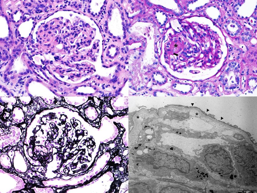
A B
C D
Figure 2 Renal biopsy. (A) Glomeruli show thickened capillary wall and fragmented red blood cells are trapped in the mesangial matrix
(hematoxylin and eosin stain, ×40). (B) The periodic acid Schiff (PAS) stained glomerulus demonstrates segmental mesangiolysis (asterisk)
and double contours in capillary basement membrane (arrow) (×40). (C) Light microscopy image of the kidney biopsy specimen shows
double contours of the glomerular basement membrane with Jones methenamine silver stain (×40). (D) Electron microscopy of the kidney
biopsy reveals irregular electron-lucent expansion of the subendothelial zone (asterisk) and diffuse foot process fusion (arrowhead) (×8,000).
no recurrence or unanticipated outcome during the follow- to the 4 th month (2). The development of HUS in the
up period. current case occurred 12 days postpartum, consistent with
All procedures performed in studies involving human previous reports. Initially, development of HUS can be
participants were in accordance with the ethical standards of detected through a decrease in platelet counts and AKI.
the institutional and/or national research committee(s) and Differentiation from other causes of AKI that may occur
with the Helsinki Declaration (as revised in 2013). Written during pregnancy, including preeclampsia, eclampsia,
informed consent was obtained from the patient. HELLP (hemolysis, elevated liver enzymes, low platelet)
syndrome, and thrombotic thrombocytopenic purpura
(TTP), is important (6). Other common causes of AKI,
Discussion
such as volume depletion or acute tubular necrosis, should
We present a case of pregnancy-associated atypical HUS also be considered. In particular, TTP is very similar to
during the postpartum period in a young woman who has the expression of HUS, which can complicate differential
two genetic mutations in the complement cascade. The diagnosis. However, even before ADAMTS13 activity test
patient had previously given birth and had no history of results are confirmed, TTP is more thrombocytopenic
diagnosis or treatment with thrombocytopenia or AKI. and has less severe renal failure than HUS. Furthermore,
Fakhouri et al. reported that approximately 20% of atypical TTP predominantly occurs in the 2nd or 3rd trimester of
HUS patients in France are related to pregnancy; ~80% of pregnancy (7).
these were reported during the postpartum period, with the Genomic rearrangement in the CFH-CFHR region
time of onset varying widely from the 3rd day postpartum resulting in copy number gain of CFHR3 and CFHR1
© Annals of Translational Medicine. All rights reserved. Ann Transl Med 2021;9(1):79 | http://dx.doi.org/10.21037/atm-20-3789Page 4 of 5 Choi et al. Postpartum atypical HUS
Patient
Control1
Control2
1.5
Relative copy number (MLPA)
1.0
0.5
Relative copy number (exome sequencing)
1.5
1.0
0.5
0.0
CFH CFHR3 CFHR1 CFHR4 CFHR2 CFHR5
196650000 196700000 196750000 196800000 196850000 196900000 196950000
Position in chromosome 1
Figure 3 The relative copy number based on the multiplex ligation-dependent probe amplification (MLPA) study (upper plot). The value
1.0 on Y axis represents two copies or copy neutral. Each “+” represents each probe in MLPA study. The relative copy number based on
the exome sequencing copy number analysis (lower plot). Each “+” represents an exon region in exome sequencing study. The grey zone
indicates 99% confidence interval of normal controls without copy number alterations.
was discovered in our patient by MLPA analysis. Several patient. The function of CFHR4 remains unclear, though
previous studies have reported atypical HUS by the it is known to bind to the C3b and to have a complement
CFHR1/CFH hybrid gene (4,8,9). Valoti et al. reported the modulatory activity in the form of a factor H cofactor
generation of CFHR1/CFH hybrid gene and an extra copy enhancing activity (10). The I536R mutation in our patients
of CFHR3 and CFHR1 in a patient with atypical HUS (4). is located in CFHR4 SCR9, and its role in protein function
Although the patient in the current case did not have a and disease association is unknown. In addition, except for a
CFHR1/CFH hybrid gene, her atypical HUS was likely deletion of CFHR1/CFHR4, no other CFHR4 mutation has
caused by a CFHR3-CFHR1 copy number gain, which is been reported to be associated with atypical HUS.
similar to a variant described by Valoti et al. (4). In addition Our study found a very rare CFHR3-CFHR1 copy
to the copy number gain of CFHR3-CFHR1, a variant of number gain as the cause of atypical HUS. However, our
uncertain significance was found in the CFHR4 gene in our study has some limitations. First, a functional study was
© Annals of Translational Medicine. All rights reserved. Ann Transl Med 2021;9(1):79 | http://dx.doi.org/10.21037/atm-20-3789Annals of Translational Medicine, Vol 9, No 1 January 2021 Page 5 of 5
not performed on the discovered mutations. In addition, original work is properly cited (including links to both the
the mechanism of how CFHR3-CFHR1 copy number gain formal publication through the relevant DOI and the license).
generated atypical HUS could not be revealed. Second, See: https://creativecommons.org/licenses/by-nc-nd/4.0/.
although no family member with similar symptoms was
found, it is a limitation of our study that genetic testing of
References
the family members was not performed because of that.
In summary, we describe a case of atypical HUS that 1. Lee H, Kang E, Kang HG, et al. Consensus regarding
occurred during the postpartum period and was likely diagnosis and management of atypical hemolytic uremic
caused by a CFHR3-CFHR1 copy number gain. This case syndrome. Korean J Intern Med 2020;35:25-40.
suggests that genetic testing of atypical HUS should include 2. Fakhouri F, Roumenina L, Provot F, et al. Pregnancy-
analysis of CFH-CFHR rearrangements as well as general associated hemolytic uremic syndrome revisited in the
screening for complement-associated genes. era of complement gene mutations. J Am Soc Nephrol
2010;21:859-67.
3. Noris M, Bresin E, Mele C, et al. Genetic Atypical
Acknowledgments
Hemolytic-Uremic Syndrome. In: Adam MP, Ardinger
Funding: This study was supported by the Korean Ministry HH, Pagon RA, et al. editors. GeneReviews(®). Seattle
of Education (2017R1D1A1B03029582) and grant (WA); 1993.
(BCRI20076 and BCRI20041) of Chonnam National 4. Valoti E, Alberti M, Tortajada A, et al. A novel atypical
University Hospital Biomedical Research Institute. hemolytic uremic syndrome-associated hybrid CFHR1/
CFH gene encoding a fusion protein that antagonizes
factor H-dependent complement regulation. J Am Soc
Footnote
Nephrol 2015;26:209-19.
Reporting Checklist: The authors have completed the CARE 5. Gagnier JJ, Kienle G, Altman DG, et al. The CARE
reporting checklist. Available at http://dx.doi.org/10.21037/ Guidelines: Consensus-based Clinical Case Reporting
atm-20-3789 Guideline Development. Glob Adv Health Med
2013;2:38-43.
Conflicts of Interest: All authors have completed the ICMJE 6. Baghli S, Abendroth C, Farooq U, et al. Atypical
uniform disclosure form (available at http://dx.doi. Presentation of Pregnancy-Related Hemolytic Uremic
org/10.21037/atm-20-3789). The authors have no conflicts Syndrome. Am J Kidney Dis 2018;72:451-6.
of interest to declare. 7. Fakhouri F. Pregnancy-related thrombotic
microangiopathies: Clues from complement biology.
Ethical Statement: The authors are accountable for all Transfus Apher Sci 2016;54:199-202.
aspects of the work in ensuring that questions related 8. Alobaidi S, AlDabbagh A, Alamoudi A, et al. Three
to the accuracy or integrity of any part of the work are months interval therapy of Eculizumab in a patient with
appropriately investigated and resolved. All procedures atypical hemolytic uremic syndrome with hybrid CFHR1/
performed in studies involving human participants were in CFH gene. CEN Case Rep 2019;8:139-43.
accordance with the ethical standards of the institutional 9. Eyler SJ, Meyer NC, Zhang Y, et al. A novel hybrid
and/or national research committee(s) and with the Helsinki CFHR1/CFH gene causes atypical hemolytic uremic
Declaration (as revised in 2013). Written informed consent syndrome. Pediatr Nephrol 2013;28:2221-5.
was obtained from the patient for publication of this 10. Skerka C, Chen Q, Fremeaux-Bacchi V, et al. Complement
manuscript and any accompanying images. factor H related proteins (CFHRs). Mol Immunol
2013;56:170-80.
Open Access Statement: This is an Open Access article
distributed in accordance with the Creative Commons
Cite this article as: Choi HS, Yun JW, Kim HJ, Oh D, Kim NI,
Attribution-NonCommercial-NoDerivs 4.0 International
Kim CS, Ma SK, Kim SW, Bae EH. Atypical hemolytic uremic
License (CC BY-NC-ND 4.0), which permits the non-
syndrome after childbirth: a case report. Ann Transl Med
commercial replication and distribution of the article with
2021;9(1):79. doi: 10.21037/atm-20-3789
the strict proviso that no changes or edits are made and the
© Annals of Translational Medicine. All rights reserved. Ann Transl Med 2021;9(1):79 | http://dx.doi.org/10.21037/atm-20-3789You can also read