Automatic Segmentation, Localization, and Identification of Vertebrae in 3D CT Images Using Cascaded Convolutional Neural Networks
←
→
Page content transcription
If your browser does not render page correctly, please read the page content below
Automatic Segmentation, Localization, and
                                            Identification of Vertebrae in 3D CT Images
                                           Using Cascaded Convolutional Neural Networks

                                           Naoto Masuzawa1 , Yoshiro Kitamura1 , Keigo Nakamura1 , Satoshi Iizuka2 , and
                                                                       Edgar Simo-Serra3
arXiv:2009.13798v1 [eess.IV] 29 Sep 2020

                                                    1
                                                   Imaging Technology Center, Fujifilm Corporation, Minato, Tokyo, Japan
                                                2
                                                  Center for Artificial Intelligence Research, University of Tsukuba, Tsukuba,
                                                                                  Ibaraki, Japan
                                            3
                                                Department of Computer Science and Engineering, Waseda University, Shinjuku,
                                                                                   Tokyo, Japan
                                                                         naoto.masuzawa@fujifilm.com

                                                        Abstract. This paper presents a method for automatic segmentation,
                                                        localization, and identification of vertebrae in arbitrary 3D CT images.
                                                        Many previous works do not perform the three tasks simultaneously even
                                                        though requiring a priori knowledge of which part of the anatomy is
                                                        visible in the 3D CT images. Our method tackles all these tasks in a
                                                        single multi-stage framework without any assumptions. In the first stage,
                                                        we train a 3D Fully Convolutional Networks to find the bounding boxes
                                                        of the cervical, thoracic, and lumbar vertebrae. In the second stage, we
                                                        train an iterative 3D Fully Convolutional Networks to segment individual
                                                        vertebrae in the bounding box. The input to the second networks have an
                                                        auxiliary channel in addition to the 3D CT images. Given the segmented
                                                        vertebra regions in the auxiliary channel, the networks output the next
                                                        vertebra. The proposed method is evaluated in terms of segmentation,
                                                        localization, and identification accuracy with two public datasets of 15
                                                        3D CT images from the MICCAI CSI 2014 workshop challenge and 302
                                                        3D CT images with various pathologies introduced in [1]. Our method
                                                        achieved a mean Dice score of 96%, a mean localization error of 8.3 mm,
                                                        and a mean identification rate of 84%. In summary, our method achieved
                                                        better performance than all existing works in all the three metrics.

                                                        Keywords: Vertebrae · Segmentation · Localization · Identification ·
                                                        Convolutional neural networks

                                           1        Introduction
                                           Automatic segmentation, localization, and identification of individual vertebrae
                                           from 3D CT (Computed Tomography) images play an important role in a pre-
                                           processing step of automatic analysis of the spine. However, many previous works
                                           are not able to perform segmentation, localization, and identification simultane-
                                           ously and require a priori knowledge of which part of the anatomy is visible in
                                           the 3D CT images.
2 N. Masuzawa et al.
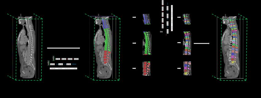
Fig. 2. a) A sagittal slice of 3D CT im-
Fig. 1. Differences in anatomy between ages which includes cervical (C1-C7),
cervical and thoracic vertebrae, and thoracic (T1-T12), and lumbar (L1-L5)
thoracic and lumbar vertebrae. vertebrae. b) Segmentation and identi-
fication of the individual vertebrae.
We overcome those drawbacks with a single multi-stage framework. More
specifically, in the first stage, we train a 3D Fully Convolutional Networks (we
call it ”Semantic Segmentation Net”), which segments cervical, thoracic, and
lumbar vertebrae so as to find the bounding boxes. As shown in Figure 1, tho-
racic vertebrae are distinguished from the cervical and lumbar vertebrae by
whether they connect to their ribs and therefore it appears that the Semantic
Segmentation Net performs well even if the field-of-view (FOV) is limited. In
the second stage, we train an iterative 3D Fully Convolutional Networks (we
call it ”Iterative Instance Segmentation Net”), which segments (i.e., predicts the
labels of all voxels in the 3D CT images), localizes (i.e., finds the centroids of
all vertebrae), and identifies (i.e., assigns the anatomical labels) the vertebrae
in the bounding box one-by-one. Figure 2 shows an example input image and
the corresponding image synthesized by the proposed method. In summary, our
contribution is as follows. 1) A two-stage coarse-to-fine approach for vertebrae
segmentation, localization, and identification. 2) In-depth experiments and com-
parisons with existing approaches.
2 Related work
The challenges associated with automatic segmentation, localization, and iden-
tification of individual vertebrae are due to the following three points. 1) High
similarity in appearance of the vertebrae. 2) The various pathologies such as the
abnormal spine curvature and vertebral fractures. 3) The variability of input 3D
CT images such as FOV, resolution, and image artifacts. To address these chal-
lenges, many methods have been proposed. Traditionally, vertebral segmentation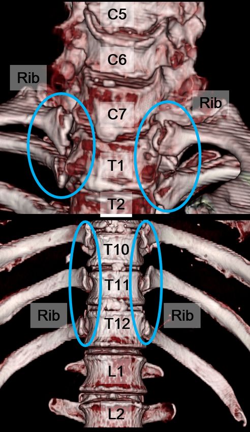
Automatic Segmentation, Localization and Identification of Vertebrae       3

has used mathematical methods such as atlas-based segmentation or deformable
models [5, 8, 9]. Speaking of localization and identification, Glocker et al. [1, 2]
proposed a method based on regression forests with a challenging dataset. They
introduced 302 3D CT images with various pathologies, the narrow FOV, and
metal artifacts. Recently, deep learning has been employed in the applications
of vertebral segmentation, localization, and identification. Yang et al. [13] pro-
posed a deep image-to-image network (DI2IN) to predict centroid coordinates
of vertebrae. On the other hand, the common way to segment vertebrae using
deep learning is to use semantic segmentation to predict the labels of all voxels
in input 3D CT images. For example, Janssens et al. [4] proposed a 3D fully con-
volutional neural networks (FCN) to segment lumbar vertebrae. However, the
way based on the semantic segmentation can segment vertebrae such as lumbar
only when whole of the vertebrae is visible in 3D CT images. This motivated
Lessmann et al. [10] to consider vertebral segmentation as an instance segmenta-
tion problem. The networks introduced by Lessman et al. [10] have an auxiliary
channel in addition to the input. Given the segmented vertebra regions in the
auxiliary channel, the networks output the next vertebra. Thus, the method
proposed by Lessmann et al. [10] is able to perform vertebral segmentation even
though whole of the vertebrae is not visible in 3D CT images and the number
of vertebra is not known a priori.
    Although the method by Lessmann et al. [10] achieves high segmentation
accuracy, it does not predict anatomical labels (i.e., cervical C1-C7, thoracic
T1-T12, lumbar L1-L5) for each vertebra and it does not handle general 3D CT
images where it is not known in advance which part of the anatomy is visible.
In fact, their method requires a priori knowledge of anatomy, such as lumbar 5.
On the other hand, our approach is able to predict anatomical labels and handle
general 3D CT images.

3   Proposed Method

                 Fig. 3. A schematic view of the present approach.

   Our method relies on a two-stage approach as shown in Figure 3. The first
stage aims to segment cervical, thoracic, and lumbar vertebrae from input 3D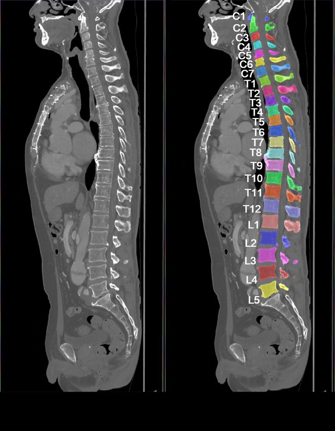
4      N. Masuzawa et al.

CT images. Individual vertebrae are segmented in the second stage. Moreover,
vertebral centroid coordinates and their labels are also obtained. Below we first
present our training dataset, followed by descriptions of the Semantic Segmen-
tation Net and the Iterative Instance Segmentation Net.

3.1   Training dataset
We prepared 1035 3D CT images (head: 181, chest 477, abdomen: 270, leg: 107)
for training which are obtained from diverse manufacturer’s equipment (e.g., GE,
Siemens, Toshiba, etc.). The leg 3D CT images were prepared for the purpose of
suppressing false positive in the first stage. The slice thickness ranges from 0.4
mm to 3.0 mm, and the in-plane resolution ranges from 0.34 mm to 0.97 mm.
They have been selected to contain the abnormal spine curvature, metal artifacts,
and the narrow FOV. Our spine model for training includes n = 25 individual
vertebrae, where the regular 19 from the cervical, thoracic, and lumbar vertebrae
consist irregular lumbar 6. Reference segmentations of the visible vertebrae were
generated by manually correcting automatic segmentations.

3.2   Stage 1: Semantic Segmentation Net
The convolutional neural networks are widely used to solve segmentation tasks in
supervised learning technique. Recent works have shown that this technique can
be successfully applied to the multi-organ segmentation in 3D CT images [11]. In
our method, we develop the Semantic Segmentation Net which segment cervical,
thoracic, and lumbar vertebrae from 3D CT images to find the bounding boxes.
    Figure 4 shows a schematic drawing of the architecture. Our architecture is
based on a 3D FCN [11]. For our Semantic Segmentation Net, the convolutions
performed in each stage use volumetric kernels having size of 3×3×3 and strides
of 1 followed by batch normalization [3] and ReLU as the activate function, the
max pooling uses volumetric kernels having size of 2×2×2 and strides of 2, and
the deconvolutions use volumetric kernels having size of 4×4×4 and strides of 2.

              Fig. 4. Architecture of the Semantic Segmentation Net.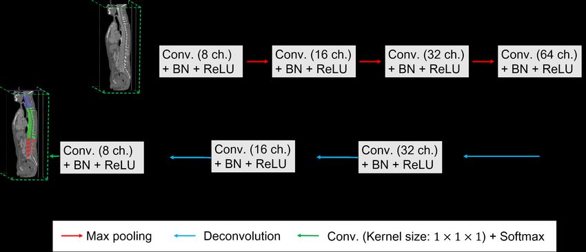
Automatic Segmentation, Localization and Identification of Vertebrae    5

Data augmentation and training In the preprocessing steps, input 3D CT
images are clipped to the [-512.0, 1024.0] range and then normalized to be in
the [-1.0, 1.0] interval. After that, input 3D CT images are rescaled to 1.0 mm
isotropic voxels. For each training iteration, we randomly crop 160×160×160
voxels from the input 3D CT images and apply data augmentation. In particular,
we apply an affine transformation consisting of a random rotation between -15
and +15 degrees, and random scaling between -20% and +20%, both sampled
from uniform distributions. In addition, we apply a Gaussian noise with µ =
0.0 and σ = [0.0, 50.0/1536.0]. In the training iteration, bootstrapped cross
entropy loss functions [6] were optimized with the Adam optimizer [7] with a
learning rate of 0.001 since the multi-class dice loss can be unstable. The idea
behind bootstrapping [6] is to backpropagate cross entropy loss not from all but
a subset of voxels that the posterior probabilities are less than a threshold. In
our experiment, 10% of total voxels are used for the backpropagation.

3.3   Stage 2: Iterative Instance Segmentation Net

              Fig. 5. Architecture of the Instance Segmentation Net.

    The goal of the second stage is segmenting, localizing, and assigning anatom-
ical labels to each vertebra. To this end, we developed the Iterative Instance
Segmentation Net inspired by Lessmann et al. [10]. The input to the Iterative
Instance Segmentation Net has an auxiliary channel in addition to the 3D CT
images. Given the segmented vertebra regions in the auxiliary channel, the net-
works output the next vertebra. The method by Lessmann et al. [10] requires
lumbar 5 region as a priori knowledge, and therefore it is not able to handle
general 3D CT images. By contrast, due to using the segmentation results in the
first stage, our method is able to handle general 3D CT images.
    Figure 5 shows a schematic drawing of the architecture. For our Iterative
Instance Segmentation Net, the convolutions performed in each stage use volu-
metric kernels having size of 3×3×3 and strides of 1 followed by batch normal-
6 N. Masuzawa et al. ization [3] and ReLU as the activate function, the max pooling uses volumetric kernels having size of 2×2×2 and strides of 2, and the deconvolutions use vol- umetric kernels having size of 4×4×4 and strides of 2. The anatomical labels of individual vertebrae are counted starting from the boundaries of cervical and thoracic vertebrae or thoracic and lumbar vertebrae. Finally, centroids of verte- brae are calculated using the segmentation results. Data augmentation and training In the preprocessing steps, similar to the first stage, input 3D CT images are clipped to the [-512.0, 1024.0] range and then normalized to be in the [-1.0, 1.0] interval. After that, input 3D CT images are rescaled to 1.0 mm isotropic voxels. For each training iteration, we randomly crop the spine region from the input 3D CT images and apply data augmentation. In particular, we apply an affine transformation consisting of a random rotation between -15 and +15 degrees, and random scaling between -20% and +20%, both sampled from uniform distributions. In addition, we apply a Gaussian noise with µ = 0.0 and σ = [0.0, 50.0/1536.0]. In the training iteration, the Dice loss of the segmented volume were optimized with the Adam optimizer [7] with a learning rate of 0.001. 4 Experimental Results We present two sets of experimental results. The first one is on vertebral seg- mentation and the second one is about vertebral localization and identification. We validate our algorithm with two public datasets of 15 3D CT images with reference segmentations from the MICCAI CSI (Computational Spine Imaging) 2014 workshop challenge and 302 3D CT images of the patients with various types of pathologies introduced in [1]. There are unusual appearances in the sec- ond dataset such as abnormal spine curvature and metal artifacts. In addition, the FOV of each volume varies widely. 4.1 Segmentation Performance We evaluated our method in terms of the segmentation accuracy with the MIC- CAI CSI 2014 workshop challenge. The CSI dataset consists of 15 3D CT images of healthy young adults, aged 2034 years. The images were scanned with either a Philips iCT 256 slice CT scanner or a Siemens Sensation 64 slice CT scanner (120 kVp, with IV-contrast). The in-plane resolution ranges from 0.31 mm to 0.36 mm and the slice thickness ranges from 0.7 mm to 1.0 mm. Each volume cover thoracic and lumbar vertebrae. We evaluate the segmentation performance using Average Symmetric Surface Distance (ASSD), Hausdorff Distance (HD), and Dice score on condition that the final segmentation masks are rescaled to the resolution of the input 3D CT images. The results on the CSI dataset is summarized in Table 1. Our method achieved slightly better performance than existing methods. The examples of the segmentations and the anatomical labels obtained with our method are shown in Figure 6. In all the 15 3D images, the
Automatic Segmentation, Localization and Identification of Vertebrae     7

Semantic Segmentation Net provided the Iterative Instance Segmentation Net
with the accurate bounding boxes. Moreover, the Iterative Instance Segmenta-
tion Net segmented the vertebrae precisely and predicted all of the anatomical
labels.

      Table 1. Comparison of Dice scores, ASSD and HD for segmentation results.

                       Method     Dice score(%)   ASSD (mm)     HD (mm)
             Janssens et al [4]     95.7 %           0.37          4.32
            Lessman et. al [10]     94.9 %           0.19            -
                 Our method         96.6 %           0.10          2.11

Fig. 6. Segmentation results and predicted anatomical labels obtained with the pro-
posed method.

4.2     Identification and Localization Performance
We evaluate localization and identification performance with 302 3D CT im-
ages introduced in [1]. This dataset is challenging since it includes wide vari-
eties of anomalies such as the abnormal spine curvature and the metal artifacts.
Furthermore, the FOV of each volume is largely different. In this dataset, the
reference centroid coordinates of the vertebrae and the anatomical labels were
given by clinical experts. We evaluate our method with the two metrics described
in [2], which are the Euclidean distance error (in mm) and identification rates
(Id.Rates) defined in [1]. On calculating these metrics, the final segmentation
8 N. Masuzawa et al.
masks are rescaled to the resolution of the input 3D CT images. Table 2 shows
a comparison between our method and previous works [12, 13]. The mean local-
ization error is 8.3 mm, and the mean identification rate is 84%. Our method
achieved better performance than the other existing methods.
Table 2. Comparison of localization errors in mm and identification rates.
Method Mean Std Id.rates
Glocker et al. [1] 12.4 11.2 70%
Suzani et al. [12] 18.2 11.4 -
Yang et al. [13] 9.1 7.2 80%
All
Yang et al. [13] (+1000) 8.5 7.7 83%
Our method 8.3 7.6 84%
Glocker et al. [1] 7.0 4.7 80%
Suzani et al. [12] 17.1 8.7 -
Yang et al. [13] 6.6 3.9 83%
Cervical
Yang et al. [13] (+1000) 5.8 3.9 88%
Our method 5.7 3.8 89%
Glocker et al. [1] 13.8 11.8 62%
Suzani et al. [12] 17.2 11.8 -
Yang et al. [13] 9.9 7.5 74%
Thoracic
Yang et al. [13] (+1000) 9.5 8.5 78%
Our method 9.3 8.3 79%
Glocker et al. [1] 14.3 12.3 75%
Suzani et al. [12] 20.3 12.2 -
Yang et al. [13] 10.9 9.1 80%
Lumbar
Yang et al. [13] (+1000) 9.9 9.1 84%
Our method 9.8 9.0 85%
5 Conclusion
In this paper, we propose a multi-stage framework for segmentation, localization
and identification of vertebrae in 3D CT images. A novelty of this framework is to
divide the three tasks into two stages. The first stage is multi-class segmentation
of cervical, thoracic, and lumbar vertebrae. The second stage is iterative instance
segmentation of individual vertebrae. By doing this, the method successfully
works without a priori knowledge of which part of the anatomy is visible in the
3D CT images. This means that the method can be applied to a wide range of
3D CT images and applications. In the experiments using two public datasets,
the method achieved the best Dice score for volume segmentation, and achieved
the best mean localization error and identification rate. As far as we know, this
is the first unified framework that tackles the three tasks simultaneously with
the state of the art performance. We hope that the proposed method will help
doctors in clinical practice.Automatic Segmentation, Localization and Identification of Vertebrae 9
References
1. Ben, G., et al.: Automatic localization and identification of vertebrae in arbitrary
field-of-view ct scans. In: Medical Image Computing and ComputerAssisted Inter-
vention. pp. 590–598. Springer Berlin Heidelberg (2012)
2. Ben, G., et al.: Vertebrae localization in pathological spine ct via dense classifica-
tion from sparse annotations. In: Medical Image Computing and ComputerAssisted
Intervention. pp. 262–270. Springer Berlin Heidelberg (2013)
3. Ioffe, S., et al.: Batch normalization: Accelerating deep network training by re-
ducing internal covariate shift. In: International Conference on Machine Learning.
vol. 37, pp. 448–456 (2015)
4. Janssens, R., et al.: Fully automatic segmentation of lumbar vertebrae from ct
images using cascaded 3d fully convolutional networks. In: IEEE 15th International
Symposium Biomedical Imaging. pp. 893–897 (2018)
5. Jianhua, Y., et al.: A multi-center milestone study of clinical vertebral ct segmen-
tation. In: Computerized Medical Imaging and Graphics 49. pp. 16–28 (2016)
6. Keshwani, D., et al.: Computation of total kidney volume from ct images in auto-
somal dominant polycystic kidney disease using multi-task 3d convolutional neural
networks. In: Medical Image Computing & Computer Assisted Intervention, Ma-
chine Learning in Medical Imaging workshop. vol. 66, pp. 90–99 (2018)
7. Kingma, D.P., et al.: Adam: A method for stochastic optimization. arXiv preprint
arXiv:1412.6980 (2014)
8. Klinder, T., et al.: Automated model-based vertebra detection, identification, and
segmentation ct images. In: Medical Image Analysis 13. pp. 471–482 (2009)
9. Korez, R., et al.: A framework for automated spine and vertebrae interpolation-
based detection and model-based segmentation. In: IEEE 15th International Sym-
posium Biomedical Imaging 34. pp. 1649–1662 (2015)
10. Lessmann, N., et al.: Iterative fully convolutional neural networks for automatic
vertebra segmentation and identification. In: Medical Image Analysis. vol. 53, pp.
142–155 (2019)
11. Roth, H.R., et al.: An application of cascaded 3d fully convolutional networks for
medical image segmentation. In: Computerized Medical Imaging and Graphics.
vol. 66, pp. 90–99 (2018)
12. Suzuani, A., et al.: Fast automatic vertebrae detection and localization in patho-
logical ct scans -a deep learning approach. In: Medical Image Computing and
Computer-Assisted Intervention. pp. 678–686 (2015)
13. Yang, D., et al.: Automatic vertebra labeling in large-scale 3d ct using deep image-
to-image network with message passing and sparsity regularization. In: Medical
Image Computing and Computer-Assisted Intervention. pp. 678–686 (2017)You can also read