Bezold's Abscess: A Case Report and Review of Cases Over 20 Years - Cureus
←
→
Page content transcription
If your browser does not render page correctly, please read the page content below
Open Access Case
                               Report                                                  DOI: 10.7759/cureus.21533

                                                 Bezold’s Abscess: A Case Report and Review of
                                                 Cases Over 20 Years
Review began 01/10/2022
                                                 Abdullah S. Alkhaldi 1 , Mohammed Alwabili 2 , Thamer Albilasi 2 , Khabti Almuhanna 2
Review ended 01/16/2022
Published 01/23/2022                             1. College of Medicine, King Saud Bin Abdulaziz University for Health Sciences, Riyadh, SAU 2. Department of
© Copyright 2022                                 Otolaryngology - Head & Neck Surgery, Prince Sultan Military Medical City, Riyadh, SAU
Alkhaldi et al. This is an open access article
distributed under the terms of the Creative      Corresponding author: Abdullah S. Alkhaldi, aabdulochik@gmail.com
Commons Attribution License CC-BY 4.0.,
which permits unrestricted use, distribution,
and reproduction in any medium, provided
the original author and source are credited.
                                                 Abstract
                                                 Bezold’s abscess (BA) is a severe and rare extracranial complication of suppurative acute mastoiditis. The
                                                 diagnosis of BA requires a high index of suspicion due to its rarity. In this study, we present a rare case of BA,
                                                 in addition to a review of literature over 20 years. We searched for all cases in English literature from 2000 to
                                                 2020 in PubMed and found 27 cases (28 cases including the current case). BA was more prevalent in males
                                                 (17/28, 60.7%) and adults (17/28, 60.7%). Of the 28 cases, six were associated with cholesteatoma and
                                                 another six cases occurred with concomitant sinus thrombosis.

                                                 Categories: Otolaryngology
                                                 Keywords: bezold abscess, suppurative mastoiditis, complications of acute mastoiditis, acute mastoiditis, bezold’s
                                                 abscess

                                                 Introduction
                                                 Bezold's abscess (BA) is caused by pus draining through the medial wall of the mastoid process and
                                                 producing a suppurative collection in the digastric sulcus [1]. This abscess is named after Friedrich von
                                                 Bezold, a German otologist who first reported a neck abscess in the sternocleidomastoid muscle in 1881 [2].
                                                 This suppurative collection might track to the digastric muscle and involve the retromaxillary fossa along
                                                 the occipital artery. If left untreated, further deep extension can occur. In case of a severe infection of the
                                                 mastoid bone, the suppurative contents of the mastoid air cells may descend along the upper insertion of the
                                                 sternocleidomastoid muscle, causing pus to accumulate between the muscle and the fascia. The contents of
                                                 BA can extend to the mediastinum if not treated appropriately and promptly, resulting in acute
                                                 mediastinitis, which has a 70% fatality rate [3]. Mastoiditis can affect people of all ages, although it is more
                                                 common in older adults [4]. The discovery of antibiotics has revolutionized the course of mastoiditis and
                                                 significantly decreased its complications. As a result, BA has become less severe and less frequent. The
                                                 clinical significance of the mastoid bone is linked to the nearby anatomical structures, such as the middle
                                                 cranial fossa, posterior cranial fossa, sigmoid and lateral sinuses, facial nerve canal, semicircular canals, and
                                                 petrous tip of the temporal bone. This study describes a case of BA, in addition to a review of literature from
                                                 2000 to 2020.

                                                 Case Presentation
                                                 A 46-year-old male presented to our emergency department complaining of left otorrhea, high fever, left
                                                 post-auricular pain, and a painful left neck swelling, which increased in size over the past four days with
                                                 decreased hearing. The patient had a history of right otorrhea periodically for three years that was treated
                                                 multiple times with oral antibiotics and local antibiotic ear drops. In addition, the patient had a history of
                                                 Bell’s palsy three years previously, which resolved with medical treatment and facial exercise. The patient
                                                 denied a history of vertigo, tinnitus, airway symptoms, dysphagia, odynophagia, or trismus. There was no
                                                 history of previous ear surgery.

                                                 On examination, the patient was febrile; he experienced pain, but his vital signs were stable. There was
                                                 post-auricular erythema and a left lateral diffuse swelling over the mastoid bone extending down to the
                                                 upper sternocleidomastoid muscle with tenderness and fluctuation (Figure 1). On otoscopy, the right ear was
                                                 unremarkable. The left ear had a clear external auditory canal and dull tympanic membrane. Rinne test was
                                                 negative for the left ear but positive for the right ear. Weber test indicated lateralization to the left side. The
                                                 eye examination was unremarkable (there was no nystagmus), and the fistula test was negative. Facial
                                                 movement was symmetrical, suggesting that the facial nerve was intact, and the examination of the other
                                                 cranial nerves was unremarkable. There were no signs of meningeal irritation or palpable lymph nodes. A
                                                 flexible scope showed a clear nasopharynx, oropharynx, and hypopharynx. Laboratory results showed
                                                 increased inflammatory markers (Table 1).

                                How to cite this article
                                Alkhaldi A S, Alwabili M, Albilasi T, et al. (January 23, 2022) Bezold’s Abscess: A Case Report and Review of Cases Over 20 Years. Cureus 14(1):
                                e21533. DOI 10.7759/cureus.21533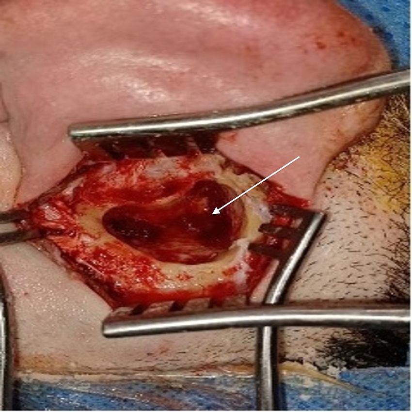
FIGURE 1: Left lateral diffuse swelling over the mastoid bone extending
                                                     down to the upper sternocleidomastoid muscle.

               Test                                                                 Result

               White blood cells                                                    12,000/mm3 (mainly neutrophils)

               C-reactive protein                                                   60 mg/L

               Erythrocyte sedimentation rate                                       80 mm/hour

               Blood sugar                                                          12 mmol

              TABLE 1: Laboratory results of the patient

                                                  Computed tomography of the head and neck with contrast
                                                  There was evidence of acute mastoiditis with bony erosive changes in the left mastoid bone and a thick
                                                  walled peripherally enhancing radiolucency measuring 3.5 x 2.6 x 3.5 cm in the left retrosternal region
                                                  abutting the sternocleidomastoid muscle posteriorly (Figures 2, 3).

2022 Alkhaldi et al. Cureus 14(1): e21533. DOI 10.7759/cureus.21533                                                                                         2 of 10
FIGURE 2: Coronal enhanced CT image of the head and neck showing
opacification of the left lateral inferior to the mastoid area with rim
enhancement, medial to the sternocleidomastoid muscle, a typical
picture commonly seen with Bezold’s abscess.
2022 Alkhaldi et al. Cureus 14(1): e21533. DOI 10.7759/cureus.21533 3 of 10FIGURE 3: Axial CT at the level of the mastoid showing posterior defect
of the left mastoid cortex with complete mastoid opacification.
Management
On arrival to the emergency department, intravenous (IV) ceftriaxone 2 g and IV paracetamol 1 g were
started immediately. On admission, ceftazidime 2 g and metronidazole 500 mg were prescribed. The next
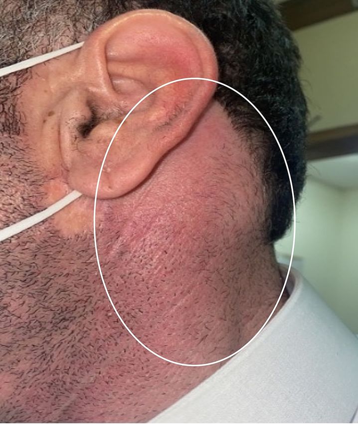
day, the patient underwent a cortical mastoidectomy, and an incision and drainage of the abscess under
general anesthesia was performed. After the incision, copious pus came out, which was sent for culture
(Figure 4). The result showed no growth. Five days after the surgery, the patient was discharged home on
ciprofloxacin and clindamycin in a good condition. The patient was followed up in the clinic one week after
discharge; he was doing well with no active complaint, and his wound was clean.
2022 Alkhaldi et al. Cureus 14(1): e21533. DOI 10.7759/cureus.21533 4 of 10FIGURE 4: Static view of intra-operative finding showing copious pus
collection in the mastoid following cortical mastoidectomy.
Discussion
BA is a severe and rare extracranial complication of suppurative acute mastoiditis. We searched English
literature for all the cases reported from 2000 to 2020 in PubMed. We found 28 cases (including ours), as
presented in Table 2. There are other reported cases, which were excluded due to not being in English and
not being registered in PubMed. BA as reported in the literature was more common in males (17/28, 60.7%)
than in females (11/28, 39.3%). It is also diagnosed more in adults (17/28, 60.7%) than in children 18 years
and younger (11/28, 39.3%). The age range was 10 weeks to 77 years. BA is extremely rare in infants and
young children due to incomplete mastoid pneumatization [5]. In the absence of complete pneumatization,
mastoid bone walls are thick and difficult to erode. Only three cases of BA have been described in patients
younger than five years.
Case Year of
Author Age Sex Management Culture Coexistence complications/comorbidities
no. publication
Marioni et al. 18
1 2001 Female IV cefotaxime Not available None
[5] months
Mastoidectomy + decompression of an epidural
Zapanta et 17 Alpha-hemolytic
2 2001 Female abscess + I and D + IV clindamycin, ceftriaxone, Multiple dural sinus thrombosis
al. [6] years streptococci
and vancomycin + myringotomy and tube
Uchida et al. 25 Staphylococcus +
3 2002 Male Mastoidectomy + I and D + IV antibiotics Cholesteatoma
[7] years Veillonella species
19 Staphylococcus
4 Jose et al. [8] 2003 Male I and D + IV flucloxacillin none
years aureus
Schöndorf et 10
5 2004 Female Mastoidectomy + IV antibiotic No growth none
2022 Alkhaldi et al. Cureus 14(1): e21533. DOI 10.7759/cureus.21533 5 of 10al. [9] weeks
Ching et al. 14 Lateral sinus thrombosis, poststreptococcal
6 2006 Male Mastoidectomy + IV ceftriaxone and metronidazole Streptococcus milleri
[10] years glomerulonephritis
Bhat and Pyogenic meningitis + cholesteatoma +
12 I and D + IV ceftazidime + temporal craniotomy + Pseudomonas
7 Manjunath 2007 Male sigmoid sinus thrombosis + cerebellar
years radical mastoidectomy aeruginosa
[11] abscess + CSF otorrhea + perilymph fistula
Mastoidectomy + insertion of grommet tube + I and
McMullan
8 2009 8 years Male D + IV cefotaxime, clindamycin, vancomycin, No growth Sigmoid sinus thrombus
[12]
meropenem
Vlastos et al. Streptococcus Sigmoid sinus thrombosis and occipital
9 2010 3 years Female Mastoidectomy + IV clindamycin and ceftriaxone
[13] pneumoniae osteomyelitis
Patel et al. 35 Mastoidectomy + I and D + IV
10 2010 Male Not available HIV
[14] years piperacillin/tazobactam and vancomycin
Sheikh et al. 26
11 2011 Male I and D of neck abscess acid-fast bacilli Prior cholesteatoma and mastoidectomy
[15] years
Mascarinas 77 Streptococcus
12 2011 Female Mastoidectomy+ I and D + IV antibiotic Postradiation
et al. [16] years viridans
Li and Ren 32
13 2012 Female Mastoidectomy and I and D Not available Cholesteatoma
[17] years
Janardhan et 60
14 2012 Male Mastoidectomy + I and D + IV antibiotics Not available Congenital cholesteatoma
al. [18] years
Secko and 32
15 2013 Male I and D + IV ceftriaxone Not available HIV
Aherne [19] years
Nelson and
12 Streptococcus
16 Jeanmonod 2013 Female IV clindamycin + dexamethasone + I and D None
years pyogenes
[20]
Lionello et al. 35 Mastoidectomy + I and D + IV ceftriaxone and
17 2013 Male No growth Cholesteatoma
[21] years metronidazole
modified radical mastoidectomy with type III
Pradhananga 14
18 2014 Female tympanoplasty + I and D + broad spectrum Not available none
[22] years
antibiotics
Al-Baharna 73 Peptostreptococcus Diabetes, hypertension, renal impairment,
19 2016 Male Mastoidectomy + I and D + IV ceftriaxone
et al. [23] years species and cardiomyopathy
Quoraishi et 44 IV cefotaxime + myringotomy with tube insertion +
20 2016 Male No growth none
al. [24] years cortical mastoidectomy + I and D
Nasir and 52 IV ampicillin/sulbactam + modified radical Klebsiella
21 2017 Male Diabetes, left facial nerve palsy grade V
Asha'ari [25] years mastoidectomy + I and D pneumoniae
Parkinson's disease + thrombosis or right
Yaita et al. 70 Streptococcus
22 2018 Female IV ampicillin/sulbactam + IV ceftriaxone transverse sinus + acute infarction of the
[26] years constellatus
right cerebellum
Methicillin-sensitive
Eswaran et 15 topical and systemic antibiotics + modified radical Staphylococcus
23 2019 Female None
al. [27] years mastoidectomy aureus + acid-fast
bacilli
Katayama et 52 Streptococcus
24 2018 Male IV ceftriaxone + metronidazole + drainage Diabetes + hyperlipidemia
al. [28] years pneumoniae
IV ceftriaxone and vancomycin + cortical
Mustafa et al. 14 Streptococcus
25 2018 Female mastoidectomy + myringotomy with tube insertion + None
[29] years pneumoniae
abscess drainage
right tympanomastoidectomy + canaloplasty +
Malik et al. 55
26 2019 Male incision and drainage of Bezold’s abscess + IV Not available Skull base osteomyelitis
[30] years
vancomycin, cefepime, and metronidazole
2022 Alkhaldi et al. Cureus 14(1): e21533. DOI 10.7759/cureus.21533 6 of 10Lyoubi et al. 62 IV ceftriaxone and moxifloxacin + wide
27 2020 Male Not available None
[31] years mastoidectomy + surgical drainage of abscess
46 IV ceftriaxone + cortical mastoidectomy + I and D
28 Our case 2021 Male No growth None
years of abscess
TABLE 2: Review of all reported cases of Bezold's abscess in the English Literature between 2000
and 2020
I and D, incision and drainage; IV, intravenous; HIV, human immunodeficiency virus; CSF, cerebrospinal fluid
The diagnosis of BA requires a high index of suspicion due to its rarity. Patients may not show any signs of
sepsis, and clinicians should be aware of this complication in patients with otitis media or acute mastoiditis.
The most prevalent reported clinical signs and symptoms were fever, otalgia, neck edema, neck pain,
otorrhea, torticollis, facial paralysis, and hearing loss [5]. Our patient was mainly complaining of otorrhea,
high fever, post-auricular pain, and a painful left neck swelling increasing in size.
The mastoid tip, which is pneumatized in adults, is composed of thin-walled air cells. The lateral wall of the
mastoid is composed of thicker bone than that of the medial wall. The lateral wall serves as an insertion site
for the sternocleidomastoid, digastric, splenius capitis, and longissimus capitis muscles. Pus in the mastoid
erodes through the mastoid tip, the area of least resistance, which is inferior and medial. The abscesses are
formed deep in the neck muscles, which delay its detection. Another factor that could contribute to the
delayed diagnosis is the unfamiliarity of the disease to the clinician.
BA has been linked to lateral sinus thrombosis [32], which is caused by the compression or thrombosis of the
internal jugular vein. We found six cases with concomitant sinus thrombosis. Similarly, six cases were
associated with either primary or recurrent cholesteatoma. The presence of cholesteatoma in mastoiditis
blocks the middle ear aditus and directs the inflammatory process to the mastoid tip. Patients with a history
of cholesteatoma appear to be at an increased risk for BA.
The diagnosis can be challenging because of rarity of this abscess and the variable signs and symptoms.
Computed tomography (CT) and magnetic resonance imaging (MRI) images can locate the abscess, as
described in Table 3. The CT scan in the current case of BA indicated an ipsilateral opacification of the
middle ear and mastoid cavity, often associated with bony erosions. The collection of pus can be detected
along the sternocleidomastoid muscle. Contrasted CT images of the temporal bone and neck are most
important for both the diagnosis and subsequent surgical treatment. In addition, the laboratory evaluation is
often supportive in diagnosis, as the leukocyte count is usually high and the erythrocyte sedimentation rate
elevated.
Author Modality Findings
Marioni et al. Non-erosive debris throughout the middle ear cavity and mastoid on the right side and thickening of
CT
[5] prevertebral and retropharyngeal spaces on the same side.
Zapanta et Left-sided coalescent mastoiditis and pansinus opacification. Left-side sigmoid sinus showing the filling defect
CT
al. [6] or “empty delta sign”.
Uchida et al. Round-shaped soft tissue mass from the mastoid process through the sigmoid sinus sulcus causing extensive
CT
[7] bony destruction of both the temporal and occipital bones.
Opacification of the left middle ear and mastoid air cells, lateral sinus thrombosis, and adjacent area of
Jose et al. [8] MRI
meningeal inflammation.
Schöndorf et Bright signal shows inflammation in mastoid bone with involvement of insertion of the left sternocleidomastoid
MRI
al. [9] muscle.
Opaque left mastoid air cell as well as a filling defect in the left sigmoid sinus in keeping with septic
Ching et al.
CT thrombophlebitis. Note also the small extradural collection adjacent to the sigmoid sinus. More inferiorly, there
[10]
is a septated Bezold’s abscess with an enhancing rim just posterior to the tip of the left mastoid process.
Bhat and
Manjunath CT Erosion of the sinus plate, the cerebellar abscess, and the dilation of the ventricles.
[11]
Bilateral otomastoiditis with associated bony destruction in the mastoid cavities, extension into the right
McMullan
CT sigmoid sinus, and extension into the neck inferiorly, consistent with an abscess related to the deep surface of
2022 Alkhaldi et al. Cureus 14(1): e21533. DOI 10.7759/cureus.21533 7 of 10[12] the right sternocleidomastoid muscle. There was also partially occlusive thrombus in the right sigmoid sinus.
Vlastos et al. Osteolytic process within the left mastoid, edema, and a small abscess formation in the left upper neck region
CT
[13] (Bezold’s abscess) and thrombosis of the ipsilateral sigmoid sinus.
Left-sided coalescent mastoiditis. A 3.6 x 1.8 cm abscess at the level of the left mastoid tip tracked deep to the
Patel et al. left sternocleidomastoid and extended medially and anteriorly into the pre-vertebral space surrounding the
CT
[14] anterior arch of C1. Contrast study showed a hypoplastic left jugular bulb and no flow in the internal jugular
bulb or sigmoid sinus.
Sheikh et al.
Not available.
[15]
On the left side, coalescent mastoiditis and an abscess within the superior aspect of the SCM muscle
Mascarinas
CT communicating with the mastoid cavity through a bony dehiscence of the mastoid tip, consistent with a Bezold’s
et al. [16]
abscess.
Two masses located in the right mastoid cavity and neck measuring 4.0 cm x 3.0 cm and 5.0 cm x 3.0 cm in
Li and Ren
MRI diameter. The masses had smooth, well-defined outlines with intermediate signal intensity on T1-weighted and
[17]
hyperintense on T2-weighted without post-contrast enhancement.
Soft tissue attenuation of the right mastoid antrum with absence of air cells. There was an absence of medial
Janardhan et
CT wall of mastoid antrum in the region of Trautmann’s triangle and sinus plate. Also, a bony deficiency was seen
al. [18]
in the posterior meatal wall.
Edema of the maxillary, sphenoid, and mastoid air cells consistent with sinusitis. CT of the neck at the level of
Secko and
CT the mandible demonstrated a small hypodense lesion in the area of the sternocleidomastoid muscle consistent
Aherne [19]
with abscess.
Extensive opacification of the left mastoid temporal bone consistent with acute otomastoiditis. In addition, there
Nelson and
was bony erosion and destruction of the mastoid tip inferiorly with extensive surrounding inflammation within
Jeanmonod CT
the adjacent soft tissues and a 1 cm peripherally enhancing, developing Bezold’s abscess with diffuse reactive
[20]
adenopathy within the left neck.
Lionello et al. At the C1-C2 level, there was soft tissue edema on the left side involving the sternocleidomastoid muscle,
CT
[21] which appeared swollen and unevenly enhanced; focal areas of necrosis.
Air fluid collection in the left mastoid and middle ear cavity. There was erosion of the mastoid cavity and sinus
Pradhananga
CT plate. A defect was noted in the medial wall of the left mastoid cavity. Fluid collection with air foci within was
[22]
also noted in soft tissue adjacent to the left mastoid cavity and extending into the neck, suggestive of abscess.
Al-Baharna
MRI Abscess collection within the sternocleidomastoid muscle continuous with mastoid collection.
et al. [23]
Right mastoiditis with bony erosion anterior to the sigmoid venous sinus. Overlying superficial abscess, tracking
Quoraishi et
CT inferiorly deep to the sternocleidomastoid muscle, and anteriorly to the margin of the styloid process and carotid
al. [24]
sheath.
Soft tissue density within the left middle ear cavity and mastoid air cells with wide erosion at the posteroinferior
Nasir and part of the mastoid, medial to the mastoid tip. There was abscess collection deep to the sternomastoid muscle
CT
Asha'ari [25] below the mastoid tip erosion. The collection extended inferiorly along the paravertebral muscles until the
seventh cervical vertebrae.
Yaita et al. Polycystic lesions (abscesses) on her right posterior and on the lateral region of her neck, thrombosis in her
CT
[26] right internal jugular vein, and multiple nodules on her bilateral lung fields (septic emboli) were present.
Soft tissue opacification in right middle ear cavity and mastoid antrum with breach in the mastoid tip. Breach in
Eswaran et the continuity of the right mastoid bone was also seen posteriorly abutting the right sigmoid sinus. There was
CT
al. [27] erosion in the posterior bony canal wall. Hypodense area with thick enhancing wall was noted medial to the
superior part of the sternocleidomastoid muscle, suggestive Bezold’s abscess.
Katayama et Revealed multiple abscesses spread from the right temporal bone to the right sternocleidomastoid muscle. It
CT
al. [28] also demonstrated osteolysis at his right mastoid process.
Mustafa et al. Irregular hypodensity below the right mastoid and right half of the occipital bone surrounded with postcontrast
CT
[29] (red circle) increase of density represent the abscess formation.
Right-sided chronic mastoiditis, erosion of the inferior mastoid cells, extension of the infection into the neck
Malik et al.
CT spaces, and formation of a Bezold’s abscess in the ipsilateral sternocleidomastoid muscle, extending into the
[30]
retropharyngeal space
Lyoubi et al. Right-sided chronic mastoiditis, erosion of the inferior mastoid cells, and cervical cellulitis collected in the right
2022 Alkhaldi et al. Cureus 14(1): e21533. DOI 10.7759/cureus.21533 8 of 10[31] CT sternocleidomastoid muscle measuring 33 × 15 mm extended to 40 mm.
TABLE 3: Radiological findings of reported cases of Bezold's abscess
CT, computed tomography; MRI, magnetic resonance imaging
Mastoiditis has a similar bacteriology to acute otitis media, with Streptococci species the major pathogens. In
our review, Streptococci were the most frequent causative organisms. However, multiple organisms, both
gram positive and negative, as well as anaerobes, were cultured. Antibiotics effective against gram-positive
organisms should be initiated since they are the most frequent causative pathogens. Subsequently, culture-
based antibiotics can be described. In addition to the IV antibiotics, a surgical intervention (mastoidectomy
and abscess drainage) is required for the effective management and prevention of further complications. Of
the 28 patients with data regarding treatment, 21 (75%) underwent a mastoidectomy (Table 2). The other
eight cases required no mastoidectomy (two were children without a fully pneumatized mastoid bone). This
suggests that the surgical treatment can be tailored to mastoid bone pneumatization and the neck abscess
extension.
Conclusions
BA is a severe and rare extracranial complication of suppurative acute mastoiditis. The diagnosis of BA
requires a high index of suspicion due to its rarity and the variable signs and symptoms. In this study, we
presented a rare case of BA that was managed by IV antibiotics, incision and drainage of the abscess, and
cortical mastoidectomy. In addition, we presented a review of BA cases over 20 years (2000-2020). In almost
all the cases, the gold standard management was IV antibiotics, drainage of the abscess, and mastoidectomy.
Additional Information
Disclosures
Human subjects: Consent was obtained or waived by all participants in this study. Conflicts of interest: In
compliance with the ICMJE uniform disclosure form, all authors declare the following: Payment/services
info: All authors have declared that no financial support was received from any organization for the
submitted work. Financial relationships: All authors have declared that they have no financial
relationships at present or within the previous three years with any organizations that might have an
interest in the submitted work. Other relationships: All authors have declared that there are no other
relationships or activities that could appear to have influenced the submitted work.
Acknowledgements
Authors would like to thank Dr. Lulwah Alalayet who has participated in this study.
References
1. Castillo M, Albernaz VS, Mukherji SK, Smith MM, Weissman JL: Imaging of Bezold's abscess. AJR Am J
Roentgenol. 1998, 171:1491-5. 10.2214/ajr.171.6.9843276
2. Bezold F: Ein neuer Weg für Ausbreitung eitriger Entzündung aus den Råumen des Mittelohrs auf die
Nachbarschaft und die in diesem Falle einzuschlagende Therapie. Dtsch Medizinische Wochenschrift. 1881,
7:381-5. 10.1055/s-0029-1196026
3. Govea-Camacho LH, Pérez-Ramírez R, Cornejo-Suárez A, et al.: Diagnosis and treatment of the
complications of otitis media in adults. Case series and literature review. Cirugía y Cir (English Ed). 2016,
84:398-404. 10.1016/j.circen.2016.08.001
4. Ongkasuwan J, Valdez TA, Hulten KG, Mason EO Jr, Kaplan SL: Pneumococcal mastoiditis in children and
the emergence of multidrug-resistant serotype 19A isolates. Pediatrics. 2008, 122:34-9. 10.1542/peds.2007-
2703
5. Marioni G, de Filippis C, Tregnaghi A, Marchese-Ragona R, Staffieri A: Bezold's abscess in children: case
report and review of the literature. Int J Pediatr Otorhinolaryngol. 2001, 61:173-7. 10.1016/s0165-
5876(01)00564-x
6. Zapanta PE, Chi DH, Faust RA: A unique case of Bezold's abscess associated with multiple dural sinus
thromboses. Laryngoscope. 2001, 111:1944-8. 10.1097/00005537-200111000-00013
7. Uchida Y, Ueda H, Nakashima T: Bezold's abscess arising with recurrent cholesteatoma 20 years after the
first surgery: with a review of the 18 cases published in Japan since 1960. Auris Nasus Larynx. 2002, 29:375-
8. 10.1016/s0385-8146(02)00057-3
8. Jose J, Coatesworth AP, Anthony R, Reilly PG: Life threatening complications after partially treated
mastoiditis. BMJ. 2003, 327:41-2. 10.1136/bmj.327.7405.41
9. Schöndorf HJ, Roth B, Streppel M: Bezold's abscess following chronic mastoiditis in a newborn . Ann Otol
Rhinol Laryngol. 2004, 113:843-5. 10.1177/000348940411301013
10. Ching HY, Ramsden JD, Bottrill I: A unique presentation: Bezold's abscess and glomerulonephritis . Eur J
Pediatr. 2006, 165:569-70. 10.1007/s00431-006-0098-z
11. Bhat V, Manjunath D: Cerebrospinal fluid otorrhea presenting in complicated chronic suppurative otitis
2022 Alkhaldi et al. Cureus 14(1): e21533. DOI 10.7759/cureus.21533 9 of 10media. Ear Nose Throat J. 2007, 86:223-5.
12. McMullan B: Bezold's abscess: a serious complication of otitis media . J Paediatr Child Health. 2009, 45:616-
8. 10.1111/j.1440-1754.2009.01575.x
13. Vlastos IM, Helmis G, Athanasopoulos I, Houlakis M: Acute mastoiditis complicated with bezold abscess,
sigmoid sinus thrombosis and occipital osteomyelitis in a child. Eur Rev Med Pharmacol Sci. 2010, 14:635-8.
14. Patel N, Goodman J, Singh A: Bezold's abscess in the setting of untreated HIV infection . Laryngoscope.
2010, 120:S134. 10.1002/lary.21598
15. Sheikh FT, Murday DC, Abbas A, Main C, King AJ, Rao S, Batty V: Bezold's abscess. Emerg Med J. 2011,
28:985. 10.1136/emj.2010.109793
16. Mascarinas CA, Singer MC, Hanson MB: Bezold's abscess in the setting of radiation induced mastoiditis .
Laryngoscope. 2010, 120:S211. 10.1002/lary.21678
17. Li L, Ren J: Aural cholesteatoma with upper neck extension . Auris Nasus Larynx. 2012, 39:534-6.
10.1016/j.anl.2011.11.001
18. Janardhan N, Nara J, Peram I, Palukuri S, Chinta A, Satna K: Congenital cholesteatoma of temporal bone
with Bezold's abscess: case report. Indian J Otolaryngol Head Neck Surg. 2012, 64:97-9. 10.1007/s12070-
011-0226-6
19. Secko M, Aherne A: Diagnosis of Bezold abscess using bedside ultrasound . J Emerg Med. 2013, 44:670-2.
10.1016/j.jemermed.2012.07.066
20. Nelson D, Jeanmonod R: Bezold abscess: a rare complication of mastoiditis . Am J Emerg Med. 2013,
31:1626.e3-4. 10.1016/j.ajem.2013.06.036
21. Lionello M, Manara R, Lord L, et al.: Case report of cholesteatoma recurrence with Bezold’s abscess
presenting as a deep neck infection. B-ENT. 2013, 9:255-8.
22. Pradhananga R: An unusual complication of chronic suppurative otitis media: bezold abscess progressing to
scapular abscess. Int Arch Otorhinolaryngol. 2014, 18:412-4. 10.1055/s-0034-1372511
23. Al-Baharna H, Al-Mubaireek H, Arora V: Bezold’s abscess: a case report and review of cases over 14 years .
Indian J Otol. 2016, 22:148-51. 10.4103/0971-7749.187978
24. Quoraishi S, George J, Farboud A, Marnane C: Atypical presentation of Bezold's and Citelli's abscesses, with
recollection following an incomplete postoperative course of antibiotics. BMJ Case Rep. 2017,
2017:bcr2016218072.. 10.1136/bcr-2016-218072
25. Nasir F, Asha'ari Z: Bezold's abscess: a rare complication of acute otitis media . Malays Fam Physician. 2017,
12:26-8.
26. Yaita K, Sugi S, Hayashi M, et al.: The co-existence of Lemierre's syndrome and Bezold's abscesses due to
Streptococcus constellatus: a case report. Medicine (Baltimore). 2018, 97:e11228.
10.1097/MD.0000000000011228
27. Eswaran S, Kumar S, Kumar P: A rare case of primary tuberculous otitis media with Bezold's abscess . Indian
J Otolaryngol Head Neck Surg. 2019, 71:1462-6. 10.1007/s12070-018-1554-6
28. Katayama K, Gomi H, Shirokawa T, Akizuki H, Kobayashi H: Bezold's abscess in a diabetic patient without
significant clinical symptoms. IDCases. 2018, 12:e1-2. 10.1016/j.idcr.2017.04.006
29. Mustafa A, Toçi B, Thaçi H, Gjikolli B, Baftiu N: Acute mastoiditis complicated with concomitant Bezold's
abscess and lateral sinus thrombosis. Case Rep Otolaryngol. 2018, 2018:8702532. 10.1155/2018/8702532
30. Malik K, Dever LL, Kapila R: Bezold's abscess: a rare complication of suppurative mastoiditis . IDCases. 2019,
17:e00538. 10.1016/j.idcr.2019.e00538
31. Lyoubi H, Berrada O, Lekhbal A, Abada RA, Mahtar M: Bezold's abscess: an extremely rare complication of
suppurative mastoiditis: xase report and literature review. Int J Surg Case Rep. 2020, 77:534-7.
10.1016/j.ijscr.2020.11.052
32. Luntz M, Brodsky A, Nusem S, et al.: Acute mastoiditis - the antibiotic era: a multicenter study . Int J Pediatr
Otorhinolaryngol. 2001, 1016:1-9. 10.1016/s0165-5876(00)00425-0
2022 Alkhaldi et al. Cureus 14(1): e21533. DOI 10.7759/cureus.21533 10 of 10You can also read