Validating a Device for Whiplash Motion Simulation in a Porcine Model
←
→
Page content transcription
If your browser does not render page correctly, please read the page content below
Validating a Device for Whiplash Motion Simulation in a Porcine
                              Model
                 N. Soltan1, P.A. Cripton1,2, M.Y. Svensson3, G.P. Siegmund4,5
1
  Department of Mechanical Engineering, University of British Columbia; 2School of Biomedical
Engineering, University of British Columbia, 3Department of Mechanics and Maritime Sciences,
     Chalmers University of Technology, 4MEA Forensic Engineers & Scientists, 5School of
                          Kinesiology, University of British Columbia

                                           ABSTRACT

Whiplash injury is a common outcome following minor automobile collisions. One theorized
mechanism for whiplash injury is that the rapid head and neck motions induced by a collision can
injure nerve cells in the dorsal root ganglia through pressure gradients developed in the spinal
canal and surrounding tissues. This injury mechanism has previously been studied in human
cadaver and porcine models. However, the whiplash motion simulation methods in the latter
studies lacked the control necessary to explore the independent effects of head rotation and
retraction on the measured spinal pressures. This project aimed to address the limitations of
previous porcine whiplash studies by developing and validating a new whiplash motion simulation
device to enable further study of this injury mechanism. The new proposed device consists of two
servomotors which can be programmed to precisely actuate a headplate through mechanical
linkages. For the current study, an inert surrogate model was used for preliminary testing of the
device using a whiplash motion profile from previous porcine studies. The time scale of the motion
profile was adjusted to incrementally increase severity. The positional accuracy and repeatability
of the device was assessed through marker tracking of the headplate and logging of the motor
encoder positions. Angular rates and linear accelerations of the plate were also measured. Testing
demonstrated the strengths of the proposed device in accurately and repeatably replicating
programmed motion profiles. Some design modifications can potentially enable simulating
whiplash motion severities commensurate with previous porcine whiplash studies. With future
testing using this device, our understanding of the pressure-induced whiplash injury mechanism
can be improved, which can inform effective treatments and preventative measures for whiplash
injury.

                                       INTRODUCTION

        Despite their prevalence and large economic/social burden, neck sprains and strains,
commonly referred to as whiplash injuries, remain one of the most poorly understood automotive
injuries (Siegmund et al., 2009). Associated symptoms of whiplash include neck and upper body
pain, headache, dizziness and other cognitive/psychological symptoms (Croft et al., 2002;
                                                  1
                     2021 The Ohio State University Injury Biomechanics Symposium
                                *This paper has not been peer-reviewed
Siegmund et al., 2009). Chronic disabling symptoms from whiplash injury account for a significant
portion of overall disability from motor vehicle injuries (Sterner et al., 2004). In the United States
alone, the cost of these injuries is estimated to exceed $19 billion annually (Croft et al., 2002).

        A current challenge with effectively treating and preventing whiplash symptoms is the
absence of observable tissue damage in patients and the lack of consensus on the mechanism of
injury (Curatolo et al., 2011). Several anatomical sites and injury models have been hypothesized
to be the source of whiplash injury. One such injury model proposes that the rapid extension-
flexion of the neck during a collision (“whiplash motions”) causes pressure gradients to develop
in the spinal canal and across the vein bridges in the intervertebral foramina. These pressure
gradients are theorized to increase tissue stress and cause nerve cell damage in the dorsal root
ganglia (DRG) (Aldman, 1986; Svensson et al., 1993; Örtengren et al., 1996; Svensson et al.,
2000).

        During a rear-end collision, occupants in the impacted vehicle initially experience a
retraction of the head relative to the torso as the head is not in contact with the head restraint. This
retraction causes the upper and lower cervical spine to enter flexion and extension, respectively.
Once the natural range of retraction is reached, the neck may transition into a fully extended
posture and subsequently upon interacting with the head rest, enter a flexed posture (Ono et al.,
1993; Svensson et al., 2000). It has been demonstrated that the volume of the spinal canal reduces
and increases in extension and flexion, respectively (Yao et al., 2016). Therefore, it is theorized
that the above-described whiplash motions and localized volume changes can cause the
incompressible blood and cerebrospinal fluid in the spinal canal to displace and consequently form
transient pressure gradients along the spinal canal and across the intervertebral foramina. This
pressure effect is postulated to stress and strain the DRG, which are structures in the dorsal nerve
roots and which are located in the intervertebral foramen. The DRG house the cell bodies of
afferent nerves, therefore, direct injury to this site can explain many of the common whiplash
symptoms (Siegmund et al., 2009).

         This pressure-induced whiplash injury mechanism has previously been studied in porcine
and human cadaver models. Through exposing anaesthetized pigs to simulated whiplash motions,
Svensson et al. (1993) observed pressure pulses in the cervical spinal canal. The initial large
pressure pulses appeared to correspond to the point of maximum neck retraction indicating the
potential relevance of this phase of whiplash motion in producing injury. Following whiplash
exposure, histopathological examination of the cervical DRG indicated plasma membrane leakage
in the nerve cells of the exposed animals demonstrating cell dysfunction (Örtengren et al., 1996).
This pressure effect was similarly demonstrated in post-mortem human subjects exposed to rear-
end collisions, though tissue damage was not observable as in the in vivo experiments (Eichberger
et al., 2000).

        Despite establishing the foundational knowledge behind this whiplash injury mechanism,
questions remain about the relative contributions of several head and neck kinematic parameters
during whiplash motions to the pressure pulse magnitudes and whiplash injury risk. In the previous
porcine whiplash studies (Svensson et al., 1993), the method of generating whiplash motions could
not be used to study the independent effect of neck retraction distance and extension angle on the
measured spinal pressure magnitudes. Understanding the influence of these kinematics parameters
                                                    2
                       2021 The Ohio State University Injury Biomechanics Symposium
                                  *This paper has not been peer-reviewed
on the pressure pulse magnitudes and whiplash injury risk can inform the design of motor vehicle
safety systems for effective prevention of this injury. Thus, the objective of this project is to test
and validate a custom-built device that aims to simulate whiplash motions with precise movement
control. Specifically, we aim to 1) assess whether the proposed device is able to replicate the
motion profiles tested in previous porcine whiplash studies, and 2) characterize the accuracy and
repeatability of the simulated whiplash motions. This assessment will inform any device design
modifications in order to enable further study of the DRG whiplash injury mechanism in an in vivo
porcine model.

                                            METHODS

Porcine Model

       The whiplash device was designed to allow for further study of whiplash in an in vivo
porcine model for several reasons. Firstly, our aim is to ultimately study the effect of whiplash
motions on the dynamics and responses of a living system. Live tissue is needed to produce the
proposed whiplash injury mechanism (cellular damage in the DRG). Additionally, it is unclear
how the absence of arterial blood pressure in a cadaveric model affects the spinal pressure
measurements (Eichberger et al., 2000).

       Furthermore, the vertebral column of the pig in particular has gross similarities to the
human vertebral column in terms of size and anatomy (Busscher et al., 2010; Sheng et al., 2010,
2016) and serves as a reasonable qualitative surrogate. The porcine model has been used in prior
biomechanical studies in the context of whiplash (Svensson et al., 1993), spinal cord (Jones et al.,
2012; Lee et al., 2013), and traumatic brain injury (Duhaime, 2006).

Device design

        The custom whiplash device was designed and built at MEA Forensic Engineers &
Scientists (Figure 1). The device consists of a frame which holds two rotary servomotors
(Yaskawa, SGMCS-2ZN, Japan) with a rated torque and speed of 200 N∙m and 150 rpm,
respectively. The motors can achieve an instantaneous maximum torque of 600 N∙m at zero speed.
Each servomotor is equipped with a 20-bit encoder which allows for position feedback control. A
series of mechanical linkages are attached to the motors on one end and a biteplate on the other
end via ball joints. Using this device, the anaesthetized animal will be placed on the operating table
with its head cantilevered off of the table edge and the biteplate will be secured in the animal’s
mouth. The combined rotation of the motors will be used to translate and rotate the biteplate to
control and simulate programmed whiplash motions.

                                                   3
                      2021 The Ohio State University Injury Biomechanics Symposium
                                 *This paper has not been peer-reviewed
Figure 1: Whiplash apparatus. A: Side view, B: Isometric view (biteplate not shown).
This device design aims to address some of the limitations of the apparatus used in the
previous porcine whiplash studies (Svensson et al., 1993). Our device and the previous apparatus
primarily differ by the method used to generate the whiplash motions. With the previous device,
whiplash motions were generated using a pre-tensed rubber-strap which transmitted force to a
headplate. The animal was placed laterally on an operating table with its heads affixed to the
headplate which was freely movable in the horizontal plane. Depending on the tension of the strap,
the pull-force on the headplate could be adjusted with an accuracy of ± 20-100 N (Svensson et al.,
1993). This design allowed for free motion of the animal’s head and maximum neck extension
angles were determined by the animal’s physiology, though in subsequent tests a rigid head
restraint was introduced to physically limit the maximum extension angle (Bostrom et al., 1996).
Despite its advantages, this device was limited in its ability to precisely control head kinematic
parameters. Servomotors were used in our device with the aim of programming and testing specific
motion profiles with good accuracy and repeatability. As a result, parameters such as head
retraction distance, extension angle, and speed/accelerations during whiplash motions can be
systematically varied and their effects on the resulting pressure pulses and tissue injury can be
independently studied.
Inert surrogate model
Prior to animal testing, an inert surrogate model was developed for the preliminary
validation of the whiplash device. The surrogate model strictly functions to mimic the physical
constraints of the biteplate and motor linkages during in vivo animal testing and does not attempt
to produce a biofidelic response.
The surrogate model (Figure 2) consists of a 2’’ diameter hose attached to a plate with an
approximately 5 kg mass added to represent the animal’s neck and head, respectively. The 5 kg
mass was conservatively selected and represents approximately 25% of the body weight of the
pigs that will eventually be tested with the device. The hose length was constrained to 30 cm from
the plate using a U-bolt screwed to a wood board which was clamped to the operating table. The
length of the hose approximates the neck length of the pig up to the T1 vertebra (Condotta et al.,
4
2021 The Ohio State University Injury Biomechanics Symposium
*This paper has not been peer-reviewed2018) as the animals will likely be constrained to the table via straps at this vertebral level similar
to previous experiments (Svensson et al., 1993).
Figure 2: Surrogate model to represent the pig head and neck.
Tested motion profile
A motion profile was obtained through video marker tracking from previous (unpublished)
porcine whiplash testing (Figure 3). These tests simulated whiplash motions using a mechanism
similar to the device described in Svensson et al. (1993). This particular motion profile was
selected as it models a relatively severe whiplash motion with a peak head extension angle of 80
degrees in approximately 70 ms. Previous human volunteer and post-mortem human subject
(PMHS) testing of whiplash injury have involved sled tests with deltaV’s up to 10 and 25 km/h,
respectively (Szabo et al., 1994; Siegmund et al., 1997; Eichberger et al., 1998; Philippens et al.,
2000; Kang et al., 2014) as most neck injuries from rear-end collisions were found to occur with
deltaV’s less than 20 km/h (Philippens et al., 2000). For comparison with our selected motion
profile, rear-end PMHS testing around 20 km/h deltaV’s produced maximum head extension
angles of 50-60 degrees at around 200-250 ms (Bertholon et al., 2000; Philippens et al., 2000;
Kang et al., 2014). Our selected motion profile represents an extreme upper end in terms of
whiplash motion severity and is used here to discern the extent of the motor and device capabilities.
If our device can reproduce this extreme motion, it will be capable of reproducing all other
intermediate motion profiles.
5
2021 The Ohio State University Injury Biomechanics Symposium
*This paper has not been peer-reviewedFigure 3: Original motion profile.
In order to incrementally test the device capabilities, the time scale of the selected motion
profile was altered using a time factor (TF). Figure 4 shows the original motion profile (TF = 1)
compared to the scaled motion profiles with TF = 8, 4, 3.5, 3, 2. A TF of 8 for example represents
the same range of motion as the original profile over 8 times the time period.
Figure 4: Scaled motion profiles.
6
2021 The Ohio State University Injury Biomechanics Symposium
*This paper has not been peer-reviewedA custom Matlab (R2019b, Mathworks Inc., Natick, MA) program was used to determine
the required motor rotations to achieve the input motion profiles. The program output included
incremental motor rotations which were then programmed and implemented using the NI-Motion
module in Labview (2013SP1, National Instruments, Austin, TX). Motion profiles were
incrementally tested starting with TF = 8 until an issue such as encoder following error or torque
overload occurred. An intermediate motion profile (TF = 4) was tested three times to quantify the
device repeatability.
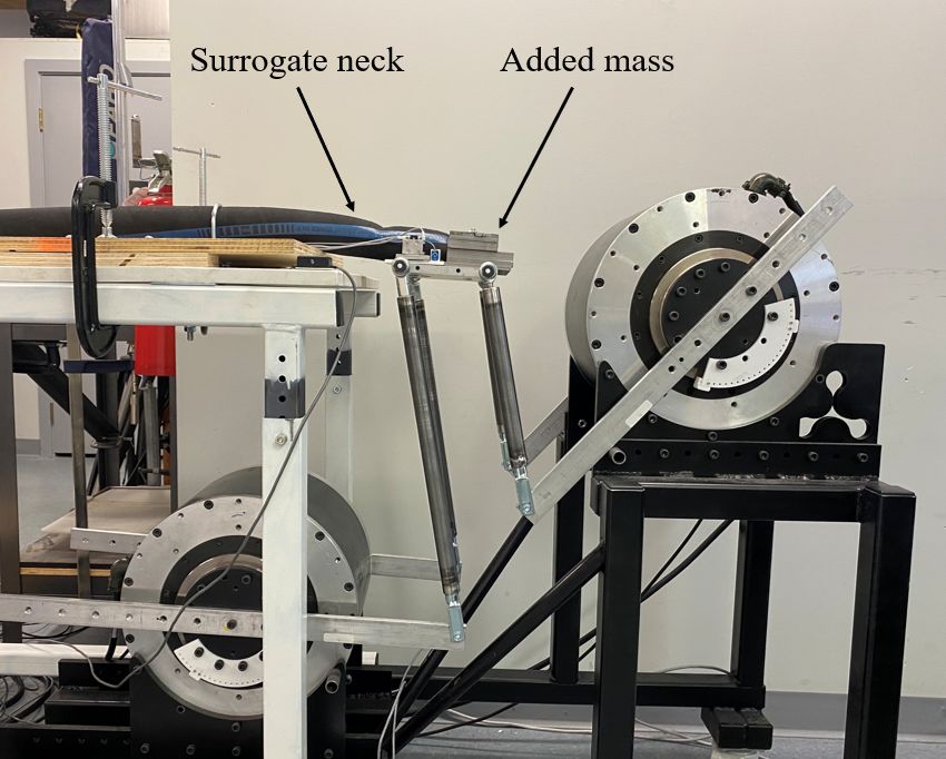
Data collection and processing
The surrogate headplate was instrumented (Figure 5) with biaxial linear accelerometers
(Endevco, 7265A, Irvine, CA) and a uniaxial angular rate sensor (DTS, ARS Pro, Seal Beach,
CA). The controller voltage signal to the Motor 1 servo drive was also recorded in order to
calculate the motor torque output. Motor 1 was selected for the torque calculations as it is the
limiting motor due to its larger required range of motion. Data was recorded for 5 seconds at 10
kHz using a 16-bit DAQ card (PXI-6221, National Instruments, Austin, TX). All data channels
conformed to SAE J211 Channel Class 1000 (SAE, 2003). Recorded instrumentation data was
digitally low-pass filtered using Matlab according to Channel Class 1000.
Figure 5: Instrumented plate.
Through Labview, the Motor 1 (M1) and Motor 2 (M2) encoder positions were logged at
100 Hz to capture the motor rotational positions. Positive motor rotation is counter clockwise for
both motors from the view in Figure 2. Additionally, two markers were placed on the bolthead
attaching the articulated arms to the headplate (white markers in Figure 5) and sagittal-view videos
were captured at 240 fps using an iPhone 11 (Apple, Cupertino, CA). Markers were digitized using
open-source software (Kinovea, https://www.kinovea.org/). Videos and instrumentation data were
synchronized using an LED which turned on when the DAQ was triggered.
7
2021 The Ohio State University Injury Biomechanics Symposium
*This paper has not been peer-reviewedRESULTS
Incremental motion profiles
The plate rotation and retraction, M1 and M2 encoder positions, and M1 torque output are
presented in Figure 6 (TF = 8 and 4), and 7 (TF = 3.5 and 3). The input values represent the
programmed motion profiles while the output values represent the video marker tracking data
(rotation and retraction), and the logged encoder positions (M1 and M2 position). Testing was
completed up to TF = 3 as the M1 rated torque limit of 200 N×m was reached at this level at
approximately 110 ms (Figure 7).
Figure 6: Position and torque data for TF = 8 and 4.
8
2021 The Ohio State University Injury Biomechanics Symposium
*This paper has not been peer-reviewedFigure 7: Position and torque data for TF = 3.5 and 3.
Figures 8 and 9 present the angular rate and acceleration data for TF = 8, 4, and TF = 3.5,
3, respectively. A peak angular rate, x acceleration and y acceleration of 16 rad/s, 9 g, and 3 g,
were achieved with the TF = 3.5 motion profile, respectively. Prior to reaching the torque overload,
the TF = 3 motion profile achieved a peak angular rate, x acceleration and y acceleration of 24
rad/s, 15 g, and 14 g, respectively
9
2021 The Ohio State University Injury Biomechanics Symposium
*This paper has not been peer-reviewedFigure 8: Angular rate and acceleration data for TF = 8 and 4.
10
2021 The Ohio State University Injury Biomechanics Symposium
*This paper has not been peer-reviewedFigure 9: Angular rate and acceleration data for TF = 3.5 and 3.
Accuracy
From the data in Figures 6 and 7, the percent error between the input and output peak values
and time-to-peak values were determined and are tabulated in Table 1. This data can be used to
quantify the accuracy of the device in producing the programmed motion profiles. Time-to-peak
and peak values for the rotation and retraction plots had percent errors up 39%. Percent errors for
both motors were no more than 5% except for the TF = 3 case where the torque overload in M1
caused a delay in the time-to-peak.
11
2021 The Ohio State University Injury Biomechanics Symposium
*This paper has not been peer-reviewedTable 1: Percent error between input and output peak and time to peak values
Rotation Retraction M1 Position M2 Position
TF Time- Peak Time- Peak Time- Peak Time- Peak
to-peak Value to-peak Value to-peak Value to-peak Value
8 8% 10% 1% 6% 0% 0% 1% 0%
4 12% 31% 12% 10% 3% 1% 0% 0%
3.5 16% 39% 15% 9% 0% 1% 5% 0%
3 33% 36% 35% 8% 19% 1% 6% 0%
Repeatability
An intermediate motion profile (TF = 4) was tested three times to assess the repeatability
of the produced motion profiles. Table 2 presents the mean, standard deviation (SD) and
coefficient of variation (CoV) for the peak and time-to peak-values of the rotation, retraction, and
M1 and M2 position traces. The time-to-peak and peak values for all traces had a CoV less than
5%.
Table 2: Mean, standard deviation and coefficient of variation between the TF = 4 trials (n = 3)
Mean SD CoV
Rotation Time-to-Peak [ms] 322 10 3%
Peak value [deg] 105 2 2%
Retraction Time-to-Peak [ms] 447 17 4%
Peak value [cm] 26 1 2%
M1 Position Time-to-Peak [ms] 350 0 0%
Peak value [deg] 50 0 0%
M2 Position Time-to-Peak [ms] 440 10 2%
Peak value [deg] 33 0 0%
DISCUSSION
This project aimed to test and validate a new device which can simulate whiplash motions
in a porcine model and attempts to address the limitations of previous designs. Our evaluation
included assessing whether the device could replicate motion profiles from previous whiplash
studies and to quantify the accuracy and repeatability of the simulated whiplash motions.
One relatively severe whiplash motion profile was selected from a previous porcine
whiplash study to be replicated (Obj. 1). To assess the device and motor capabilities, the selected
motion profile was scaled via a time factor and incrementally tested. With the current device
design, we successfully replicated the selected motion profile when scaled with a TF = 3.5. This
motion profile consisted of a peak extension angle of 80 degrees in approximately 250 ms which
is representative of previous PMHS sled tests investigating cervical spine injuries from rear-end
collisions (Bertholon et al., 2000; Philippens et al., 2000; Kang et al., 2014). With this motion
profile, a peak angular rate of 16 rad/s and x and y accelerations of 15 and 30 g were achieved,
12
2021 The Ohio State University Injury Biomechanics Symposium
*This paper has not been peer-reviewedrespectively. Peak resultant plate accelerations exceed the range of previous low-speed human
volunteer rear-end collision studies (Siegmund et al., 1997) and are within range of the
approximately 20 g head accelerations reached in previous porcine whiplash studies (Svensson et
al., 1993).
A TF = 3 motion profile was also tested; however, M1 reached its rated torque limit within
the first 110 ms where an initial rapid ramp up is required. This indicates a limitation of the current
device design which prevents testing of higher severities. Several potential solutions can be
implemented to address this limitation. Currently, the motor servo system is programmed to output
a maximum current corresponding to the rated torque limit (200 N×m). However, the motors are
capable of an instantaneous maximum torque up to 600 N×m. This maximum torque limit can be
achieved for intermittent periods which is adequate for the short period in our whiplash motion
profiles. To harness the maximum torque limit, the motor servo system can be reprogrammed.
Another potential solution can be to optimize the lengths of the linkages attached to the motors to
reduce the torque load on M1 at the initial ramp up period of the motion profile. Additionally, with
the current setup, most of the added mass on the plate is biased towards M1. Redistributing the
mass to better represent the position of the porcine head center of gravity may help offload M1
and improve the device’s ability to generate the extreme pulse selected. These potential solutions
will be explored in future work.
Several measurements were made to assess how accurately programmed motion profiles
could be replicated with our device (Obj. 2). Marker tracking from digitized videos were used to
assess the rotation and retraction of the headplate while logged encoder positions were used to
assess the angular positions of the motors. Accuracy and repeatability assessments were conducted
using the magnitude of the peak values and time-to-peak points of the motion profile traces as
these quantities represent the fundamental characteristics of the motion profiles. The plate rotation
and retraction had percent errors up to 39% which increased with decreasing TF. Additionally, in
all cases for rotation, the peak value percent error was larger than the time-to-peak percent error.
These large percent errors for the plate rotation and retraction can be attributed to several factors.
First, the behavior of the plate is largely dependent on the behavior of the hose used as the surrogate
neck. From the recorded videos, we visually observed that at the lower TF cases, the hose exhibited
buckling near the plate attachment point which resulted in larger rotation angles. This limitation
will be addressed in subsequent work where the surrogate head and neck model will be replaced
with cadaver pigs which will elicit a more biofidelic response. Additionally, some discrepancy in
the plate rotation and retraction measurements can be attributed to lens distortion error which was
not corrected for as well as errors in spatial calibration and marker tracking. However, as expected,
at the motor level, motor angular positions had percent errors no more than 5% for tests with TF
= 3.5 and larger.
An intermediate motion profile (TF = 4) was tested three times to assess the repeatability
of the produced motion profiles (Obj. 2). Despite the overshoot of the plate rotation and retraction
from the buckling of the hose which affected accuracy, motion profiles were produced with good
repeatability. Coefficient of variation values were below 5% for all measured quantities. The good
repeatability of this proposed device is a significant strength compared to previous designs.
13
2021 The Ohio State University Injury Biomechanics Symposium
*This paper has not been peer-reviewedThis new proposed device aims to simulate whiplash motions in a porcine model and
address the limitations of previous work. Our preliminary assessment of this device indicates that
it has potential to enable further study of the DRG whiplash injury mechanism by producing
specific programmed motion profiles with good accuracy and repeatability. With some additional
improvements, this device can be used in future studies to systematically investigate the effect of
various head and neck kinematic parameters on the measured spinal pressures during simulated
whiplash. Subsequent validation and testing of this device will involve design changes to achieve
the whiplash severities tested in previous studies. Additionally, the device will be further validated
using cadaver porcine testing prior to in vivo experiments.
CONCLUSIONS
Preliminary assessment of a new whiplash simulation device indicates promising potential
for it to address specific limitations of previous methods used to study whiplash injury in a porcine
model. The current device enables more accurate and repeatable control of specific programmed
motion profiles. This capability can be used in future porcine studies to further elucidate the DRG
whiplash injury mechanism by correlating head and neck kinematics parameter to measured spinal
pressures and tissue damage. Through subsequent design modifications, the device’s capabilities
will be optimized to reach the severe whiplash motion severities tested in previous porcine
whiplash studies. Improving our understanding of the whiplash injury mechanism is an important
step to treating and ultimately preventing this poorly understood affliction.
ACKNOWLEDGEMENTS
Thank you to Jeff Nickel and Mircea Oala-Florescu of MEA Forensic Engineers &
Scientists for their technical support and contributions to the whiplash device development. The
authors also acknowledge funding support from the Natural Sciences and Engineering Research
Council of Canada and the Insurance Institute for Highway Safety.
14
2021 The Ohio State University Injury Biomechanics Symposium
*This paper has not been peer-reviewedREFERENCES
ALDMAN, B. (1986). An analytical approach to the impact biomechanics of head and neck injury,
30th Annual Proceedings American Association for Automotive Medicine, pp. 439–454.
BERTHOLON, N., ROBIN, S., LE-COZ, J., POTEIR, P., LASSAU, J. AND SKALLI, W. (2000).
Human head and cervical spine behaviour during low-speed rear-end impacts: PMHS tests
with a rigid seat, IRCOBI Conference.
BOSTROM, O. ET AL. (1996). A New Neck Injury Criterion Candidate Based on Injury Findings
in the Cervical Spine Ganglia after Experimental Sagittal Whiplash, Ircobi, pp. 123(119)-
136(133).
BUSSCHER, I., PLOEGMAKERS, J. J. W., VERKERKE, G. J. AND VELDHUIZEN, A. G.
(2010). Comparative anatomical dimensions of the complete human and porcine spine,
European Spine Journal, 19(7), pp. 1104–1114. doi: 10.1007/s00586-010-1326-9.
CONDOTTA, I., BROWN-BRANDL, T., STINN, J. AND DAVIS, J. D. (2018). Dimensions of
the Modern Pig, American Society of Agricultural and Biological Engineers, (February
2019). doi: 10.13031/trans.12826.
CROFT, A. C., HERRING, P., FREEMAN, M. D. AND HANELINE, M. T. (2002). The neck
injury criterion: Future considerations, Accident Analysis and Prevention, 34(2), pp. 247–
255. doi: 10.1016/S0001-4575(01)00020-3.
CURATOLO, M., BOGDUK, N., IVANCIC, P. C., MCLEAN, S. A., SIEGMUND, G. P. AND
WINKELSTEIN, B. A. (2011). The role of tissue damage in whiplash-associated disorders,
Spine, 36(25), pp. S309–S315. doi: 10.1097/BRS.0b013e318238842a.
DUHAIME, A. C. (2006). Large animal models of traumatic injury to the immature brain,
Developmental Neuroscience, 28(4–5), pp. 380–387. doi: 10.1159/000094164.
EICHBERGER, A., STEFFAN, H., GEIGLE, B., SVENSSON, M. Y., BOSTRÖM, O.,
LEINZINGER, P. E. AND DAROK, M. (1998). Evaluation of the applicability of the neck
injury criterion in rear end impacts on the basis of human subject tests, IRCOBI
Conference, pp. 321–333. doi: 10.1016/B978-0-323-07980-8.00006-0.
EICHBERGER, A., DAROK, M., STEFFAN, H., LEINZINGER, P. E., BOSTRÖM, O. AND
SVENSSON, M. Y. (2000). Pressure measurements in the spinal canal of post-mortem
human subjects during rear-end impact and correlation of results to the neck injury
criterion, Accident Analysis and Prevention, 32(2), pp. 251–260. doi: 10.1016/S0001-
4575(99)00097-4.
JONES, C. F., LEE, J. H. T., KWON, B. K. AND CRIPTON, P. A. (2012). Development of a
large-animal model to measure dynamic cerebrospinal fluid pressure during spinal cord
injury: Laboratory investigation, Journal of Neurosurgery: Spine, 16(6), pp. 624–635. doi:
15
2021 The Ohio State University Injury Biomechanics Symposium
*This paper has not been peer-reviewed10.3171/2012.3.SPINE11970.
KANG, Y. S., MOORHOUSE, K., ICKE, K., HERRIOTT, R. AND BOLTE, J. (2014). Head and
cervical spine responses of post mortem human subjects in moderate speed rear impacts,
IRCOBI Conference, pp. 268–285.
LEE, J. H. T. ET AL. (2013). A Novel porcine model of traumatic thoracic spinal cord injury,
Journal of Neurotrauma, 30(3), pp. 142–159. doi: 10.1089/neu.2012.2386.
ONO, K. AND KANNO, M. (1993). Influences of the physical parameters on the risk to neck
injuries in low impact speed rear-end collisions, Accident Analysis and Prevention, 28(4),
pp. 493–499. doi: 10.1016/0001-4575(96)00019-X.
ÖRTENGREN, T., HANSSON, H. A., LÖVSUND, P., SVENSSON, M. Y., SUNESON, A. AND
SALJÖ, A. (1996). Membrane leakage in spinal ganglion nerve cells induced by
experimental whiplash extension motion: A study in pigs, Journal of Neurotrauma, 13(3),
pp. 171–180. doi: 10.1089/neu.1996.13.171.
PHILIPPENS, M., WISMANS, J., CAPPO, H., YOGANANDAN, N. AND PINTAR, F. (2000).
Whole body kinematics using post mortem human subjects in experimental rear impact,
IRCOBI Conference, (4), pp. 363–378.
SAE. (2003). Instrumentation for Impact Test-Part1-Electronic Instrumentation (SAE J211-1 Dec
03), SAE International.
SHENG, S. R., WANG, X. Y., XU, H. Z., ZHU, G. Q. AND ZHOU, Y. F. (2010). Anatomy of
large animal spines and its comparison to the human spine: A systematic review, European
Spine Journal, 19(1), pp. 46–56. doi: 10.1007/s00586-009-1192-5.
SHENG, S. R., XU, H. Z., WANG, Y. L., ZHU, Q. A., MAO, F. M., LIN, Y. AND WANG, X. Y.
(2016). Comparison of cervical spine anatomy in calves, pigs and humans, PLoS ONE,
11(2), pp. 1–10. doi: 10.1371/journal.pone.0148610.
SIEGMUND, G. P., KING, D. J., LAWRENCE, J. M., WHEELER, J. B., BRAULT, J. R. AND
SMITH, T. A. (1997). Head / Neck Kinematic Response of Human Subjects in Low-Speed
Rear-End Collisions, Society of Automotive Engineers.
SIEGMUND, G. P., WINKELSTEIN, B. A., IVANCIC, P. C., SVENSSON, M. Y. AND
VASAVADA, A. (2009). The Anatomy and biomechanics of acute and chronic whiplash
injury, Traffic Injury Prevention, 10(2), pp. 101–112. doi: 10.1080/15389580802593269.
STERNER, Y. AND GERDLE, B. (2004). Acute and chronic whiplash disorders - A review,
Journal of Rehabilitation Medicine, 36(5), pp. 193–210. doi:
10.1080/16501970410030742.
SVENSSON, M. Y., ALDMAN, B., HANSSON, H. A, LÖVSUND, P., SEEMAN, T.,
16
2021 The Ohio State University Injury Biomechanics Symposium
*This paper has not been peer-reviewedSUNESON, A. AND ÖRTENGREN, T. (1993). Pressure Effects in the Spinal Canal
during Whiplash Extension Motion: A Possible Cause of Injury to the Cervical Spinal
Ganglia, IRCOBI Conference, pp. 189–200.
SVENSSON, M. Y., BOSTRÖM, O., DAVIDSSON, J., HANSSON, H. A., HÅLAND, Y.,
LÖVSUND, P., SUNESON, A. AND SÄLJÖ, A. (2000). Neck injuries in car collisions -
A review covering a possible injury mechanism and the development of a new rear-impact
dummy, Accident Analysis and Prevention, 32(2), pp. 167–175. doi: 10.1016/S0001-
4575(99)00080-9.
SZABO, T. J., WELCHER, J. B., ANDERSON, R. D., RICE, M. M., WARD, J. A., PAULO, L.
R. AND CARPENTER, N. J. (1994). Human occupant kinematic response to low speed
rear-end impacts, SAE Technical Papers, 103, pp. 630–642. doi: 10.4271/940532.
YAO, H. D., SVENSSON, M. Y. AND NILSSON, H. (2016). Transient pressure changes in the
vertebral canal during whiplash motion - A hydrodynamic modeling approach, Journal of
Biomechanics, 49(3), pp. 416–422. doi: 10.1016/j.jbiomech.2016.01.005.
17
2021 The Ohio State University Injury Biomechanics Symposium
*This paper has not been peer-reviewedYou can also read