Biliary Tract and Pancreatic Disease - Clinical - Journal of ...
←
→
Page content transcription
If your browser does not render page correctly, please read the page content below
Clinical
Upper Gastrointestinal Disorders in Urgent Care, Part 2:
Biliary Tract and Pancreatic
Disease
Urgent message: Upper abdominal pain is a common presentation in
urgent care practice. Narrowing the differential diagnosis is sometimes
difficult. Understanding the pathophysiology of each disease is the key to
making the correct diagnosis and providing the proper treatment.
TRACEY Q. DAVIDOFF, MD
art 1 of this series focused on disorders of the stom-
P ach—gastritis and peptic ulcer disease—on the left side
of the upper abdomen. This article focuses on the right
side and center of the upper abdomen: biliary tract dis-
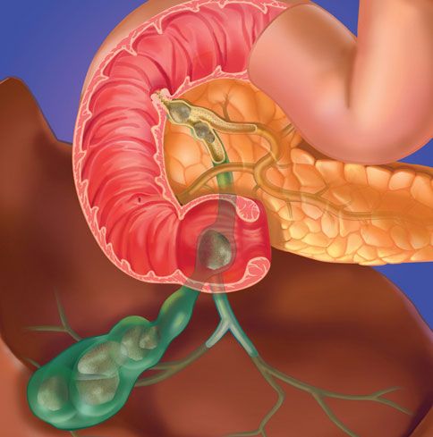
ease and pancreatitis (Figure 1). Because these diseases
are regularly encountered in the urgent care center, the
urgent care provider must have a thorough understand-
ing of them.
Biliary Tract Disease
The gallbladder’s main function is to concentrate bile by
the absorption of water and sodium. Fasting retains and
concentrates bile, and it is secreted into the duodenum
by eating. Impaired gallbladder contraction is seen in
pregnancy, obesity, rapid weight loss, diabetes mellitus,
and patients receiving total parenteral nutrition (TPN).
About 10% to 15% of residents of developed nations
©Phototake.com
will form gallstones in their lifetime.1 In the United
States, approximately 6% of men and 9% of women
have gallstones.2 Stones form when there is an imbal-
ance in the chemical constituents of bile, resulting in
precipitation of one or more of the components. It is
unclear why this occurs in some patients and not others,
Tracey Q. Davidoff, MD, is an urgent care physician at Accelcare Medical
although risk factors do exist. Gallstones can occur in
Urgent Care in Rochester, New York, is on the Board of Directors of the
Urgent Care College of Physicians, and is a member of the JUCM Editorial all age groups, but the incidence increases with age.
Board. Everyone remembers the 4 F’s from medical school: “fat,
8 JUCM T h e J o u r n a l o f U r g e n t C a r e M e d i c i n e | M a y 2 0 1 5 w w w. j u c m . c o m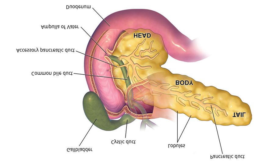
B I L I A RY T R A C T A N D PA N C R E AT I C D I S E A S E

 Figure 1. Anatomy of the pancreas, gallbladder, and duodenum.

 Modified with permission under a Creative Commons BY 3.0 US license from Blausen.com staff. Blausen gallery 2014. Wikiversity Journal of Medicine.
 doi:10.15347/wjm/2014.010. ISSN 20018762. Original figure available from: http://upload.wikimedia.org/wikipedia/commons/thumb/7/7e/Blausen_0699_
 PancreasAnatomy2.png/1280px-Blausen_0699_PancreasAnatomy2.png

fertile, female, and forty.” Estrogens cause more choles-                               surgical illness in pregnancy, with an incidence of symp-
terol to be excreted in bile, increasing risk. Obesity is cer-                          tomatic gallstones of about 0.1%.3 Surgical intervention
tainly a factor, and family history is also contributory.                               should be delayed until after delivery if at all possible,
   Biliary sludge is a mixture of particulate matter and                                although laparoscopic cholecystectomy has been
bile that may turn into tiny gallstones. It is common in                                proven safe in pregnancy.
patients who are or have been pregnant or who are
receiving TPN. Persistent sludge may become gallstones,                                 Biliary Colic
or the sludge itself may cause cholecystitis. Most patients                             The most common illness related to gallstones is biliary
with sludge are asymptomatic.                                                           colic. The patient experiences right upper quadrant or
   The majority of patients found to have incidental                                    midepigastric abdominal pain lasting for 2 to 6 hours,
gallstones are asymptomatic and are not at risk for                                     which then remits spontaneously. The pain is intense,
developing gallbladder disease. Less than 20% will                                      sharp, and stabbing in nature and may be associated
develop symptoms over 15 years, and their illness will                                  with nausea, vomiting, fever, and diaphoresis. It may
not be severe. The risk of developing a severe compli-                                  radiate around (not through) to the back, specifically
cation from these stones is much less than the risk of a                                the right shoulder blade. It is not exacerbated by move-
complication from a cholecystectomy, and therefore it                                   ment, and not relieved by position, movement, or pas-
is not recommended that patients with asymptomatic                                      sage of flatus or stool. Symptoms occur because of
gallstones undergo cholecystectomy. Once a patient                                      increasing intraluminal pressure and distention of the
develops symptoms, however, their subsequent risk of                                    biliary tract as 1 or more stones migrate from the gall-
developing complications is much higher, and prophy-                                    bladder into the cystic or common bile duct. Com-
lactic cholecystectomy is advisable.2                                                   monly the symptoms begin between 9 p.m. and 4 a.m.
   Biliary tract disorders are the second most common                                   Dyspepsia, belching, and bloating are nonspecific symp-

10   JUCM T h e J o u r n a l o f U r g e n t C a r e M e d i c i n e | M a y 2 0 1 5                                                       w w w. j u c m . c o m
B I L I A RY T R A C T A N D PA N C R E AT I C D I S E A S E

toms that are just as common in people with gallstones              Table 1. Complications of Gallstones
as those without. A common trigger is a fatty meal,
although association with meals is not universal. If the            • Dehydration                                  • Sepsis
                                                                    • Acute cholecystitis                          • Perforation
initial obstruction is not relieved by persistent contrac-
                                                                    • Choledocholithiasis                          • Gallstone ileus
tions, or if the stone does not fall back into the gallblad-        • Acute pancreatitis                           • Mirizzi syndrome
der, an inflammatory response will occur by mechani-                • Acute or ascending cholangitis               • Gallbladder cancer
cal, chemical, or infectious means. If pain lasts more
than 5 to 6 hours, think complications, such as acute
cholecystitis or acute pancreatitis.                              acute cholecystitis (see the section “Acute Cholecystitis”).
   These patients look uncomfortable, but findings on             It should be noted, however, that the absence of stones
the physical examination are usually benign. There may            on ultrasonography does not rule out their existence;
be some right upper quadrant tenderness, which is often           small stones and sludge may be missed,1 and the sensi-
underwhelming compared with the amount of pain the                tivity of ultrasonography may be operator dependent.
patient appears to be in. There are no peritoneal signs.             Once a patient has developed symptoms related to gall-
There should be no jaundice. There may be some signs              stones, definitive treatment with surgery is recom-
of dehydration, such as dry mouth or poor skin turgor             mended, because the risk of subsequent symptoms or
if vomiting has been severe.                                      complications is high.4 Stones can occur and become
   Although the diagnosis of biliary colic is frequently          symptomatic even after the gallbladder has been
made by medical history, ultrasonography is necessary             removed, months to even years after cholecystectomy.
to identify the presence of stones and to rule out other          Therefore, a history of cholecystectomy does not rule out
illnesses or complications that may be occurring such as          the possibility of biliary colic. The incidence of this is

w w w. j u c m . c o m                                         JUCM T h e J o u r n a l o f U r g e n t C a r e M e d i c i n e | M a y 2 0 1 5   11

                                   ®

                                         ®
B I L I A RY T R A C T A N D PA N C R E AT I C D I S E A S E

 Table 2. Laboratory Studies to Rule Out Complications of Biliary Colic                                       should be given if dehydration is sus-
 Test                                                     May Rule Out                                        pected. Patients having symptoms for
                                                                                                              more than 6 hours or who have evi-
 Liver biochemical markers (AST, ALT,                       Hepatitis, biliary tract obstruction,
                                                                                                              dence of complications should be
 ALK, bilirubin)                                            acute cholecystitis
                                                                                                              referred to an emergency department
 Serum amylase and lipase                                   Acute pancreatitis                                (ED). Patients whose condition
 CBC count                                                  Acute cholecystitis or acute cholangitis          improves and who are asymptomatic
 Urinalysis                                                 UTI, renal colic                                  after an episode of biliary colic can be
                                                                                                              referred to surgery for outpatient, elec-
 Upper endoscopy                                            PUD, gastritis                                    tive cholecystectomy.
 Endoscopic ultrasonography                                 Chronic pancreatitis                                 Red-flag symptoms and signs indi-
 ERCP with sphincter of Oddi manometry Sphincter of Oddi dysfunction                                          cating impending complications
                                                                                                              (Table 1) include pain lasting more
                                                            Acute cholecystitis and functional
 Cholescintigraphy with or without CCK                                                                        than 6 to 8 hours, intractable vomiting,
                                                            gallbladder disorder
                                                                                                              fever, jaundice, elevated white blood
 ECG                                                        Ischemic heart disease                            cell (WBC) count, elevated enzyme lev-
 Esophageal manometry                                       Esophageal spasm                                  els on liver function tests, and elevated
                                                                                                              amylase and/or lipase levels.2 Findings
 ALK = anaplastic lymphoma kinase; ALT = alanine aminotransferase; AST = aspartate aminotransferase; CBC =
 complete blood cell; CCK = cholecystokinin; ECG = electrocardiography; ERCP = endoscopic retrograde          on all laboratory studies (Table 2)
 cholangiopancreatography; PUD = peptic ulcer disease; UTI = urinary tract infection.                         should be normal in patients with
                                                                                                              uncomplicated gallbladder disease or
                                                                                                              biliary colic. Patients suspected of hav-
 Table 3. Differential Diagnosis of Upper Abdominal                                      ing a complication, or in whom the diagnosis is unclear,
 Pain Not Related to Biliary or Pancreatic Disease                                       should undergo ultrasonography, if available, or should
 • Esophagitis or gastroesophageal reflux disease                                        be referred to the nearest ED for further evaluation.
 • Acute hepatitis                                                                           Atypical symptoms should prompt an investigation
 • Peptic ulcer disease                                                                  for other diagnoses (Table 3), even if the patient has
 • Gastritis or duodenitis                                                               known or documented gallstones. These include
 • Dyspepsia                                                                                 ! Chest pain
 • Irritable bowel disease                                                                   ! Nonspecific abdominal pain
 • Disorders of the right kidney, including renal colic                                      ! Belching
 • Right lower lobe pneumonia
 • Fitz-Hugh-Curtis syndrome                                                                 ! Fullness after meals or early satiety
 • Subhepatic or intra-abdominal abscess                                                     ! Fluid regurgitation
 • Perforated viscus                                                                         ! Abdominal distention or bloating
 • Cardiac ischemia                                                                          ! Epigastric or retrosternal burning sensation
 • Black widow spider envenomation                                                           ! Nausea or vomiting without typical biliary colic
 • Retrocecal appendicitis                                                                       pain

unknown, but it is probably low.5 Stones occurring imme-                                Acute Cholecystitis
diately after surgery are called retained stones, and those                             Acute cholecystitis is an inflammation of the gallbladder
occurring months to years after are recurrent stones.                                   most commonly caused by gallstones. Approximately
   Patients with biliary colic should be treated for pain                               1% to 3% of patients with symptomatic gallstones will
with opioids and nonsteroidal anti-inflammatory drugs:                                  develop acute cholecystitis.3
oral if possible, but parenteral if nausea and vomiting pre-                               In most cases, acute cholecystitis is caused by to
vent the use of oral medications. All narcotics increase                                obstruction of the cystic duct of the gallbladder by
biliary pressure and spasm of the sphincter of Oddi and                                 a stone or sludge that has impacted at the neck of the gall-
theoretically could make biliary obstruction worse; how-                                bladder. This obstruction causes the pressure in the gall-
ever, proof of this has never been reported in the literature.5                         bladder to build and, in combination with bile, causes an
Pain relief is more important than any theoretical risk.                                inflammatory response. Secondary bacterial infection
Antiemetics may be required. Intravenous hydration                                      may occur in up to 20% of patients,3 usually with enteric

12   JUCM T h e J o u r n a l o f U r g e n t C a r e M e d i c i n e | M a y 2 0 1 5                                               w w w. j u c m . c o m
BILIARY TRACT AND PANCREATIC DISEASE
organisms such as Escherichia coli, Klebsiella, and Entero-
coccus faecalis. The overall mortality of a single episode
of acute cholecystitis is approximately 3%, depending
on the patient’s underlying health and surgical risk.4
In acute cholecystitis, pain is localized to the right
upper quadrant and lasts for more than 4 to 6 hours,6
as opposed to biliary colic, in which the pain is more
intermittent. The pain may radiate to the right shoulder
or around to the back. Nausea and vomiting are com-
mon. There is abdominal tenderness in the right upper
quadrant, there is voluntary and involuntary guarding,
and there will be positive findings for Murphy sign (pain
with palpation on deep inspiration in the right upper
quadrant with associated inspiratory arrest). The sen-
sitivity and specificity of Murphy sign are 97% and 48%,
respectively.6 If septicemia is present, there may be fever.
Mild jaundice may be present.
Typically patients will have a leukocytosis with
increased band cells. Liver enzymes and bilirubin levels
will more than likely not be elevated, because the
obstruction is limited to the gallbladder. Think com-
plications if alkaline phosphatase and bilirubin levels
are elevated.6 Amylase and lipase levels may be slightly
elevated because of the passage of smaller stones or
sludge prior to obstruction. It should be noted that lab-
oratory findings are nonspecific and cannot defini-
tively distinguish colic from cholecystitis.
All patients suspected of having acute cholecystitis should
be referred to a hospital.3
The diagnosis of acute cholecystitis can be made on
the basis of medical history, but it must be supported
by the characteristic findings on ultrasonography,
which is the test of choice. Typically there is perichole-
cystic fluid around the gallbladder, edema of the gall-
bladder wall, gallstones, and a sonographic Murphy
sign. The sensitivity and specificity of ultrasonography
for cholecystitis are 88% and 80%, respectively; for gall-
stones, the respective values are 84% and 99%.6 Distin-
guishing biliary colic from acute cholecystitis can be
difficult. Murphy sign is the most sensitive (65%) and
specific (87%) element of the history and physical that
can distinguish the two. In the sonographic Murphy
sign, the ultrasound probe is used for palpation and
visualizing the stones during the test.
Plain abdominal films show gallstones in only 10%
of cases.3 Gas within the gallbladder may be seen in
emphysematous cholecystitis, which is rare. Plain films
should not be routinely ordered for suspected gallblad-
der disease.
When the diagnosis remains in question after ultra-
JUCM The Journal of Urgent Care Medicine | M a y 2 0 1 5 13Urgent Care BILIARY TRACT AND PANCREATIC DISEASE
Medicine
Table 4. Diagnostic Criteria for Acute Cholangitis (2013 Tokyo
Medical Guidelines)
Professional Diagnosis should be suspected if patient has one of the following:
• Fever and/or shaking chills
Liability • Laboratory evidence of an inflammatory response (elevated WBC count,
elevated CRP level, etc.)
Insurance And one of the following:
• Jaundice
• Abnormal liver enzyme levels (ALK, ALT, AST, GGTP)
The Wood Insurance Group, a leading Diagnosis should be definite if the patient also has
national insurance underwriter, offers • Biliary dilation on imaging
• Evidence of an etiology on imaging (stricture, stone, or stent)
significantly discounted, competitively
priced Medical Professional Liability ALK = anaplastic lymphoma kinase; ALT = alanine aminotransferase; AST = aspartate
aminotransferase; CRP = C-reactive protein; GGTP = !-glutamyl transpeptidase; WBC = white blood
Insurance for Urgent Care Medicine. cell.
Data from Kiriyama S, Takada T, Strasberg SM, et al; Tokyo Guidelines Revision Committee. TG13
We have been serving the Urgent Care guidelines for diagnosis and severity grading of acute cholangitis (with videos). J Hepatobiliary
Pancreat Sci 2013;20:24–34.
community for over 25 years, and our
UCM products were designed specif-
sonography, biliary scintigraphy or a hydroxyiminodiacetic acid
ically for Urgent Care Clinics. (HIDA) scan can be performed. The patient is injected with radiola-
beled HIDA. In healthy gallbladders, uptake can be seen in 1 to 2
Our Total Quality hours, but in inflamed gallbladders, there is no uptake seen. The
Approach includes: sensitivity and specificity of HIDA scans for acute cholecystitis are
approximately 97% and 90%, respectively.6
" Preferred Coverage Features The treatment of acute cholecystitis is ultimately surgical. In most
– Per visit rating (type & number) patients, the disease is managed initially with medical treatment,
– Prior Acts Coverage including fasting, rehydration, and pain medications. The impacted
– Defense outside the limit stone falls back into the gallbladder, and then it can be removed
– Unlimited Tail available electively. If this does not happen, gangrenous cholecystitis,
empyema, or perforation may result. Nonsteroidal anti-inflamma-
– Exclusive “Best Practice”
tory drugs, specifically ketorolac,6 are recommended as first-line
Discounts treatment for pain because they also provide an anti-inflammatory
– Protects the Clinic and Providers effect. Some patients may require narcotic pain medications as well.
" Exceptional
Antiemetics may also be required. If an infectious process is sus-
Service Standards
pected, treatment with a second- or third-generation cephalosporin
– Easy application process and metronidazole is also recommended.
– Risk Mgmt/Educational support Twenty percent of patients with acute cholecystitis need emer-
– Fast turnaround on policy gency surgery when peritonitis or emphysematous cholecystitis (air
changes in the gallbladder) is present.3,4 The remaining 80% can be observed
– Rapid response claim service with medical management to have the inflammation “cool down,”
and then laparoscopic cholecystectomy can be performed in several
days, usually within 72 hours of presentation.3,4
Choledocholithiasis
A stone lodged in the common bile duct may cause typical biliary
colic pain, however it will last longer and eventually lead to more
complicated disease. Serum aspartate aminotransferase (AST) and ala-
nine aminotransferase (ALT) levels are typically elevated. If the stone
8201 North Hayden Road does not pass, serum anaplastic lymphoma kinase (ALK), bilirubin,
Scottsdale, Arizona 85258
(800) 695-0219 • Fax (602) 230-8207
E-mail: davidw@woodinsurancegroup.com
14 JUCM T h e J o u r n a l o f U r g e n t C a r e M e d i c i n e | M a y 2 0 1 5BILIARY TRACT AND PANCREATIC DISEASE and !-glutamyl transpeptidase (GGTP) levels may become elevated out of proportion to the ALT and AST levels. Acute cholangitis may subsequently develop causing fever, leukocytosis, and sepsis syn- drome. Ultrasonography may be helpful, but visualizing stones in the distal portion of the duct may be difficult. Magnetic resonance cholangiopancreatography, endoscopic ultrasonography, or ERCP may be useful in these cases. Ascending Cholangitis Ascending or acute cholangitis occurs as a result of infection and stasis of infected bile in the biliary tract. Its severity can range from mild to life-threatening. The patient will present with fever, abdom- inal pain, and jaundice (often called Charcot triad), then progress to confusion and hypotension. Septic shock can occur, progressing to multisystem organ failure. Patients taking glucocorticoids, espe- cially the elderly, may have no symptoms until hypotension occurs. Laboratory findings include a cholestatic pattern of abnormal liver enzymes and an elevated WBC count. Bacteria enter the biliary tract because of gallstones, sludge, or manipulation of the sphincter of Oddi, usually as an iatrogenic effect of ERCP, stent placement, or percutaneous drainage, but bacterial ingress can be spontaneous. E. coli is the most common gram-neg- ative bacteria, followed by Klebsiella and Enterobacter species. Entero- coccus species may also be responsible. Occasionally a mixed picture with anaerobes occurs (Table 4). Ascending cholangitis is a medical emergency that requires immediate evaluation in an ED. Pancreatitis Acute Pancreatitis Acute pancreatitis is an inflammatory process that can range from mild to severe. Mild cases can resolve with minimum treatment and have a mortality rate of
B I L I A RY T R A C T A N D PA N C R E AT I C D I S E A S E

 Table 5. Causes of Pancreatitis                                                        again after the pancreatitis has resolved.
 • Gallstones                             • Viral infections (mumps,                        Hypercalcemia is also a recognized etiology of pan-
 • Alcohol                                  coxsackievirus,                             creatitis, and it can be seen in malignancy, TPN, sar-
 • Idiopathic                               cytomegalovirus,                            coidosis, vitamin D toxicity, and as a complication of
 • Hypertriglyceridemia                     echovirus, hepatitis B                      cardiopulmonary bypass.
 • Endoscopic retrograde                    virus)                                          Because of the strong relation of drugs to pancreatitis,
   cholangiopancreatography               • Hypercalcemia                               it is important to obtain a thorough medication history
 • Drugs                                  • Hyperparathyroidism
                                                                                        for all patients presenting with pancreatitis, especially
 • Autoimmune disease                     • Ischemia
 • Genetic disorders                      • Penetrating peptic ulcer                    those whose disease is of an unclear etiology (Table 6).
 • Abdominal trauma                       • Scorpion venom                              To be considered drug-induced, the pancreatitis must
 • Postoperative                          • Organophosphates                            occur while the patient is taking the drug and resolve
   complications                          • Pancreatic tumor                            when the drug is stopped.
 • Bacterial infections                   • Pancreas divisum                                Rare causes of pancreatitis include autoimmune dis-
   (Legionella, Leptospira,               • Sphincter of Oddi                           ease, trauma, ampullary carcinoma, intraductal papillary
   Mycoplasma, Salmonella)                  dysfunction
                                                                                        mucinous neoplasm of the pancreas, pancreas divisum,
 • Parasitic infections
   (Ascaris, Cryptosporidium,                                                           and other congenital anomalies of the pancreas.
   Toxoplasma)                                                                              If the underlying cause of pancreatitis is not identified
                                                                                        and treated, the risk of recurrent pancreatitis within
                                                                                        6 years may be as high as 40%.7 If gallstone pancreatitis
   Gallstone pancreatitis is caused by passage of gall-                                 is recognized and the gallbladder is removed, the risk is
stones through the cystic duct into the distal common                                   reduced to 10%.
bile duct, where they obstruct the biliary and pancreatic                                   Patients presenting with pancreatitis will give a med-
ducts. This obstruction and resulting dilation of the ducts                             ical history that is based on the underlying disorder. For
causes inflammation, which activates the process of pan-                                example, a patient with gallstone pancreatitis will pre-
creatitis. Incidence is highest in patients with small gall-                            sent with symptoms of biliary colic. A patient with iatro-
stones or sludge because these materials are more likely                                genic pancreatitis may present after having undergone
to proceed as far as the common bile duct. Gallstone pan-                               ERCP the day before. A patient with drug-induced pan-
creatitis is more common in Caucasians, Hispanics, and                                  creatitis will have recently started taking a new medica-
American Indians. Obesity is not only a risk factor; obese                              tion or an increased dose. Alcoholic pancreatitis may be
patients are more likely to have more severe disease. Preg-                             preceded by a binge or even a single alcoholic drink.
nancy is also a risk factor for gallstone pancreatitis.7                                Pancreatitis is heralded by a constant, boring5 mid-
   The incidence of alcohol-related acute pancreatitis                                  epigastric abdominal pain radiating through to the back,
peaks between the ages of 35 and 44 years. The risk of                                  frequently accompanied by nausea and vomiting. The
death is also higher in this group, perhaps because of                                  pain may radiate to other areas of the abdomen, and its
poor baseline nutrition.                                                                intensity can vary from minor to severe. Abdominal dis-
   Idiopathic pancreatitis is more common in patients                                   tention or bloating is a common symptom, because
of African heritage.                                                                    patients can often develop ileus. Patients usually feel
   Pancreatitis occurring after ERCP, or iatrogenic pan-                                better sitting up with the knees bent than they do when
creatitis, occurs in about 3.7% of patients undergoing                                  lying flat.5
the procedure, although individual risk factors also play                                   On examination, the patient may be in moderate dis-
a role. Iatrogenic pancreatitis may also occur after                                    tress from pain. Low-grade fever, tachycardia, and
abdominal surgery, cardiac surgery, liver biopsy, and                                   hypotension are often present. Midepigastric abdominal
abdominal procedures performed by interventional radi-                                  tenderness is usually present. Peritonitis, if present, is
ologists. Hypercalcemia from TPN may also be an iatro-                                  generally a late finding because of the retroperitoneal
genic etiology of pancreatitis.                                                         location of the organ. Bowel sounds may be diminished
   Elevated triglyceride levels can be seen in familial dis-                            if ileus is present. Cullen sign is a traditional finding of
orders, the third trimester of pregnancy, poorly con-                                   bluish discoloration around the umbilicus, and Grey
trolled type 2 diabetes, alcoholism, and hypothyroidism.                                Turner sign is a bluish discoloration of the flanks rarely
Triglyceride levels can be falsely elevated during an                                   seen with hemorrhagic pancreatitis. As the patient
episode of acute pancreatitis, so levels should be tested                               becomes sicker, signs of multiorgan system failure, such

16   JUCM T h e J o u r n a l o f U r g e n t C a r e M e d i c i n e | M a y 2 0 1 5                                                w w w. j u c m . c o m
B I L I A RY T R A C T A N D PA N C R E AT I C D I S E A S E
as respiratory distress, third-spacing, and decreased urine Table 6. Drugs Commonly Associated with
output, may occur. Pancreatitis
The mainstays of laboratory testing for pancreatitis
are amylase and lipase levels. These are both digestive • Azathioprine • Sulfasalazine
• Valproic acid • Furosemide
enzymes normally found in the pancreas whose serum
• Mercaptopurine • Rifampin
levels increase as the pancreas becomes inflamed. Unfor- • Mesalamine • Carbamazepine
tunately, low levels of amylase may also be found in • Estrogens • Acetaminophen
numerous other tissues and neoplasms, making a find- • Opiates • Enalapril
ing of an elevated amylase level less specific for pancre- • Tetracyclines • Hydrochlorothiazide
atitis. Amylase levels rise quickly and return to normal • Steroids • Erythromycin
within 3 to 4 days of resolution of pancreatitis. Lipase • Trimethoprim-sulfamethoxazole
level, however, is much more specific and more accurate
in the diagnosis of pancreatitis. Its rise is also rapid, but graphy is useful if it is thought that the pancreatitis is
it may take 7 to 14 days to return to baseline. It is the due to gallstones, whereas it is less useful in nonbiliary
preferred test for pancreatitis. There is no benefit to etiologies. Computed tomography scanning provides
ordering both tests when evaluating a patient for pan- superior anatomic definition, especially when grading
creatitis. It should be noted that the levels of amylase disease and when looking for abscess. It may be less use-
and lipase do not correlate to the severity of disease. ful in mild or early disease. ERCP may be useful in those
Plain radiographs in the setting of pancreatitis are use- patients in whom the etiology is unclear.
ful only for excluding other disease processes that may The Ranson criteria (Table 7) for determining the
be responsible for the patient’s symptoms. Ultrasono- prognosis of pancreatitis have long been used to attempt
w w w. j u c m . c o m JUCM T h e J o u r n a l o f U r g e n t C a r e M e d i c i n e | M a y 2 0 1 5 17B I L I A RY T R A C T A N D PA N C R E AT I C D I S E A S E
Table 7. The Ranson Criteria for Prediction of Severity Chronic Pancreatitis
in Acute Pancreatitis Chronic pancreatitis is a relapsing and remitting disease
process that in most cases is not a complication of acute
Criteria Values
pancreatitis. Alcohol abuse is the most common etiol-
On admission ogy, with an idiopathic etiology accounting for most of
the remaining cases. Patients may present with acute
• Age (y) >55
exacerbation of chronic pain. The pain is similar to that
• WBC count >16,000/mm3 in acute pancreatitis: midepigastric abdominal pain radi-
ating to the back. Patients may have nausea and vom-
• Blood glucose level >200 mg/dL
iting. The episode may be precipitated by alcohol inges-
• LDH level >350 U/L tion. Patients who have had pancreatitis for some time
• AST level >250 U/L may appear chronically ill, with wasting, clubbing, and
possibly stigmata of chronic liver disease. Abdominal
At 48 hours tenderness may be less prominent. Amylase and lipase
• Hematocrit Decrease by ≥10% levels may be elevated, but they may also be normal in
long-term disease.
• BUN level Increase by >5 mg/dL When patients with chronic pancreatitis present with
• Serum calcium level 6 L assessment in an ED for possible admission.
The presence of 1 to 3 criteria indicates mild pancreatitis; mortality rate increases
when ≥4 criteria are met. AST = aspartate aminotransferase; BUN = blood urea Conclusion
nitrogen; LDH = lactate dehydrogenase; WBC = white blood cell.
Adapted from Ranson JH, Rifkind KM, Roses DF, et al. Prognostic signs and the
Upper abdominal pain is a common presentation in
role of operative management in acute pancreatitis. Surg Gynecol Obstet urgent care centers. Assessing for and ruling out the more
1974;139:69–81.
serious illnesses is the key to successful management.
Patients with red-flag symptoms should be sent to an
to predict poor outcome, but research has shown them ED in a timely fashion. Pain management and hydration
to have a poor predictive value that does not improve status are important in all causes and should be evaluated
on clinical judgment.5 Many other scoring systems have and treated accordingly. ■
been developed, but similar to Ranson, they have not
References
shown any clinical usefulness.8 1. Sanders G, Kingsnorth AN. Gallstones. BMJ 2007;335:295–300.
The majority of patients with suspected or confirmed pan- 2. Zakko SF. Uncomplicated gallstone disease in adults. Waltham (MA): Wolters Kluwer
creatitis seen in an urgent care center should be sent to an ED Health, UpToDate. © 2015 [updated 2015 February 17; cited 2015 March 25]. Available
from: http://www.uptodate.com/contents/uncomplicated-gallstone-disease-in-adults
for further evaluation. 3. Indar AA, Beckingham IJ Acute cholecystitis. BMJ 2002;325:639–643.
Ninety percent of all patients with pancreatitis recover 4. Vollmer CM, Zakko SF, Afdhal NH. Treatment of acute calculous cholecystitis. Waltham
(MA): Wolters Kluwer Health, UpToDate. © 2015 [updated 2015 February 3; cited 2015
without complications and with the use of only sup- March 2]. Available from: http://www.uptodate.com/contents/treatment-of-acute-calcu-
portive measures. The mainstay of treatment is to rest lous-cholecystitis
5. Atilla R, Oktay C. Pancreatitis and cholecystitis. In: Tintinalli J, Stapczynski JS, Cline DM,
the pancreas by withholding oral intake, although in Ma OJ, Cydulka RK, Meckler GD, eds. Tintinalli’s Emergency Medicine: A Comprehensive
mild cases, patients can be given clear liquids. Adequate Study Guide. 7th edition. New York, New York: McGraw-Hill; 2011:558–566.
6. Zakko SF, Afdhal NH. Acute cholecystitis: pathogenesis, clinical features, and diagnosis.
intravenous hydration should be provided to keep urine Waltham (MA): Wolters Kluwer Health, UpToDate. © 2015 [updated 2013 November 19;
output at about 100 mL/h. Patients in unstable condi- cited 2015 March 2]. Available from: http://www.uptodate.com/contents/acute-chole-
cystitis-pathogenesis-clinical-features-and-diagnosis
tion should be treated with aggressive supportive meas- 7. VanWoerkom R, Adler DG. Acute pancreatitis: review and clinical update. Hospital Physi-
ures and may require treatment in an intensive care cian 2009;January:9–19. Available from: http://www.turner-white.com/memberfile.
php?PubCode=hp_ jan09_acute.pdf
unit. Adequate pain medication and antiemetics should 8. Ranson JH, Rifkind KM, Roses DF, et al. Prognostic signs and the role of operative man-
be provided. Patients with biliary disease should be agement in acute pancreatitis. Surg Gynecol Obstet 1974;139:69–81.
9. Kiriyama S, Takada T, Strasberg SM, et al; Tokyo Guidelines Revision Committee. TG13
treated accordingly. Empiric antibiotics are not recom- guidelines for diagnosis and severity grading of acute cholangitis (with videos). J Hepa-
mended except in severe disease. tobiliary Pancreat Sci 2013;20:24–34.
18 JUCM T h e J o u r n a l o f U r g e n t C a r e M e d i c i n e | M a y 2 0 1 5 w w w. j u c m . c o mYou can also read