Biomechanics of Dromaeosaurid Dinosaur Claws: Application of X-Ray Microtomography, Nanoindentation, and Finite Element Analysis
←
→
Page content transcription
If your browser does not render page correctly, please read the page content below
THE ANATOMICAL RECORD 292:1397–1405 (2009)

                 Biomechanics of Dromaeosaurid
               Dinosaur Claws: Application of X-Ray
              Microtomography, Nanoindentation, and
                     Finite Element Analysis
                        PHILLIP L. MANNING,1,2* LEE MARGETTS,1,3 MARK R. JOHNSON,4
                    PHILIP J. WITHERS,4 WILLIAM I. SELLERS,5 PETER L. FALKINGHAM,1
                       PAUL M. MUMMERY,4 PAUL M. BARRETT,6 AND DAVID R. RAYMONT7
              1
                School of Earth, Atmospheric and Environmental Sciences, and The Manchester Museum,
                                    University of Manchester, Manchester, United Kingdom
                     2
                       Department of Earth and Environmental Sciences, University of Pennsylvania,
                                                   Philadelphia, Pennsylvania
                     3
                       School of Computer Science, University of Manchester, Manchester, United Kingdom
                        4
                          School of Materials, University of Manchester, Manchester, United Kingdom
                 5
                   Faculty of Life Sciences, The Mill, University of Manchester, Manchester, United Kingdom
                                      6
                                        Natural History Museum, London, United Kingdom
                         7
                           School of Engineering, Computing and Mathematics, University of Exeter,
                                                    Exeter, United Kingdom

                                                          ABSTRACT
                               Dromaeosaurid theropod dinosaurs, such as Velociraptor, possess
                          strongly recurved, hypertrophied and hyperextensible ungual claws on the
                          pes (digit II) and manus. The morphology of these unguals has been linked
                          to the capture and despatching of prey. However, the mechanical proper-
                          ties or, more importantly, the mechanical potential of these structures
                          have not been explored. Generation of a 3D finite element (FE) stress/
                          strain contour map of a Velociraptor manual ungual has allowed us to eval-
                          uate quantitatively the mechanical behavior of a dromaeosaurid claw for
                          the first time. An X-ray microtomography scan allowed construction of an
                          accurate 3D FE mesh. Analogue material from an extant avian theropod,
                          the pedal digit and claw of an eagle owl (Bubo bubo), was analyzed to pro-
                          vide input data for the Velociraptor claw FE model (FEM). The resultant
                          FEM confirms that dromaeosaurid claws were well-adapted for climbing as
                          they would have been resistant to forces acting in a single (longitudinal)
                          plane, in this case due to gravity. However, the strength of the unguals
                          was limited with respect to forces acting tangential to the long-axis of the
                          claw. The tip of the claw functioned as the puncturing and gripping ele-
                          ment of the structure, whereas the expanded proximal portion transferred
                          the load stress through the trabeculae and cortical bone. Enhanced climb-
                          ing abilities of dromaeosaurid dinosaurs supports a scansorial phase in the
                          evolution of flight. Anat Rec, 292:1397–1405, 2009. V  C 2009 Wiley-Liss, Inc.

                          Key words: dromaeosaur; theropod; ungual; finite element
                                     analysis

 Grant sponsor: Engineering and Physical Sciences Research       M139PL, United Kingdom. Fax: þ44(0)161 306 9360.
Council in the United Kingdom (finite element modeling);          E-mail: phil.manning@manchester.ac.uk
Grant numbers: EP/F055595/1, EP/D037867/1.                         Received 9 June 2009; Accepted 9 June 2009
  *Correspondence to: Phillip L. Manning, School of Earth,       DOI 10.1002/ar.20986
Atmospheric, and Environmental Sciences, and The Manchester      Published online in Wiley InterScience (www.interscience.wiley.
Museum, University of Manchester, Oxford Road, Manchester,       com).

V
C 2009 WILEY-LISS, INC.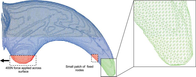
1398                                                MANNING ET AL.

                   INTRODUCTION                                  Claw form and function varies widely among verte-
                                                              brates, however, claw sheath composition does not. Claw
   Predatory dinosaurs (Theropoda) were diverse and suc-
                                                              sheaths, nails, and hooves are composed of keratin, a
cessful animals during the Mesozoic Era and are cur-
                                                              strong, fibrous protein (Raven and Johnson, 1992). Kera-
rently represented in modern ecosystems by their direct
                                                              tin protects the bone of the terminal phalanx and assists
descendants, the birds (Holtz and Osmólska, 2004; Var-
                                                              in providing traction during climbing, prey capture, and
gos and Fallon, 2004; Pennisi, 2005). Nonavian theropods      occasionally killing [brief review in Manning et al.,
ranged in size from the tiny Microraptor zhaoianus (with      (2006)]. The morphology of dromaeosaurid unguals, com-
a trunk length of only 47 mm) to the huge Tyrannosaurus       bined with rare soft tissue preservation (Clark et al.,
rex (up to 14 m in length) (Holtz and Osmólska, 2004).       1999), indicates that theropod claws would have been
The theropod clade that forms the focus of this study,        similarly protected. Extant Phylogenetic Bracketing
Dromaeosauridae (Matthew and Brown, 1922), was an             [EPB: Witmer (1995)] can be used to reconstruct the
obligatory bipedal group of lightly-built cursors (esti-      structure and properties of the keratin claw sheaths pos-
mated live weight from 20 to 80 kg) that were close to        sessed by nonavian dinosaurs. Mammal claw sheaths
avian ancestry (Norell and Makovicky, 2004). Dromaeo-         are composed of a-keratin (helical), but bird and reptile
saurid limbs exhibit numerous anatomical specializations      claws are composed of b-keratins (pleated-sheet) (Fraser
including long raptorial hands with three functional dig-     and MacRae, 1980). As nonavian dinosaurs fall within
its, highly mobile hand-wrist elements, unique caudal         the EPB formed by birds and crocodilians and leave sim-
vertebrae with highly elongate prezygapophyses adapted        ilar osteological traces (attachment grooves) it is likely
to assist balance, and a recurved, hypertrophied and          that the claw sheaths of these animals were also com-
hyperextensible claw on pedal digit II (Ostrom, 1969,         posed of b-keratin (Manning et al., 2006).
1990). The functional and palaeobiological significance of        The keratinous claw sheath is strongest mechanically
the strongly recurved manual ungual of the Late Creta-        along its parasagittal plane (Vincent and Owers, 1986;
ceous Asian dromaeosaurid Velociraptor (Norell and            Bonser, 2000), representing a function of the microstruc-
Makovicky, 1999) forms the subject of this article.           tural arrangements of the individual keratin fibers
   Dromaeosaurid unguals are commonly interpreted as          (Beaupre and Carter, 1992). A requirement of all biologi-
weapons for disemboweling (with pedal unguals) or cap-        cal materials subject to loading is that stresses are kept
turing/manipulating (with manual unguals) large-bodied        within safe limits (Alexander, 1981), and it is likely that
prey, such as iguanodontian ornithopods (Ostrom, 1969).       the claw sheath and ungual phalanx of dromaeosaurid
The effectiveness of the enlarged pedal digit II ungual       manual claws would have had similar safety limits to
as a disemboweling implement has been challenged by           those of extant raptorial falconiform and strigiform
recent experiments using a hydraulic reconstruction of a      birds. Similarities between dromaeosaurid and owl claw
dromaeosaurid hind limb (Manning et al., 2006). How-          morphology are striking, in terms of both internal and
ever, the mechanical behavior of dromaeosaurid claws          external anatomy. Comparable distributions of cortical
while under load has not been satisfactorily explored.        and trabecular bone in these taxa allow confident recon-
   Homologous structures in the feet of extant perching/      struction of the mechanical properties of dromaeosaurid
climbing birds provide useful insights into the palaeobi-     claws.
ology, evolution, form and function of dromaeosaurid
claws. However, an understanding of the microarchitec-
ture and 3D properties of dromaeosaurid unguals would                  MATERIALS AND METHODS
provide a more robust interpretation of their biomechan-      Fossil Material
ical function with the potential to provide new insights
into behavior. To determine the displacements (deforma-          A manual ungual of Velociraptor mongoliensis (Man-
                                                              chester Museum, University of Manchester, specimen
tions), strains (displacement gradients), and stresses
(force intensities) that result from subjecting dromaeo-      LL.12392) was Computed tomography (CT) scanned to
                                                              provide a 3D dataset for the finite element (FE) mesh
saurid claws to loads, it was necessary to generate a fi-
nite element model (FEM).                                     (see below for details). V. mongoliensis is known primar-
   Finite element analysis (FEA) provides a useful tech-      ily from the Djadokhta Formation (Campanian) of Mon-
                                                              golia (Osborn, 1924). Velociraptor is closely related to
nique to quantitatively analyze complex 3D biological
systems. However, the physical properties of the mate-        Deinonychus and Dromaeosaurus; however, it is distin-
                                                              guished from all other dromaeosaurids by its relatively
rial being modeled must be known if realistic FEMs are
to be generated. Mineralized fossil material cannot be        long, low skull and depressed muzzle (Sues, 1977).
used to determine the original material properties of a
bone (Young’s modulus, Poisson’s ratio, etc.) so it is nec-
                                                              Extant Material
essary to identify suitable modern analogue material.
Moreover, specifying biomaterial properties for FEA is          A subadult/juvenile eagle owl (Bubo bubo) from the bi-
difficult as they can vary within the structure under          ological sciences collections, University of Manchester,
investigation according to location (inhomogeneous            was used in this study. The bird had a frozen weight of
materials), the directions in which forces are applied        1996 g. Bubo bubo is a member of the family Strigidae,
(anisotropic behavior), the loading rate (as occurs in        which consists of all the typical owls (Penteriani et al.,
poroelastic and viscoelastic materials), and the magni-       2002).
tude of the load (which can lead to nonlinear effects)          A fully-intact terminal ungual phalanx and part of the
(Beaupre and Carter, 1992). We constrained the biomate-       adjacent phalanx, both from pedal digit III of the right
rial specifications used in this study using both evolu-       pes, were amputated from the Eagle Owl (Fig. 1). The
tionary and functional cues.                                  bones, along with all the associated soft tissue were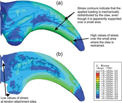
DROMAEOSAUR CLAW AND FINITE ELEMENT MODELING 1399
(2D) cross-sectional slices can be extracted along the
three orthogonal planes of the object (Elliott and Dover,
1982; Marrow et al., 2004; Babout et al., 2005). In con-
ventional radiography, the image plane is approximately
normal to the X-ray beam, and the image represents
total X-ray attenuation through the object. CT computes
a 3D digital representation from a series of 2D X-ray ra-
diographs taken around a single axis of rotation, or a 2D
slice perpendicular to the axis of rotation for 1D line de-
tector (fan beam geometry). The digital image is stored
as an array of numbers representing local X-ray attenu-
ation values for each of the small volumetric pixels (vox-
els) that make up the slice, these attenuation data are
represented in the reconstructed image as a series of
grey scale values. By thresholding the volume so as to
associate different grey levels with different materials
the 3D object can be converted into a microstructurally
faithful 3D mesh suitable for FEM describing the geome-
try of each constituent. Once each phase has been
ascribed mechanical properties, it is possible to predict
the loads experienced throughout a body according to its
exact size, shape and volume.
Measurements were carried out using a high-resolu-
tion computerized tomography and digital radiography
system (HMXST 225) from X-Tek Systems Ltd., employ-
ing a microfocus X-ray source (5 lm focal spot size) capa-
ble of tube potentials up to 225 kV. The imaging
arrangement is based on cone beam geometry. In such
an arrangement, the voxel resolution of the recon-
structed 3D volume depends on the source-to-object dis-
tance. The object was placed on a manipulator situated
between the X-ray source and the detector system (con-
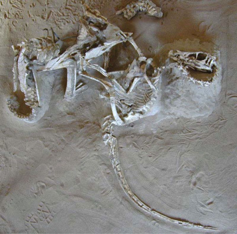
Fig. 1. High-resolution x-ray CT of owl terminal ungual phalanx and sisting of image intensifier and CCD), providing magnifi-
pedal claw (bone core and keratin sheath). Bone is white, keratin, and cation and rotation for collection of 470 radiographs over
soft-tissue are grey. 180 . The radiographs were collected using a tube poten-
tial of 50 kV with a tungsten anode target giving an
approximately 5 lm point source. To shape the energy
spectrum of the X-ray beam, and thus improve the
nano-indented to provide input data for the FE mesh of image quality in the reconstructed slices (i.e., increasing
the CT scanned Velociraptor claw (Table 1). the signal-to-noise ratio), a 0.25 mm thick copper X-ray
filter was placed just behind the point source and in
front of the sample during image acquisition. This filter
removes low-energy photons from the polychromatic
Claw Geometry beam, thus minimizing artefacts (noise) in the recon-
The arc for both the Velociraptor and owl claws were structed slices and the distribution of voxels with differ-
measured using Feduccia’s (1993) method (Fig. 2). ent intensities. The 3D tomographic volumes were
A perpendicular (CD) is drawn to bisect the chord reconstructed using a cone beam extension of the filtered
(AB) of the inner arc, which is bisected at point X. Per- backprojection algorithm developed for fan beams (Kak
pendiculars are drawn to bisect the chords AX and XB. and Slaney, 1987; Youssef et al., 2005) 32 frames were
These perpendiculars when extended, meet at the center averaged for each of the 470 projections (using an expo-
(E0 ) of the circle of which the arc is part. The radii are sure time for each frame of 120 ms). The raw 3D volume
then drawn to each end of the arc (AE0 and BE0 ). The had a pixel resolution of 17 lm, reconstructed at a quar-
angle (Y) between these radii is a measure of the ter of maximum resolution for easier data manipulation
degrees of arc (Feduccia, 1993). during the subsequent meshing routine.
X-ray Microtomography In situ Compression
X-ray micro-tomography (XMT) is a nondestructive Considering the EPB method (Witmer, 1995), bone
evaluation technique that allows the internal structure and keratin samples were collected from the pes of an
of an object to be imaged by reconstructing the spatial eagle owl (Bubo bubo). The micromechanical properties
distribution of the local linear X-ray absorption coeffi- of the keratin layer and cortical bone of the eagle owl
cients of the materials/phases contained within. This claw were determined from force displacement curves
provides a virtual 3D representation of the internal generated by an MTS XP nanoindenter with a Berkovich
architecture of an object from which two-dimensional diamond indenter (Kanari et al., 1997a,b).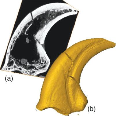
1400                                                       MANNING ET AL.

                   TABLE 1. Experimental imaging parameters for scanning the Velociraptor claw
  X-ray tube: energy,
       intensity                                  Radiograph acquisition                                 Volume reconstruction
Voltage          Current           Rotation            Exposure             Number of             Voxel              Reconstructed
 (kV)             (mA)             (degrees)          time (msec)          frames/angle         size (lm)           volume (voxels)
50                0.135               0.4                  120                 32                   17             256  256  470

     Fig. 2. Claw curvature measurements (after Feduccia, 1993).

                                                                      Fig. 3. XMT cross-section of the Velociraptor (LL. 12392) manual
Finite Element Model                                                claw showing the main biomaterial phases (trabecular and cortical
                                                                    bone).
  A microstructurally faithful FE mesh of the Velocirap-
tor ungual was created in 3D directly from the raw to-
mography images using the ScanIPTM and ScanFETM,                    the middle and the two pieces have been glued together,
commercial packages from Simpleware Ltd. The image                  slightly out of alignment. Before the FE mesh was gen-
threshold values used for segmentation (the process of              erated, the two pieces of the claw were digitally real-
discriminating between the various structures from the              igned, and the fracture was removed. The latter was
image) were selected according to estimates for the vol-            achieved by inflating the volume so that the fractured
ume fractions of cortical bone, trabecular bone, and po-            surfaces healed. The volume was then returned it to its
rosity. The CT imaging conditions used are summarized               original shape by reversing the inflation procedure,
in Table 1.                                                         while algorithmically enforcing the continuity of the sur-
  In FEA, individual FEs can be of any shape. However,              face across the original fracture zone, preventing the
with voxel image data, it is most convenient to use brick           fracture from reappearing.
elements which, when they are perfect cubes, are the FE                Figure 4 shows where the loads and boundary condi-
analogue of voxels. A one-to-one correlation between                tions were applied to the mesh. The aim was to recreate
brick elements and voxels would lead to an FE mesh                  the likely forces experienced by the claw in use, during
several orders of magnitude larger than can be proc-                prey capture, climbing, etc. A distributed force was
essed using the Abaqus software. Therefore, the model               applied over a surface area representative of the attach-
was down-sampled to allow the analyses to run on a                  ment site for the flexor tendon. The initial force of 400N
desktop computer. The resultant FE mesh contains                    was calculated using the approximate weight of Velocir-
500,000 elements rather than 30 million voxels. Care               aptor as a guide [Manning et al., (2006)]. The FE mesh
was taken to ensure that the simplified mesh still cap-              was subjected to a linear elastic analysis using ABAQUS
tured the detail of the claw microstructure. A further              CAE 6.6 with the input data being determined by nano-
simplification for this preliminary model was to ignore              indentation and from literature (above).
the keratin layer on the outer surface of the claw, with
the focus being on the modeling of the cortical and tra-
becular bone, with a true representation of porosity in
the structure (Fig. 3).
                                                                                              RESULTS
                                                                    Claw Geometry
Loading: Stress/Strain Analysis
                                                                      The Velociraptor manual ungual modeled in this study
  A FE mesh was created to represent the entire ungual              (minus its keratinous sheath) possessed a claw arc mea-
(Fig. 4). The original fossil (Fig. 3) is fractured through         surement of 127 .
DROMAEOSAUR CLAW AND FINITE ELEMENT MODELING 1401
Fig. 4. 3D finite element mesh of the Velociraptor claw, showing boundary conditions and close-up
detail of the mesh (inset).
TABLE 2. Input data derived from the eagle owl displays a distinct structural orientation that is perpen-
claw (*Values taken from literature: dicular to the ungual long-axis that can be modeled
(Van Ruijven et al., 2002) using high element count FEA (Fig. 5). Currently there
Number Mean modulus is no way of modeling element-by-element anisotropy
Region indented of indents (GPa) to 1.d.p. and within each FE isotropy has been assumed.
Model values for the Young’s modulus (summarized in
Keratin layer 10 6.8 (1.5) Table 2) and Poisson’s ratio (Kitchener and Vincent,
Cortical bone 11 21.1 (2.3)
*Trabecular bone – 19.0 1987; van Ruijven et al., 2002) were assigned isotropi-
cally to the elements that represented the cortical and
trabecular phases of FE meshes. Upon applying the
In situ Compression loading conditions shown in Fig. 4, the stress contour
plots illustrated in Fig. 6 were obtained.
Reliable indentation properties could not be obtained The initial force of 400N applied to the ungual was
for the trabecular bone due to its highly porous nature; deduced from the approximate weight of the Velociraptor
values for this were obtained from the literature (Van (Manning et al., 2006). It would be fair to assume that
Ruijven et al., 2002). The indenter tip was moved at a the stresses generated at this load provide a reasonable
rate of 5 nm s1 to a chosen cross-sectional area of the baseline for assessing the loads that would be encoun-
eagle owl claw and contact between the two determined tered when a dromaeosaur used its claws to grasp, lift,
at a specific applied load. The indentation was conducted and climb. In the living animal, several factors would
at a load range from 0 to 60 lN. Ten different points affect the intensity and distribution of load over the claw/
were indented at each load for the keratin layer and substrate contact and its transmission through the inter-
eleven different points indented for the cortical bone. nal microstructure of the claw. For example, the mechani-
The results derived from the loading–unloading cycles cal behavior of living tissues (flexibility, strength at
are shown in Table 2. different strain rates) is more complicated than has been
Note: the Poisson’s ratios used were: 0.3, 0.32, and 0.2 used here. That coupled with the rate of ascent when
(Kitchener and Vincent, 1987; Van Ruijven et al., 2002) climbing would also affect the values of peak loads.
for the keratin, cortical and trabecular phases, respec- The maximum value of stress generated by the FEM
tively. These values were employed for subsequent FEM under the assumed loading conditions, with a force of
using a commercial FEM package (Abaqus). 400N, is around 60 MPa. This is much less than the typ-
ical range of values reported in the literature for the
failure stress of extant claws of 150–200 MPa, indicating
XMT and Finite Element Model
that it is reasonable to assume that the Velociraptor
Variation in bone density and microstructure (trabecu- would have been able to support its weight on a very
lar versus cortical bone) has an important influence on a small contact surface while climbing.
system’s stiffness and strength (Carter and Hayes, It should be noted that the model chosen to represent
1977). Differences in density and microstructure, when material behavior in the current study does not include
coupled with the anisotropic properties of bone (trabecu- a failure criterion. Therefore, it may be of interest for
lar and cortical) and keratin, leads to a complex compos- future work to carry out experimental tests such as
ite system that is difficult to model. However, the three-point bending of the eagle owl claw. This would
cortical and trabecular bone in the Velociraptor ungual give an estimate of the failure strength (i.e., ultimate1402 MANNING ET AL.
Fig. 6. Contour map of Mises stress (units in GPa) on (a) the outer
surface of the claw and (b) through the mid-section.
Fig. 5. Velociraptor claw comprized of cortical and trabecular bone
and true porosity (a) 2D tomographic slice (b) 3D representation using The central hypothesis regarding claw function in this
AMIRA software. study was based upon both internal and external pedal
morphology. The role of the FE analysis was to test the
relationship of form and function, from an engineering
limit load) of the Velociraptor ungual over a range of dif-
point of view. The preliminary study was carried out
ferent scenarios.
using the microtomographic scan of the Velociraptor
manual claw, given limited access to complete pes
unguals. The findings from this study can be compared
with pes claws, on the basis of geometric and architec-
DISCUSSION tural similarity (Manning et al., 2006), with only slight
The claw geometry of mammals and birds (both pedal variation in morphology. Pes claws are typically larger
and manual) correlates well with arboreal and terres- than those of the manus, meaning that it could sustain
trial habitats. In a study of over 500 species of birds, larger loads than those subjected to the model used in
Feduccia (1993) demonstrated that modern ground and the current simulation. The biomechanical function of
tree-dwelling birds could be distinguished on the basis of morphologically comparable structures of similar geome-
pedal claw curvature. The claw arc measurements for try, but dissimilar size, should behave in a dynamically
ground birds ranged from 52.2 to 77.6 , those of perch- similar fashion (Alexander, 1989). Future work will
ing birds from 101.8 to 125.3 , and those of trunk- apply the methods from this study, by using a pedal
climbers form 129.5 to 161.6 (Feduccia, 1993). The ungual phalanx of Deinonychus (Ostrom, 1969) to test
ungual phalanges on pedal digit II of most dromaeosaur- this hypothesis further.
ids are large, narrow, pointed, and strongly curved with Many birds are successful climbers, using their feet,
lateral grooves for the claw sheath. That of Deinonychus tails, beaks, and, in the case of the South American
antirrhopus (YPM 5205) possesses an inner arc mea- hoatzin (Opisthocomus hoazin), their forelimbs while
surement of 160 (Ostrom, 1969). Comparisons with fledglings (Dominguez-Bellom, 1994). The tail feathers of
extant birds therefore support a climbing function for some birds (woodpeckers, woodhoopoes, woodcreepers,
this structure. Although a climbing/grappling function treecreepers, and some species of ovenbirds) can provide
for the dromaeosaurid pedal digit II ungual during pre- additional support (McFarland et al., 1979). Ostrom
dation has been suggested, it may also have provided as- (1969) suggested that dromaeosaurs possessed a
sistance during arboreal activity (Chatterjee, 1997; reversed hallux, but more recent work (Norell and
Manning et al., 2006). The Velociraptor manual ungual Makovicky, 1999) has indicated that this reduced digit
modeled in this study (minus the keratinous sheath) was positioned craniolaterally and high on the
possessed a claw arc measurement of 127 . It is likely metatarsus.
that this claw arc angle would increase by 10–15% with Ostrom (1969) noted that the recurved shape of the D.
the addition of a keratinous sheath, meaning the claw antirrhopus ‘‘killing claw’’ coincided with the axis of
would fall comfortably within Feduccia’s (1993) perching maximum force that could be delivered and concluded
and trunk-climber bracket. This adds further support to that it was adapted for penetrating the flesh of prey.
the proposal that dromaeosaurids might have used their Carpenter (1998) suggested that this morphology was
claws to assist climbing behavior (Chatterjee, 1997; able to provide a piercing function. This study supports
Manning et al., 2006). both interpretations, but suggests this piercing abilityDROMAEOSAUR CLAW AND FINITE ELEMENT MODELING 1403
Fig. 7. The ‘fighting pair’ Velociraptor and Protoceratops (IGM 100/25).
did not facilitate the disemboweling of prey, but was tant in dromaeosaurids, a functional switch leading to a
instead involved in scansorial behavior. more important role for the flexor muscles (particularly
The force generation of a limb is always primarily to around the foot, ankle, and hip) would have occurred, as
oppose gravity. Thus a dromaeosaur on a horizontal has been observed in extant scansorial birds (Stolpe,
surface needs to generate a primarily vertical force by 1932; Moreno, 1991). Myological analyses of extant
generating an extensor moment at the ankle (plantar- climbing birds have demonstrated that the hindlimb
flexion), knee, and hip. As the surface becomes more ver- flexors are stronger and better-developed than the exten-
tically oriented, the force required is still vertical but sors (Raikow, 1994). The caudodorsal shift of the pubis
because of the orientation change of the animal this coupled with a knee-based flexor system (Gatesy, 1990)
becomes a progressively more caudally directed force. in maniraptoran theropods likely marks this switch from
This is achieved by fixing the foot to the surface and extensor to flexor muscles in the hind limb. Adaptation
generating a flexor moment at the ankle (dorsiflexion) in hind limb flexor muscles would have also assisted
and hip but an extensor moment at the knee. Thus, it deployment of the recurved claws in climbing and prey
might be expected that if climbing behavior was impor- capture.1404 MANNING ET AL.
The evidence suggests that the form and resultant propulsion. The medial plantar crest of the tarsometa-
function of the ‘‘killer claws’’ was adapted to assist prey tarsus in some species of raptorial bird has become so
capture. All predators have to be adept at hunting, cap- enlarged that it almost encases the powerful flexor ten-
turing, and killing (McFarland et al., 1979). Dromaeo- dons. If the longitudinal ridge observed on the caudal
saur claw geometry appears functionally adapted for face of the dromaeosaur metatarsus correlates to the
prey capture, not for killing. In the light of earlier exper- medial plantar crest of birds, it seems likely to have
imental results (Manning et al., 2006) and this study, it served a similar function.
is possible to review the interpretation of the unique fos- Farlow et al., (2000) noted that theropods more closely
sil (Fig. 7) of a Velociraptor and Protoceratops (Kielan- related to birds (including dromaeosaurs) show skeletal
Jaworoska and Barsbold, 1972). features indicative of a reduction in caudofemoral mus-
The remarkable fossil of the dromaeosaur Velociraptor culature. If the function of the recurved claw on the sec-
entangled with an herbivorous dinosaur, Protoceratops ond toe was to disembowel prey, it seems illogical that
(IGM 100/25), often called the ‘‘fighting pair’’ (Carpenter, limbs evolve to become progressively weaker. However,
1998), may give some insight into claw function. The Norell and Makovicky (1999) noted that two specimens
pair was collected from the Djadokhta Formation (Late of V. mongoliensis possessed a fourth trochanter (proxi-
Cretaceous) of Mongolia (Kielan-Jaworoska and Bars- mal ridge of bone projecting from the medial shaft of the
bold, 1972) and has caused much debate as to how the femur). Given that this is the attachment site for the
two came to be preserved together (Barsbold, 1974; main limb retractor, the caudofemoralis, this suggests
Osmólska, 1993; Unwin et al., 1995; Carpenter, 1998). that V. mongoliensis still employed this muscle.
This unique fossil possibly shows a fatal stalemate, with
the predators right leg pinned under the prey’s body, the
CONCLUSIONS
left foot hooked into the throat region and its right arm
held in the jaws of its prey. The relative position of the The maximum value of stress generated by the FEM
predator’s ensnared limbs meant it was pulled close to under the assumed loading conditions (400N) was
its prey and trapped. Unable to reposition its limbs, it around 60 MPa. The failure stress of extant claws (150–
was impossible for the Velociraptor to deploy its jaws 200 MPa) was significantly higher, suggesting that Veloc-
and finish the attack, like two boxers hugging each other iraptor would have been able to support its weight on a
and unable to throw a punch. small contact area while climbing.
Flexor tendon adaptations in the foot of Velociraptor The reversed hallux (Ostrom, 1969) of dromaeosaurs
might have contributed to the fatal embrace. The tendons is now reconstructed so that this reduced digit was
(medius flexor digitorum longus) that flex the toes of positioned craniolaterally and high on the metatarsus
extant perching birds are modified so that they lock the (Norell and Makovicky, 1999), a position that would
foot in a tight grip (McFarland et al., 1979). This grip is a functionally assist in climbing behavior.
result of specialized tendons that possess a ratchet-like We suggest that the anatomy, form, and function of
mechanism in their sheaths, whereby numerous small digit II and manus claws of dromaeosaurs support the
projections on each tendon engage the ridges on the adja- hypothesis of a prey capture/grappling/climbing function.
cent walls of the sheath (Welty, 1975). This allows birds This is supported by the homologous structures observed
to relax and sleep whilst perched, without releasing their in the feet of modern perching/climbing birds. The geom-
grip and falling. The weight of the bird is enough to etry of dromaeosaur claws would have caused the claw
engage this elegant system. The underlying flexor ten- to rotate as it was pushed deeper into prey which would
dons on the feet of dromaeosaurs might have possessed a have resulted in a maximum depth of trauma equal to
similar ratchet-like ‘‘locking’’ mechanism on the second the radius of the inside arch of the claw. The feet and
toe. The possession of such flexor tendons would have hands of dromaeosaurs functioned both for locomotion
been an energy-efficient way of countering the effect of (walking, running, and climbing) and as prey capture/
the claw’s retractor ligament. They would have provided grappling devices. We speculate that a ratchet-like ‘‘lock-
a useful adaptation for climbing and holding onto their ing’’ ligament might have provided an energy-efficient
prey, using gravity and limb position to assist the claw way for dromaeosaurs to hook their recurved digit II
flexor and gripping function. The mechanism would then claws into their prey, using body weight to lock the
rely upon the dromaeosaur lifting its foot to release the claws passively and allowing the jaws to dispatch prey.
underlying tendons to allow the retractor ligament to free
the recurved claw from its prey; a dromaeosaur trapped/ ACKNOWLEDGMENTS
hugged by its prey would be unable to do so.
There is evidence for a large flexor system in D. anti- The X-ray tomography described in this article was
rrhopus (AMNH 2136), Velociraptor mongoliensis carried out within the Stress and Damage Characterisa-
(AMNH 6518), and Sinornithoides youngi (Norell and tion Unit within the School of Materials. They thank Dr
Makovicky, 1997). Halfway down the caudo-ventral sur- Jonathon Codd (University of Manchester) for the use of
face of metatarsal II, a robust longitudinal ridge (dis- the image in Fig. 7. The authors wish to thank Eric
placed slightly to the medial side) closely resembles the Snively, Peter Dodson, Eric Morschhauser and one anon-
medial plantar crest of the avian metatarsus. Norell and ymous reviewer for the helpful comments on the
Makovicky (1997) suggest this ridge might have func- manuscript.
tioned as an attachment site for the flexors and adduc-
tors of digits I and II, or possibly as a border that
channeled flexor tendons. The plantar crest on the tarso- LITERATURE CITED
metatarsus is well developed in raptorial and some spe- Alexander, R. McN. 1981. Factors of safety in the structure of ani-
cies of swimming birds, which use their feet for mals. Sci Prog 67:109–130.DROMAEOSAUR CLAW AND FINITE ELEMENT MODELING 1405 Alexander RM. 1989. Dynamics of dinosaurs and other extinct Matthew WD, Brown B. 1922. The family Deinodontidae, with giants. New York: Columbia University Press. p 167. notice of a new genus from the cretaceous of Alberta. Bull Am Babout L, Mummery PM, Marrow TJ, Tzelepi A, Withers PJ. 2005. Mus Nat Hist 46:367–385. The effect of thermal oxidation on polycrystalline graphite studied McFarland WN, Pough FH, Cade TJ, Heiser JB. 1979. Vertebrate by x-ray tomography. Carbon 43:765–774. life. New York: MacMillan Publishing. p 875. Barsbold R. 1974. Duelling dinosaurs. Priroda 2:81–83. [in Moreno E. 1991. Musculature of the pelvic appendages of the tree Russian]. creepers (Passeriformes: Certhiidae): mycological adaptations for Beaupré GS, Carter DR. 1992. Finite element analysis in biome- tail-supported climbing. Can J Zool 17:191–209. chanics. In: Biewener AA, editor. Biomechanics: structure and Norell M, Makovicky PJ. 1997. Important features of the dromaeo- systems: a practical approach. New York: IRL Press. p 149–174. saur skeleton: information from a new specimen. Am Mus Novit Bonser RHC. 2000. The Young’s modulus of ostrich claw keratin. 3215:1–28. J Mater Sci Lett 19:1039–1040. Norell M, Makovicky PJ. 1999. Important features of the dromaeo- Carpenter K. 1998. Evidence of predatory behaviour by carnivorous saur skeleton. II. Information from newly collected specimens of dinosaurs. In: Pérez-Moreno BP, Holtz TR, Sanz JL, Moratalla JJ, Velociraptor mongoliensis. Am Mus Novit 3282:1–45. editors. Aspects of theropod palaeobiology. Gaia 15:135–144. Norell M, Makovicky PJ. 2004. Dromaeosauridae. In: Weishampel Carter DR, Hayes WC. 1977. The compressive behaviour of bone as B, Dodson P, Osmólska H, editors. The Dinosauria. 2nd ed. Berk- a two-phase porous structure. J Bone Joint Surg 59A:954–962. ley: University of California Press, 862. pp 196–209. Chatterjee S. 1997. The rise of birds: 225 million years of evolution. Osborn HF. 1924. Three new theropoda, Protoceratops zone, central London: The John Hopkins Press Ltd. p 312. Mongolia. Am Mus Novit 144:1–12. Clark JM, Norell MA, Chiappe LM. 1999. An oviraptorid skeleton Ostrom JH. 1969. Osteology of Deinonychus antirrhopus, an un- from the late cretaceous of Ukhaa Tolgod, Mongolia, preserved in usual theropod from the lower cretaceous of Montana. Bull Pea- an avianlike brooding position over an oviraptorid nest. Am Mus body Mus Nat Hist 30:165. Novit 3265:1–35. Ostrom JH. 1990. Dromaeosauridae. In: Weishampel B, Dodson P, Dominguez-Bellom G, Michelangeli F, Ruiz, MC, Garcia A, Rodri- Osmólska H, editors. The Dinosauria. First edition. Berkley: Uni- guez E. 1994. Ecology of the folivorous hoatzin (Opisthocomas versity of California Press, pp 269–279, 733. hoazin) on the Venezuelan plains. Auk 111:643–651. Osmólska H. 1993. Were the Mongolian ‘‘fighting dinosaurs’’ really Elliott JC, Dover SD. 1982. X-ray microtomography. J Microsc fighting? Paléobiology (Spec Vol) 7:161–162. 126:211–213. Pennisi E. 2005. Birds wings really are like dinosaurs’ hands. Sci- Farlow JO, Gatesy SM, Holtz TR, Hutchinson JR, Robinson JM. ence 307:194–195. 2000. Theropod locomotion. Am Zool 40:640–663. Penteriani V, Gallardo M, Roche P. 2002. Landscape structure and Feduccia A. 1993. Evidence from claw geometry indicating arboreal food supply affect eagle owl (Bubo bubo) density and breeding habits of Archaeopteryx. Science 259:790–793. performance: a case of intra-population heterogeneity. J Zool Fraser RDB, MacRae TP. 1980. Molecular structure and mechanical 257:365–372. properties of keratins. In: Vincent JFV, Currey JD, editors. The Raikow RJ. 1994. Climbing adaptations in the hindlimb musculature mechanical properties of biological materials. Cambridge: Cam- of the wood creepers (Dedrocolaptinae). Condor 96:1103–1106. bridge University Press. 1968. pp. 211–246. Raven PH, Johnson GB. 1992. Biology. 3rd ed. St. Louis, Missouri: Gatesy S. 1990. Caudofemoral musculature and the evolution of Mosby Year Book. p 1217. theropod locomotion. Paleobiology 16:170–186. Stolpe M. 1932. Physiologisch-anatomische untersuchungen über Holtz TR, Osmólska H, Saurischia. 2004. In: Weishampel B, Dodson die hintere extremität der vögel. J Ornithol 80:161–247. P, Osmólska H, editors. The Dinosauria. 2nd ed. Berkley: Univer- Sues HD. 1977. The skull of Velociraptor mongoliensis, a small Cre- sity of California Press. pp. 21–24. taceous theropod dinosaur from Mongolia. Palaeontologische Kak AC, Slaney M. 1987. Principles of computerized tomographic Zeitschrift 51:173–184. imaging. New York: IEEE Press. Unwin DM, Perle A, Trueman C. 1995. Protoceratops and Velocirap- Kanari M, Tanaka K, Baba S, Eto M. 1997a. Nanoindentation tor preserved in association: evidence for predatory behaviour in behavior of a two-dimensional carbon-carbon composite for nu- dromaeosaurid dinosaurs. J Vertebr Palaeontol (Abstracts of clear applications. Carbon 35:1429–1437. papers, fifty-fifth Annual Meeting) 15 (suppl 3):57A. Kanari M, Tanaka K, Baba S, Eto M, Nakamura N. 1997b. Nanoin- van Ruijven LJ, Giesen EBW, van Eijden TMGJ. 2002. J Dent Res dentation test on electron beam-irradiated boride layer of carbon- 81:706–710. carbon composite for plasma facing component of large Tokamak Vargos AO, Fallon JF. 2004. Birds have dinosaur wings: the molecu- device. J Nucl Mater 244:168–172. lar evidence. J Exp Zool 304:1–5. Kielan-Jaworoska Z, Barsbold R. 1972. Results of the Polish- Vincent JFV, Owers P. 1986. Mechanical design of hedgehog spines Mongolian paleontological expedition, Part IV. Acta Paleontol Pol and porcupine quills. J Zool 210:55–75. 27:5–13. Welty JC. 1975. The life of birds. 2nd ed. Philadelphia: Saunders Kitchener A, Vincent JFV. 1987. Composite theory and the effect of WB. 623 p. water on the stiffness of horn keratin. J Mater Sci 22:1385–1389. Witmer LM. 1995. The extant phylogenetic bracket and the impor- Manning PL, Payne D, Pennicott J, Barrett P. 2006. Dinosaur killer tance of reconstructing soft tissue in fossils. In: Thomason JJ, claws or climbing crampons? Royal Soc Biol Lett 2:110–112. editor. Functional morphology in vertebrate palaeontology. Marrow TJ, Buffiere JY, Withers PJ, Johnson G, Engelberg D. Cambridge: Cambridge University Press. pp 19–33. 2004. High resolution x-ray tomography of short fatigue crack Youssef S, Maire E, Gaertner R. 2005. Finite element modelling of nucleation in austempered ductile cast iron. Int J Fatigue the actual structure of cellular materials determined by x-ray to- 26:717–725. mography. Acta Mater 53:719–730.
You can also read