Speckled and plain regions of avian eggshells differ in maternal deposition of calcium and metals: A hitherto overlooked chemical aspect of egg ...
←
→
Page content transcription
If your browser does not render page correctly, please read the page content below
Volume 134, 2017, pp. 721–731
                                                                                                            DOI: 10.1642/AUK-17-7.1

RESEARCH ARTICLE

Speckled and plain regions of avian eggshells differ in maternal
deposition of calcium and metals: A hitherto overlooked chemical aspect
of egg maculation
Grzegorz Orłowski,1* Przemysław Pokorny,2 Wojciech Dobicki,2 Ewa Łukaszewicz,3 and Artur Kowalczyk3
1
  Institute for Agricultural and Forest Environment, Polish Academy of Sciences, Poznań, Poland
2
  Department of Hydrobiology and Aquaculture, Wrocław University of Environmental and Life Sciences, Wrocław, Poland
3
  Division of Poultry Breeding, Wrocław University of Environmental and Life Sciences, Wrocław, Poland
* Corresponding author: orlog@poczta.onet.pl
Submitted January 9, 2017; Accepted April 4, 2017; Published June 16, 2017

    ABSTRACT
    Although it has been strongly implied that studies on structural function might resolve certain conflicting theories
    regarding the huge variability of coloration in avian eggs, including maculation, the fundamental physiological
    question about the functional role of eggshell speckling in the maternal deposition of micronutrients into maculated
    (pigment spots) and plain (unpigmented) eggshell regions remains largely unanswered. We measured (within the
    same egg) the concentrations of 2 micronutrients, the staple components of avian eggshells (calcium and
    magnesium), and 6 trace elements (copper, manganese, iron, cobalt, cadmium, and lead) in maculated (a superficial
    layer of brown pigment) and unpigmented areas of eggshells in 2 groups of Japanese Quail (Coturnix coturnix
    japonica) eggs representing 2 extremes of eggshell coloration: those with darker eggshells and more extensive
    pigment spots vs. those with bright eggshells and small, more clearly demarcated pigment spots. We found evidence
    that the concentrations of calcium and magnesium and of 2 trace elements (copper and cadmium) in both types of
    egg were significantly higher in speckled eggshell areas (where the shells were also significantly thicker) than in
    unpigmented ones. Conversely, lead (a toxic element) peaked markedly in the plain areas of eggshells compared with
    the speckled ones, whereas the concentrations of the 3 other trace elements (manganese, iron, and cobalt) measured
    in the speckled and unpigmented eggshell regions were variable and dependent on egg coloration. Our results give a
    clear indication that egg maculation can play a functional role in the chemistry of avian eggs, one that enables females
    to distribute micronutrients and trace elements into pigment spots and unpigmented regions of an eggshell to
    varying extents. This presumably represents a mechanism permitting prompt physiological adjustment to varying
    levels of maternal resources. It is possible that elements sequestered into protoporphyrin speckles are not further
    utilized by developing embryos; this might be a form of physiological chemoprotection through the disposal or
    deactivation of certain elements in pigment speckles, which do not (or only marginally) contribute to the biochemical
    processes of embryogenesis.
    Keywords: calcium, egg maculation, eggshell thickness, maternal resources, micronutrient allocation, pigments,
    protoporphyrin pigment spots, trace elements

    Las regiones manchadas y no pigmentadas de las cáscaras de los huevos de aves difieren en la
    sedimentación materna de calcio y metales: un aspecto quı́mico hasta ahora ignorado del proceso de
    machado de los huevos
    RESUMEN
    Aunque se ha sugerido fuertemente que estudios sobre la función estructural podrı́an resolver algunas teorı́as
    contradictorias sobre la enorme variabilidad en la coloración de los huevos de aves, incluyendo el manchado, la
    pregunta fisiológica fundamental sobre el papel funcional de las manchas de las cáscaras de los huevos en la
    sedimentación materna de micronutrientes en regiones manchadas (áreas pigmentadas) y sin manchas (áreas no
    pigmentadas) aún no ha sido resuelta. En este estudio medimos (dentro del mismo huevo) las concentraciones de dos
    micronutrientes, los principales componentes de las cáscaras de huevos de aves (calcio y magnesio) y seis elementos
    traza (cobre, manganeso, hierro, cobalto, cadmio y plomo) en áreas manchadas (con una capa superficial de pigmento
    café) y no manchadas de cáscaras de huevo. Hicimos las mediciones en dos grupos de Coturnix coturnix japonica que
    representan dos extremos de coloración de las cáscaras: un grupo con cáscaras más oscuras y manchas de pigmento
    más extensas vs. un grupo con cáscaras claras y manchas de pigmento más pequeñas y definidas. Encontramos
    evidencia, en ambos tipos de huevos, de que las concentraciones de calcio y magnesio y de dos elementos traza
    (cobre y cadmio) fueron significativamente más altas en las áreas manchadas (en donde las cáscaras también fueron

Q 2017 American Ornithological Society. ISSN 0004-8038, electronic ISSN 1938-4254
Direct all requests to reproduce journal content to the AOS Publications Office at pubs@americanornithology.org
722 Chemistry of pigmented and plain areas of eggshells G. Orłowski, P. Pokorny, W. Dobicki, et al.
significativamente más gruesas) que en áreas sin pigmentos. Por el contrario, el plomo (elemento tóxico) fue
marcadamente más abundante en las áreas sin pigmentos de la cáscara que en las áreas manchadas, mientras que las
concentraciones de los otros tres elementos traza (manganeso, hierro y cobalto) variaron dependiendo de la
coloración de los huevos. Nuestros resultados dan una indicación clara de que el manchado de los huevos puede jugar
un papel funcional en la quı́mica de los huevos de aves ya que permite a las hembras distribuir micronutrientes y
elementos traza en las áreas pigmentadas y no pigmentadas de las cáscaras en grados variables. Presumiblemente,
esto representa un mecanismo que permite un ajuste fisiológico rápido a los niveles variables de recursos maternos. Es
posible que los elementos secuestrados en manchas de protoporfirinas no sean utilizados más adelante por los
embriones en desarrollo: esta podrı́a ser una forma de quimioprotección fisiológica mediante el desecho o
desactivación de ciertos elementos en las manchas de pigmento, que no contribuyen a los procesos bioquı́micos de la
embriogénesis o lo hacen solo marginalmente.
Palabras clave: asignación de micronutrientes, calcio, elementos traza, espesor de las cáscaras de huevo,
manchado de los huevos, manchas de pigmentos de protoporfirina, pigmentos, recursos maternos
INTRODUCTION tetrapyrrole) is an immediate precursor of the heme
molecule, whereas biliverdin (a linear tetrapyrrole) is a
There is a growing body of evidence suggesting that no product of hemoglobin breakdown. Protoporphyrin and
single signaling or structural hypothesis can adequately biliverdin pigments can bind ions of trace metals, primarily
explain the adaptive significance of eggshell pattern iron (Fe) or zinc (Zn), but almost all other metals can be
variability among birds, including the presence of macu- incorporated into their molecules (Ostfeld and Tsutsui
lation (reviewed in Reynolds et al. 2009, Cassey et al. 2011, 1974, Mikšı́k et al. 1996, Casini et al. 2003, Williams et al.
Deeming 2011, Gosler et al. 2011, Brulez et al. 2016). 2016). Protoporphyrin and biliverdin occur at varying
However, the contents of chemical elements in speckled levels in the shells of most birds’ eggs, irrespective of their
(pigment spots) and plain (unpigmented) areas of avian coloration (Mikšı́k et al. 1996, Cassey et al. 2011, Wang et
eggs, as well as the relationship between eggshell al. 2011, Brulez et al. 2016), and their distribution varies
pigmentation and trace-element content, remain virtually between the speckled and plain areas of the shells (Cassey
unknown (see Mikšı́k et al. 1996). This is surprising, given et al. 2012). The presence of some trace elements at high
the fundamental physiological importance of eggshell levels, such as lead (Pb), copper (Cu), or Fe, can inhibit the
micronutrients such as calcium (Ca) and a variety of trace biochemical synthesis of both calcite (a staple mineral
elements, which are maternally transferred into eggs and component of eggshells; cf. Rodriguez-Navarro et al. 2002)
are indispensable for developing avian embryos (Simkiss and heme (Casini et al. 2003). Female birds in poor
1961, Reynolds 1997, Richards 1997, Miles 2000). It is physiological condition and suffering a high level of
assumed that eggshells provide female birds an excretion oxidative stress, such as those exposed to pollution, may
pathway for some trace elements (Burger 1994); conse- lay eggs with higher concentrations of protoporphyrin
quently, avian eggshells (often only their fragments of (Moreno and Osorno 2003; see Hargitai et al. 2016a,
various sizes) are used as a tool in biomonitoring studies of 2016b, and references therein) or biliverdin, or both these
environmental pollutants, including some trace elements pigments simultaneously (Jagannath et al. 2008, Hanley
(Mora 2003, Espı́n et al. 2014, Hashmi et al. 2015). and Doucet 2012), which translates into the darker
However, no studies have yet assessed whether Ca and pigmentation and greener background color of such eggs,
other metals are distributed evenly between pigmented respectively.
and plain eggshell regions, an aspect hitherto wholly Several previous studies have shown that the bioaccu-
overlooked in methodological recommendations for study- mulation of various macrominerals and microminerals, in
ing the chemistry of birds’ eggs (see Klein et al. 2012, Espı́n particular Ca, increases with the intensity of pigmentation
et al. 2014). (derived from various porphyrin and melanin pigments) of
Eggshell coloration in birds involves a background base avian feathers (Niecke et al. 1999, Chatelain et al. 2016)
color that is due to 2 pigments (Gorchein et al. 2009), and of human hair and skin (reviewed in McGraw 2003). It
namely protoporphyrin (responsible for ‘‘brown’’ and ‘‘red’’ is therefore assumed that reservoirs of melanin pigments
colors and maculation) and biliverdin (responsible for serve as a sink for potentially harmful transition metal
‘‘blue-green’’ colors), incorporated into the calcitic layer, ions, adsorbing and harboring the very minerals that
and maculation (formed primarily as a layer or layers of helped produce them and thereby offering chemoprotec-
protoporphyrin pigment located in different shell areas) tion to adjacent cells and tissues (Larsson 1993, McGraw
that may lie among Ca-carbonate crystals or be associated 2003). A recent investigation into the composition of
with accessory shell material (Kennedy and Vevers 1976, pigments present in the shells of molluscs and birds’ eggs
Deeming 2011, Brulez et al. 2016). Protoporphyrin (a cyclic revealed that protoporphyrin pigments occurred in both
The Auk: Ornithological Advances 134:721–731, Q 2017 American Ornithological SocietyG. Orłowski, P. Pokorny, W. Dobicki, et al. Chemistry of pigmented and plain areas of eggshells 723
these evolutionarily distant groups of animals but that feature distinguishing other bird species studied in the
biliverdin was present only in birds’ eggshells (Verdes et al. context of the relationship between eggshell thickness
2015). Another recent investigation of molluscs showed and brown speckling; see above), which occurs immedi-
that the more intensive dark pigmentation due to the ately (~3 hr) before oviposition and is derived from
presence of protoporphyrin increased the content of some pigment granules in the apical cells of the shell gland
trace elements (measured only by scanning electron (Woodard and Mather 1964, Tamura et al. 1965, Baird et
microscope) like Cu (Williams et al. 2016). We therefore al. 1975). Quail eggs have been used as a model in many
hypothesize that an analogous relationship between the previous experimental investigations to examine the
presence of protoporphyrin pigments and the content of maternal and environmental effects of variation in egg
macrominerals and microminerals (trace elements) should coloration in the context of the deposition of 2 basic
also apply to the maculation of birds’ eggs, as suggested eggshell pigments and to analyze the sources of variation
earlier by Mikšı́k et al. (1996). To date, this presumed in egg coloration patterns and pigment spot distributions.
relationship between the chemical composition and the Female quails can promptly adapt the coloration of their
role of egg color and presence of maculation appears to eggs both to body condition (more heavily maculated
tally with the structural function of eggshell coloration (cf. eggs are laid by females with a poorer body condition;
Hanley 2012, Hanley and Doucet 2012). Assuming this Duval et al. 2013, 2016) and to darkly colored substrates
relationship to have been generally overlooked to date, we (Lovell et al. 2013); such eggs are characterized by a
explore it in an attempt to resolve some of the conflicting greater hatchability and lower embryonic mortality (Taha
theories regarding the variation in eggshell coloration in 2011, Hassan et al. 2013). Very recently, Duval et al.
birds (cf. Reynolds et al. 2009, Gosler et al. 2011, Maurer et (2016) reported some conflicting results on the role of
al. 2011b, Sparks 2011). Of equal importance, although speckling patterns in the accumulation of brown pigment
there is a fairly substantial body of knowledge on variation in quail eggshells; they found that eggshell spot coverage
in overall eggshell thickness in different species and orders was negatively correlated with the protoporphyrin
of birds (Maurer et al. 2012; see Appendix), including concentration measured in entire eggshells, which
embryo-induced eggshell thinning (reviewed in Orłowski suggests that protoporphyrin deposition does not in-
and Hałupka 2015), there is no comprehensive information crease the amount of visible brown spotting on the
available on how the presence of speckling affects eggshell eggshell. It must be noted, however, that despite such
thickness (including regional variation in a single egg) in multidimensional scientific interest in describing the
different bird species, especially in those whose eggs have a causes or effects of variation in quail egg coloration, no
thick surface (¼ cuticular) layer of pigmentation (Baird et data are available on variability in eggshell thickness in
al. 1975). relation to the presence of maculation. This confirms our
Based on the above framework, the present study inference regarding the functional role of speckling, such
addresses the fundamental physiological question, unex- as a presumed thickening of the shell at speckles due to
plored in avian biology, of the functional role of accumulation of dark pigment.
maculation or coloration in the maternal deposition of
micronutrients and trace elements in eggshells. We aimed METHODS
to explore 2 major potential effects associated with the
presence of eggshell speckling: (1) How does the eggshell Study Bird Species, Housing, and Diet
thickness in different egg regions vary when speckling is The quail eggs we used were collected from a commercial
present? (2) Do differences exist in the contents of 2 flock bred at the Department of Poultry Breeding,
micronutrients, staple components of avian eggshells (Ca Wrocław University of Environmental and Life Sciences.
and magnesium [Mg]), and 6 trace elements (Cu, During the laying period, the females were kept indoors in
manganese [Mn], Fe, cobalt [Co], and 2 toxic metals, a 3-tier colony cage, under controlled environmental
cadmium [Cd] and Pb) between the speckled and conditions (temperature 21–228C; 16 hr light:8 hr dark).
unpigmented areas of an eggshell? To attempt to find There were 5 cages at each level, with 16 quails in every
an answer to these questions, we used the brown-spotted cage (240 birds in all). The 14% slope (88 below horizontal
cryptic eggs of the Japanese Quail (Coturnix coturnix plane) of the cages’ wire mesh floors allowed the eggs to
japonica; hereafter ‘‘quail’’). Quail eggs represent a roll down immediately after oviposition. Water and
coloration typical of the egg crypsis of ground-nesting commercial feed for the laying quails were available ad
birds, with apparently distinct brown (protoporphyrin) libitum. The basic chemical composition of the feed was as
cuticular pigment spots or blotches on the eggshells follows: 88% dry matter; 190.1 g kg1 raw protein; 35.1 g
(Baird et al. 1975, Soh et al. 1993, Duval et al. 2013). It kg1 crude fiber; 64.4 g kg1 raw fat; 11.4 MJ total
should be stressed that in quails, egg speckling is the metabolic energy; and mineral contents comprising Ca
result of a superficial layer of brown pigmentation (a (33.1 g kg1), phosphorus (7.88 g kg1), selenium (0.103 g
The Auk: Ornithological Advances 134:721–731, Q 2017 American Ornithological Society724 Chemistry of pigmented and plain areas of eggshells G. Orłowski, P. Pokorny, W. Dobicki, et al.
We selected 2 smaller subsamples of eggs for the
chemical analysis of the pigment spots and unpigmented
areas of the eggshells, reflecting the 2 extremes of color
and extent of maculation of the quail eggs: (1) poorly
pigmented eggs (n ¼ 30), defined as those with light brown
shells, with relatively small, better-demarcated pigment
spots (hereafter ‘‘bright eggs’’); and (2) heavily pigmented
eggs (n ¼ 30), defined as those with dark brown shells and
more extensive pigment spots (hereafter ‘‘dark eggs’’). The
rationale for selecting these 2 distinct groups was based on
the possibility that more marked differences (both in
morphological features and chemical composition) existed
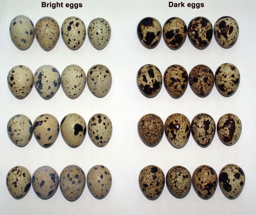
between them (Figure 1).
General description of size-related traits of eggs. In
September 2015, each egg (n ¼ 60) was weighed to the
nearest 0.001 g (Ohaus No. CT600-S); in addition, the
length, the breadth at the equator (or at the shoulder, the
broadest part of the shell), and the breadth at the geometric
FIGURE 1. Japanese Quail eggs used in our analysis of the halfway point between the sharp and blunt poles were
chemical composition of eggshells at pigment spots and in
unpigmented areas. The eggs were classified into 2 groups based measured. Then each egg was opened and its contents
on (only) visual estimates of eggshell color and the size of the poured out. Using a micrometer gauge, one of the authors
black–brown protoporphyrin pigment spots, representing 2 (P.P.) measured (to the nearest 10 lm) the thickness of the
extremes of eggshell coloration: (1) ‘‘bright eggs,’’ those with eggshells along with the adjacent shell membrane at
light brown shells with relatively small and more sharply randomly chosen point(s) in 3 egg regions: the sharp pole
demarcated pigment spots; and (2) ‘‘dark eggs,’’ those with dark
brown shells and more extensive pigment spots. Note that dark and the blunt pole (each measured only once) and the
eggs were nearly 7% heavier and 2% wider, on average, at the equator (4 measurements, subsequently averaged for further
equator than bright eggs (a significant difference; see Table 1). calculations). These measurements are listed in Table 1.
Eggshell thickness at pigment spots and in adjacent
unpigmented areas. Further measurements were made
kg1), and vitamin E (70.9 mg kg1). The daily consump- (by P.P.) to precisely assess the relationship between the
tion of feed was 25–30 g bird1. presence of pigment spots and eggshell thickness. The
eggshell thicknesses at the pigment spot and in the
Egg Selection adjacent unpigmented area (the latter always located in
No special breeding or feeding (dietary) condition was the immediate vicinity up to ~1 mm from the pigment
applied before the eggs used in the study were collected. spot) were measured in pairwise fashion in 3 egg regions:
The eggs were selected from a large sample (~600) that the sharp pole, the blunt pole (2 paired measurements for
had been laid by different females (n ¼ 240) between July 3 each, i.e. at the pigment spot and the adjacent unpigment-
and 5, 2015. The eggs were collected 3 or 4 times a day and ed area), and the equator (4 paired measurements,
immediately transferred to a refrigerator (at 38C), where subsequently averaged for further calculations). Figure 2
they were stored prior to further processing. and Table 1 summarize these measurements.
TABLE 1. Comparison of size-related traits (means 6 SE) of dark eggs (n ¼ 30) and bright eggs (n ¼ 30) of Japanese Quail used in the
chemical composition of eggshell maculation (pigment spots) and adjacent unpigmented areas.
a
Test of difference
Egg-size trait (unit) Dark eggs Bright eggs t P
Mass (g) 11.16 6 0.13 10.47 6 0.19 2.94 0.005
Length (mm) 32.70 6 0.19 32.16 6 0.24 1.78 0.081
Breadth at equator (mm) 25.46 6 0.13 24.98 6 0.16 2.38 0.021
Breadth in geometric middle (mm) 24.95 6 0.12 24.58 6 0.16 1.87 0.067
Eggshell thickness at equator (mm) 0.234 6 0.003 0.229 6 0.005 0.98 0.333
Eggshell thickness at sharp pole (mm) 0.261 6 0.007 0.248 6 0.008 1.22 0.227
Eggshell thickness at blunt pole (mm) 0.249 6 0.005 0.252 6 0.008 0.32 0.747
a
Independent-samples t-test.
The Auk: Ornithological Advances 134:721–731, Q 2017 American Ornithological SocietyG. Orłowski, P. Pokorny, W. Dobicki, et al. Chemistry of pigmented and plain areas of eggshells 725
Eggshell Treatment
For the chemical analysis, each eggshell (with the adjacent
membrane) was air dried to constant mass, and from each
one all the areas of pigment spots were accurately cut off from
the remaining unpigmented area. This laborious task was
performed by the technical staff of the Department of
Hydrobiology and Aquaculture, Wrocław University of
Environmental and Life Sciences, using surgical instruments
(tweezers, scissors, and scalpel). We tried to separate all
visible pigment spots larger than ~1 mm in diameter. In the
case of the less strongly maculated (i.e. bright) eggs, such a
procedure in practice yielded homogeneous and distinct
eggshell samples containing unpigmented areas and dark,
pigmented areas (spots). With regard to some heavily
maculated (dark) eggs, however, the samples from the
unpigmented eggshell region also contained some tiny
pigment spots less than ~1 mm in diameter. Consequently,
from each group of eggs (i.e. dark eggs and bright eggs; n ¼ FIGURE 2. Results (means 6 SE) of statistical analysis
(dependent-samples t-test) comparing eggshell thickness at
60), we obtained 2 paired samples of eggshells from the pigment spots and in adjacent unpigmented (plain) areas of
pigmented and unpigmented regions (n¼120 in total). These ‘‘dark eggs’’ (n ¼ 30) and ‘‘bright eggs’’ (n ¼ 30) of Japanese Quail,
were then chemically analyzed. measured in 3 egg regions: (A) equator (dark: t ¼ 12.1, P , 0.0001;
bright: t ¼ 6.74, P , 0.0001); (B) sharp pole (dark: t ¼ 0.86, P ¼
Chemical Analysis 0.398; bright: t ¼ 1.95, P ¼ 0.064); and (C) blunt pole (dark: t ¼ 3.68,
P , 0.001; bright: t ¼ 3.95, P , 0.001). No significant differences
Two of the authors (W.D. and P.P.) performed the chemical were found between dark and bright eggs in eggshell thickness
analysis at the Department of Hydrobiology and Aquacul- at the pigment spots or in the unpigmented eggshell areas in the
ture, Wrocław University of Environmental and Life 3 egg regions (independent-samples t-test, P 0.068).
Sciences. The eggshell samples were mineralized in nitric
acid in a high-pressure microwave digestion system
(MARS-5; CEM, Matthews, North Carolina, USA). Flame pairwise comparisons of the concentration differences of the
atomic absorption spectroscopy (SpectrAA FS220; Varian, 8 elements in the pigment spots and the unpigmented areas
Palo Alto, California, USA) was then used to determine the in each of the 2 egg types, as well as in pooled samples of dark
content of 2 micronutrients (Ca and Mg) and 6 trace and bright eggs. We also performed an analogous pairwise
elements (Cu, Mn, Fe, Co, Cd, and Pb). The measurement comparison for the eggshell thickness data determined for
process was validated using reference material DORM-3 pigment spots and adjacent unpigmented areas measured in
(fish protein) provided by the National Research Council 3 egg regions: equator, sharp pole, and blunt pole.
of Canada Institute for National Measurement Standards Secondly, using the independent-samples t-test, we
(Ottawa, Ontario, Canada). The mean recoveries in 6 assessed the differences between dark eggs and bright
replicates of certified material (0.9 g) expressed the eggs with respect to (1) the concentrations of elements
precision of the method, or the difference between the measured in eggshell pigment spots, (2) the concentrations
mean value obtained by analyzing a certified reference of elements measured in unpigmented eggshell areas, and
material and its certified value for Cu, Fe, Cd, and Pb (3) general size-related morphological traits of eggs.
performed on the same sample; this was within 5% of the All the concentrations of elements, eggshell thickness
concentrations stated for the reference material. All metal data, and size-related traits of eggs met the assumption of
concentrations were expressed in milligrams per kilogram normality; the use of parametric statistics was therefore
(mg kg1) or parts per million (ppm) of dry mass. justified. The statistical analyses were done using Statistica
7.0 (StatSoft, Tulsa, Oklahoma, USA) and Excel software.
Statistical Analysis The statistical significance level was 0.05.
Our first aim was to assess the differences between the
concentrations of elements measured in the pigment spots RESULTS
and unpigmented areas of eggshells in the 2 predefined egg
types representing 2 extremes in eggshell coloration: those The shells of the dark eggs (Figure 1) were significantly (7%
with dark shells and more extensive pigment spots vs. those and 6% on average, respectively) thicker at pigment spots
with bright shells and small but more sharply demarcated than in adjacent unpigmented areas at their equators and
pigment spots. We used the dependent-samples t-test for blunt poles (Figure 2). In bright eggs (Figure 1), however,
The Auk: Ornithological Advances 134:721–731, Q 2017 American Ornithological Society726 Chemistry of pigmented and plain areas of eggshells G. Orłowski, P. Pokorny, W. Dobicki, et al. FIGURE 3. Concentrations (means 6 SE) of 8 chemical elements measured in the pigment spots of eggshells and unpigmented (plain) eggshell areas of ‘‘dark eggs’’ (n ¼ 30) and ‘‘bright eggs’’ (n ¼ 30) of Japanese Quail. See Figure 1 for examples of eggs in both groups; Appendix Table 3 lists the concentrations for both types of eggs. these differences were even more pronounced; eggshells The concentrations of the other 3 elements (Mn, Fe, Co) were, on average, 14% and 13% thicker at pigment spots measured in pigment spots and unpigmented areas were than in adjacent unpigmented areas at their equators and variable and depended on the egg type. In dark eggs, Mn blunt poles, respectively (Figure 2). and Fe levels were 19% and 20% lower, respectively, at Chemical analysis showed that the concentrations of Ca pigment spots than in unpigmented regions; but in bright and Mg (and also of Cu and Cd) in the shells of both dark eggs, Mn and Fe levels were 10% and 31% higher, and bright eggs were significantly higher in the pigment respectively, at pigment spots than in unpigmented areas spots than in unpigmented areas (Figure 3; Appendix Table (Figure 3). Co levels varied significantly only in dark eggs, 3). Specifically, Cu levels were 10% and 85% higher, on pigment spots containing 106% more Co than unpigment- average, in the pigment spots than in unpigmented areas of ed areas (Figure 3; Appendix Table 3). dark and bright eggs, respectively; the corresponding Furthermore, we found significant differences between average values for the other 3 of these elements in each dark and bright eggs with regard to the concentrations of group of eggshells were 30% and 148% for Cd, 21% and all the elements measured in unpigmented regions (Table 33% for Mg, and 9% and 21% for Ca (Figure 3). The 2). The levels of 6 (Cu, Mn, Fe, Cd, Pb, and Ca) were higher opposite pattern was observed in the case of Pb, which in the dark eggs than in the bright ones, whereas the appeared to prevail in the plain areas of eggshells concentrations of the other 2 (Co and Mg) peaked in compared with the pigment spots: 213% and 817% lower bright eggs (Figure 3). in the pigment spots of dark and bright eggs, respectively Similarly, there were significant differences between (Figure 3). dark and bright eggs in the concentrations of all elements The Auk: Ornithological Advances 134:721–731, Q 2017 American Ornithological Society
G. Orłowski, P. Pokorny, W. Dobicki, et al.                              Chemistry of pigmented and plain areas of eggshells   727

TABLE 2. Results of the statistical comparison (independent-         layer, compared to some other birds’ eggs that have a thin
samples t-test) of concentrations of 8 chemical elements (ppm        internalized or superficial layer (cf. Duval et al. 2016).
dry mass) between pigment spots (maculation) on the shells of
                                                                     However, it seems that a thick superficial pigment layer
dark and bright eggs, and between unpigmented shell areas of
dark and bright eggs determined for 60 eggs of Japanese Quail.       (such as in the quail) does not contradict the concept that
For the element concentrations, see Figure 3 and Appendix            maculation strengthens the eggshell (Gosler et al. 2005).
Table 3.                                                                It is highly probable that higher levels of 4 elements (Ca,
             Plain area (n ¼ 60)        Pigment spot (n ¼ 60)        Mg, Cu, and Cd) in maculated eggshell areas indicate a
                                                                     close association of these metals with the presence of the
Element        t             P                t            P         protoporphyrin or dark pigmentation responsible for the
Cu            7.83       ,0.0001        4.83           ,0.0001      brown coloration. On the other hand, our chemical
Mn            8.96       ,0.0001        1.16            0.251       analysis involved both the pigment layer (approximately
Fe            6.91       ,0.0001        3.12            0.003       10–20% of the eggshell thickness) and the other inner
Co           3.18        0.002          6.81           ,0.0001      layers of eggshell plus the shell membrane without visible
Cd            3.39        0.001         3.90           ,0.001
Pb            2.97        0.004          4.84           ,0.0001      brown pigmentation, showing that protoporphyrin pig-
Mg           2.73        0.008         4.05           ,0.001       ment was present in different concentrations in all these
Ca            2.21        0.031         3.08            0.004       layers (Tamura and Fujii 1967). Hence, this is probably
                                                                     itself a confounding factor and would have an influence on
                                                                     the levels of certain elements in an entire eggshell sample.
                                                                     This applies in particular to Ca and Mg, whose levels are
except Mn measured at pigment spots (Table 2). Thus, the             not homogeneous throughout the shell thickness (Abdel-
pigment spots of bright eggs had higher Cu, Fe, Cd, Mg,              Salam et al. 2006). Conversely, another toxic element (Pb)
and Ca concentrations but lower Co and Pb levels in                  peaked markedly in unpigmented eggshell areas (where its
comparison with dark eggs (Table 2). Notably, we found               level was nearly 93 higher than in speckled areas), and this
that the plain areas in dark eggs contained 4% more Ca               metal may well be more closely bound to biliverdin or,
than the same areas in bright eggs, a significant difference;        alternatively, to the deeper unpigmented and more
conversely, the pigment spots of bright eggs contained 7%            calcified eggshell region.
more Ca than pigment spots of dark eggs (Figure 3), also a              We obtained conflicting results for 3 other trace metals
significant difference (Table 2).                                    (Mn, Fe, and Co), the concentrations of which were
                                                                     variable and dependent on egg coloration. In quail
DISCUSSION                                                           eggshells, a relatively large amount of protoporphyrin is
                                                                     also deposited in the unpigmented shell region (Tamura
Our results demonstrate for the first time that eggshells of         and Fujii 1967), particularly in those eggs with a darker
maculated avian eggs are not chemically homogeneous and              background color (Duval et al. 2013), and/or is visible as a
that there are significant differences between speckled and          small pigment inclusion on the outer shell surface (Lovell
unpigmented eggshell regions in the maternal deposition              et al. 2013). In females, the concentrations of both
of several chemical elements. In particular, we found                protoporphyrin (measured in the uterus) and biliverdin
evidence that the concentrations of 2 micronutrients (Ca             (measured in the shell gland) were independent of egg
and Mg) and 2 trace elements (Cu and Cd) were                        coloration (Baird et al. 1975, Zhao et al. 2006, Liu et al.
significantly higher in speckled eggshell regions (which             2010; see Sparks 2011). Hence, there may be an
were also significantly thicker) than in their unpigmented           equilibrium (i.e. a balanced distribution of a female’s
counterparts. Some earlier studies showed a strong                   resources) between speckled and plain eggshell regions.
association between the availability of dietary or environ-          Such an explanation is supported by our findings that the
mental Ca and eggshell maculation, implying that Ca                  maculation of bright eggs (significantly lighter and
shortages resulted in localized eggshell thinning (see               narrower at the equator than dark eggs) has dispropor-
Cherry and Gosler 2010, Hargitai et al. 2016b), but no               tionately thicker eggshells (presumably due to the greater
analyses of Ca concentrations in speckled eggshell regions           accumulation of pigment; Figure 2) than the speckles of
have previously been undertaken. We found that the                   dark eggs. This is a novel and important finding with
maculated eggshell regions were thicker, and they con-               regard to the developmental biology of the quail (and
tained significantly more Ca than the unpigmented areas,             presumably other Galliformes), suggesting that protopor-
which suggests parallel cellular transport of protoporphy-           phyrin may also be accumulated by way of an increase in
rin at high Ca levels (but see Maurer et al. 2011a). Our             eggshell thickness. Presumably, therefore, the absolute
results contradict those of previous investigations, prob-           pigment content cannot be evaluated correctly by any
ably because we selected a species with a different eggshell         attempt at a visual description of an eggshell’s external
pigmentation structure—quail eggs have a thick superficial           pigmentation (e.g., Brulez et al. 2014, Pike 2015). It thus

                                              The Auk: Ornithological Advances 134:721–731, Q 2017 American Ornithological Society
728 Chemistry of pigmented and plain areas of eggshells G. Orłowski, P. Pokorny, W. Dobicki, et al.
appears that this overlooked mechanism of pigment allocations of co-occurring eggshell elements and pig-
deposition through a thickening of its layer can better ments.
explain some conflicting results published earlier—for
example, that the greater number of visible brown spots on ACKNOWLEDGMENTS
quail eggshells is not due to enhanced protoporphyrin
deposition (Duval et al. 2016). It should also be borne in L. Hałupka and anonymous reviewers kindly provided
mind that, apart from the elements or contaminants valuable comments, and P. Senn corrected the English, which
deposited in eggshells, a portion of these maternal improved the manuscript.
resources is accumulated in the egg contents; a positive Funding statement: We received no funding for this
relationship has been reported between the level of research. The publication was supported by Wrocław Centre
pesticide residues measured in egg contents and the of Biotechnology, programme the Leading National Research
greenness of the ground color related to biliverdin pigment Centre (KNOW) for years 2014–2018.
(Jagannath et al. 2008, Hanley and Doucet 2012). Ethics statement: All procedures for this study were
Our findings imply that speckling can play a functional conducted in compliance with Polish legislation.
Author contributions: G.O. and E.Ł. conceived the study.
role in the chemistry of avian eggs by enabling females to
G.O., P.P., W.D., A.K., and E.Ł. designed the methods. P.P.,
variously distribute micronutrients and trace elements into
W.D., A.K., and E.Ł. performed the experiments. G.O.
pigment spots and unpigmented regions of eggshells; this analyzed the data. G.O. and E.Ł. wrote the paper. E.Ł., P.P.,
presumably represents a mechanism by which they can A.K., and W.D. contributed substantial materials, resources,
make prompt physiological adjustments to varying levels and/or funding.
of maternal resources. There is an urgent need to
investigate how maculation affects eggshell thickness and
the levels of micronutrients or trace elements in eggs LITERATURE CITED
varying in the location or thickness of the pigment layer, Abdel-Salam, Z. A., A. M. Abdou, and M. Harith (2006). Elemental
especially in the context of the embryonic depletion of and ultrastructural analysis of the eggshell: Ca, Mg and Na
eggshell micronutrients and trace elements. We strongly distribution during embryonic development via LIBS and SEM
recommend studies aimed at resolving the still open techniques. International Journal of Poultry Science 5:35–42.
question of how maternal resources (i.e. micronutrients, Baird, T., S. E. Solomon, and D. R. Tedstone (1975). Localisation
trace elements, and pigments) allocated in pigment and characterisation of egg shell porphyrins in several avian
speckles and in different eggshell layers contribute to species. British Poultry Science 16:201–208.
Brulez, K., P. Cassey, A. Meeson, I. Mikšı́k, S. L. Webber, A. G.
embryonic development during their depletion (Orłowski
Gosler, and S. J. Reynolds (2014). Eggshell spot scoring
et al. 2016), especially in view of the fact that embryo- methods cannot be used as a reliable proxy to determine
induced shell thinning at pigment speckles is minimal pigment quantity. Journal of Avian Biology 45:94–102.
compared to that in plain eggshell regions (Maurer et al. Brulez, K., I. Mikšı́k, C. R. Cooney, M. E. Hauber, P. G. Lovell, G.
2011c) and that the innermost (mammillary) layer of an Maurer, S. J. Portugal, D. Russell, S. J. Reynolds, and P. Cassey
eggshell is eroded or decalcified during embryonic (2016). Eggshell pigment composition covaries with phylog-
development (Karlsson and Lilja 2008, Igic et al. 2017). eny but not with life history or with nesting ecology traits of
This suggests that certain elements sequestered into British passerines. Ecology and Evolution 6:1637–1645.
eggshell pigment speckles (especially in those located in Bulla, M., M. Šálek, and A. G. Gosler (2012). Eggshell spotting
the upper eggshell layers not subject to decalcification) does not predict male incubation but marks thinner areas of
a shorebird’s shells. The Auk 129:26–35.
may not be further utilized by developing embryos. This
Burger, J. (1994). Heavy metals in avian eggshells: Another
might be a form of physiological chemoprotection through excretion method. Journal of Toxicology and Environmental
the disposal or deactivation of certain metals, transferred Health 41:207–220.
from females’ bodies to egg speckles, which do not (or only Casini, S., M. C. Fossi, C. Leonzio, and A. Renzoni (2003). Review:
marginally) contribute to embryogenesis. Finally, because Porphyrins as biomarkers for hazard assessment of bird
the speckling or coloration of eggshells, a consequence of populations: Destructive and non-destructive use. Ecotoxi-
putative pigment deposition, affects Ca and metal levels in cology 12:297–305.
eggshells with an outer pigment layer, follow-up studies Cassey, P., G. Maurer, P. G. Lovell, and D. Hanley (2011).
are necessary on another species that varies in the position Conspicuous eggs and colourful hypotheses: Testing the role
of multiple influences on avian eggshell appearance. Avian
of pigment layers. In this respect, we strongly recommend
Biology Research 4:185–195.
the use of analytical methods that enable elemental Cassey, P., I. Mikšı́k, S. J. Portugal, G. Maurer, J. G. Ewen, E. Zarate,
concentrations in eggshell micro-samples to be deter- M. A. Sewell, F. Karadas, T. Grim, and M. E. Hauber (2012).
mined; isolated pigment spots or speckles or even Avian eggshell pigments are not consistently correlated with
powdered pigment scratched off an egg permits an colour measurements or egg constituents in two Turdus
unequivocal relationship to be found between maternal thrushes. Journal of Avian Biology 43:503–512.
The Auk: Ornithological Advances 134:721–731, Q 2017 American Ornithological SocietyG. Orłowski, P. Pokorny, W. Dobicki, et al. Chemistry of pigmented and plain areas of eggshells 729
Chatelain, M., J. Gasparini, and A. Frantz (2016). Do trace metals Sherameti and A. Varma, Editors). Springer International,
select for darker birds in urban areas? An experimental Cham, Switzerland. pp. 127–143.
exposure to lead and zinc. Global Change Biology 22:2380– Hassan, H. A., S. S. El-Nesr, A. M. R. Osman, and G. A. Arram
2391. (2013). Ultrastructure of eggshell, egg weight loss and
Cherry, M. I., and A. G. Gosler (2010). Avian eggshell coloration: hatching traits of Japanese Quail varying in eggshell color
New perspectives on adaptive explanations. Biological and pattern using image analysis. Egyptian Poultry Science
Journal of the Linnean Society 100:753–762. Journal 34:1–17.
Deeming, D. C. (2011). A review of the relationship between Igic, B., M. E. Hauber, C. Moskát, T. Grim, M. D. Shawkey, P.
eggshell colour and water vapour conductance. Avian Procházka, and M. Honza (2017). Brood parasite and host
Biology Research 4:224–230. eggshells undergo similar levels of decalcification during
Duval, C., P. Cassey, P. G. Lovell, I. Mikšı́k, S. J. Reynolds, and K. A. embryonic development. Journal of Zoology 301:165–173.
Spencer (2016). Maternal influence on eggshell maculation: Jagannath, A., R. F. Shore, L. A. Walker, P. N. Ferns, and A. G.
Implications for cryptic camouflaged eggs. Journal of Gosler (2008). Eggshell pigmentation indicates pesticide
Ornithology 157:303–310. contamination. Journal of Applied Ecology 45:133–140.
Duval, C., P. Cassey, I. Mikšı́k, S. J. Reynolds, and K. Spencer (2013). Karlsson, O., and C. Lilja (2008). Eggshell structure, mode of
Condition-dependent strategies of eggshell pigmentation: An development and growth rate in birds. Zoology 111:494–502.
experimental study of Japanese Quail (Coturnix coturnix Kennedy, G. Y., and H. G. Vevers (1976). A survey of avian
japonica). Journal of Experimental Biology 216:700–708. eggshell pigments. Comparative Biochemistry and Physiolo-
Espı́n, S., A. J. Garcı́a-Fernández, D. Herzke, R. F. Shore, B. van gy B 55:117–123.
Hattum, E. Martı́nez-López, M. Coeurdassier, I. Eulaers, C. Klein, R., M. Bartel-Steinbach, J. Koschorreck, M. Paulus, K.
Fritsch, P. Gómez-Ramı́rez, V. L. B. Jaspers, et al. (2014). Tarricone, D. Teubner, G. Wagner, T. Weimann, and M. Veith
Sampling and Contaminant Monitoring Protocol for Raptors. (2012). Standardization of egg collection from aquatic birds
Research Networking Programme-EURAPMON. http://www. for biomonitoring—a critical review. Environmental Science
eurapmon.net/results/contaminant-monitoring-protocol and Technology 46:5273–5284.
Garcı́a-Navas, V., J. J. Sanz, S. Merino, J. Martı́nez-de la Puente, E. Larsson, B. S. (1993). Interaction between chemicals and
Lobato, S. del Cerro, J. Rivero, R. de Castañeda, and J. Moreno melanin. Pigment Cell Research 6:127–133.
(2011). Experimental evidence for the role of calcium in Liu, H. C., M. C. Hsiao, Y. H. Hu, S. R. Lee, and W. T. K. Cheng
eggshell pigmentation pattern and breeding performance in (2010). Eggshell pigmentation study in blue-shelled and
Blue Tits Cyanistes caeruleus. Journal of Ornithology 152:71– white-shelled ducks. Asian-Australasian Journal of Animal
82. Sciences 23:162–168.
Gorchein, A., C. K. Lim, and P. Cassey (2009). Extraction and Lovell, P. G., G. D. Ruxton, K. V. Langridge, and K. A. Spencer
analysis of colourful eggshell pigments using HPLC and (2013). Egg-laying substrate selection for optimal camouflage
HPLC/electrospray ionization tandem mass spectrometry. by quail. Current Biology 23:260–264.
Biomedical Chromatography 23:602–606. Maurer, G., S. J. Portugal, I. Boomer, and P. Cassey (2011a). Avian
Gosler, A. G., O. R. Connor, and R. H. C. Bonser (2011). embryonic development does not change the stable isotope
Protoporphyrin and eggshell strength: Preliminary findings composition of the calcite eggshell. Reproduction, Fertility
from a passerine bird. Avian Biology Research 4:214–223. and Development 23:339–345.
Gosler, A. G., J. P. Higham, and S. J. Reynolds (2005). Why are Maurer, G., S. J. Portugal, and P. Cassey (2011b). Review: An
birds’ eggs speckled? Ecology Letters 8:1105–1113. embryo’s eye view of avian eggshell pigmentation. Journal of
Hanley, D. (2012). The function and evolution of egg colour in Avian Biology 42:494–504.
birds. Electronic Theses and Dissertations. Ph.D. dissertation, Maurer, G., S. J. Portugal, and P. Cassey (2012). A comparison of
University of Windsor, Windsor, ON, Canada. indices and measured values of eggshell thickness of
Hanley, D., and S. M. Doucet (2012). Does environmental different shell regions using museum eggs of 230 European
contamination influence egg coloration? A long-term study bird species. Ibis 154:714–724.
in Herring Gulls. Journal of Applied Ecology 49:1055–1063. Maurer, G., S. Portugal, I. Mikšı́k, and P. Cassey (2011c). Speckles
Hargitai, R., G. Nagy, M. Herényi, Z. Nyiri, M. Laczi, G. Hegyi, Z. of cryptic Black-headed Gull eggs show no mechanical or
Eke, and J. Török (2016a). Darker eggshell spotting indicates conductance structural function. Journal of Zoology 285:194–
lower yolk antioxidant level and poorer female quality in the 204.
Eurasian Great Tit (Parus major). The Auk: Ornithological McGraw, K. J. (2003). Melanins, metals, and mate quality. Oikos
Advances 133:131–146. 102:402–406.
Hargitai, R., G. Nagy, Z. Nyiri, L. Bervoets, Z. Eke, M. Eens, and J. Mikšı́k, I., V. Holán, and Z. Deyl (1996). Avian eggshell pigments
Török (2016b). Effects of breeding habitat (woodland versus and their variability. Comparative Biochemistry and Physiol-
urban) and metal pollution on the egg characteristics of ogy B 113:607–612.
Great Tits (Parus major). Science of the Total Environment Miles, R. D. (2000). Trace minerals and avian embryo develop-
544:31–38. ment. Ciência Animal Brasileira 2:1–10.
Harrison, C. J. O. (1966). Variation in the distribution of pigment Mora, M. A. (2003). Heavy metals and metalloids in egg contents
within the shell structure of birds’ eggs. Journal of Zoology and eggshells of passerine birds from Arizona. Environmental
148:526–539. Pollution 125:393–400.
Hashmi, M. Z., N. A. Abbasi, X. Tang, and R. N. Malik (2015). Egg Moreno, J., and J. L. Osorno (2003). Avian egg colour and sexual
as a biomonitor of heavy metals in soil. In Heavy Metal selection: Does eggshell pigmentation reflect female condi-
Contamination of Soils: Monitoring and Remediation (I. tion and genetic quality? Ecology Letters 6:803–806.
The Auk: Ornithological Advances 134:721–731, Q 2017 American Ornithological Society730 Chemistry of pigmented and plain areas of eggshells G. Orłowski, P. Pokorny, W. Dobicki, et al.
Niecke, M., M. Heid, and A. Krüger (1999). Correlations between (decycling) 1 is associated with blue egg formation. Poultry
melanin pigmentation and element concentration in feathers Science 90:836–841.
of White-tailed Eagles (Haliaeetus albicilla). Journal of Williams, S. T., S. Ito, K. Wakamatsu, T. Goral, N. P. Edwards, R. A.
Ornithology 140:355–362. Wogelius, T. Henkel, L. F. C. de Oliveira, L. F. Maia, S.
Orłowski, G., and L. Hałupka (2015). Embryonic eggshell Strekopytov, T. Jeffries, et al. (2016). Identification of shell
thickness erosion: A literature survey re-assessing embryo- colour pigments in marine snails Clanculus pharaonius and C.
induced eggshell thinning in birds. Environmental Pollution margaritarius (Trochoidea; Gastropoda). PLOS One 11:
205:218–224. e0156664.
Orłowski, G., L. Hałupka, P. Pokorny, E. Klimczuk, H. Sztwiertnia, Woodard, A. E., and F. B. Mather (1964). The timing of ovulation,
and W. Dobicki (2016). The effect of embryonic development movement of the ovum through the oviduct, pigmentation
on metal and calcium content in eggs and eggshells in a and shell deposition in Japanese Quail (Coturnix coturnix
small passerine. Ibis 158:144–154. japonica). Poultry Science 43:1427–1432.
Ostfeld, D., and M. Tsutsui (1974). Novel metalloporphyrins: Zhao, R., G. Y. Xu, Z. Z. Liu, J. Y. Li, and N. Yang (2006). A study on
Syntheses and implications. Accounts of Chemical Research eggshell pigmentation: Biliverdin in blue-shelled chickens.
7:52–58. Poultry Science 85:546–549.
Pike, T. W. (2015). Modelling eggshell maculation. Avian Biology
Research 8:237–243.
Reynolds, S. J. (1997). Uptake of ingested calcium during egg APPENDIX
production in the Zebra Finch (Taeniopygia guttata). The Auk
114:562–569.
Reynolds, S. J., G. R. Martin, and P. Cassey (2009). Is sexual The Structural Function of Eggshell Maculation: A
selection blurring the functional significance of eggshell Short Review and Research Problems
coloration hypotheses? Animal Behaviour 78:209–215.
Richards, M. P. (1997). Trace mineral metabolism in the avian Previous studies have suggested that the function of
embryo. Poultry Science 76:152–164. eggshell maculation is, first and foremost, a structural
Rodriguez-Navarro, A. B., K. F. Gaines, C. S. Romanek, and G. R.
Masson (2002). Mineralization of Clapper Rail eggshell from a
one related to Ca availability (Solomon 1991, Gosler et al.
contaminated salt marsh system. Archives of Environmental 2005, 2011, Garcı́a-Navas et al. 2010), in that pigment
Contamination and Toxicology 43:449–460. spots (i.e. the maculated eggshell area) demarcate thinner
Simkiss, K. (1961). Calcium metabolism and avian reproduction. areas of the shell (reviewed by Cherry and Gosler 2010).
Biological Reviews 36:321–359. Such localized eggshell thinning at pigment spots has been
Soh, T., N. Fujihara, and O. Koga (1993). Observations of pigment reported in a few species in the Passeriformes (Gosler et al.
accumulation in the epithelium of the shell gland and
2005), Charadriiformes (Maurer et al. 2011a, 2011c, Bulla
superficial pigmentation on the egg shell in Japanese Quail.
Journal of the Faculty of Agriculture, Kyushu University 38: et al. 2012), and Falconiformes (Jagannath et al. 2008). This
73–80. phenomenon has been invoked as evidence for the
Solomon, S. E. (1991). Egg and Eggshell Quality. Wolfe, London, structural function of maculation in the mechanism
UK. compensating for Ca deficiency and/or strengthening the
Sparks, N. H. C. (2011). Eggshell pigments—from formation to eggshell (see Cherry and Gosler 2010). However, there is
deposition. Avian Biology Research 4:162–167. an important, hitherto overlooked, issue directly linked to
Taha, A. E. (2011). Analyzing of quail eggs hatchability, quality,
this physiological association between eggshell thinning
embryonic mortality and malpositions in relation to their
shell colors. Egyptian Poultry Science Journal 31:807–815. and the presence of maculation—namely, the distribution
Tamura, T., and S. Fujii (1967). Comparative observations on the and volume or thickness of the protoporphyrin layer over
distribution of fluorescent pigments (porphyrins) in the egg the eggshell surface (see Harrison 1966, Tyler 1969;
coverings of chicken and quail. Journal of the Faculty of discussed in Maurer et al. 2011b, Sparks 2011), a generality
Fisheries and Animal Husbandry, Hiroshima University 7:35–41. that must be universally applicable to all birds (cf.
Tamura, T., S. Fujii, H. Kunisaki, and M. Yamane (1965). Reynolds et al. 2009). Maurer et al. (2011b) summarized
Histological observations on the quail oviduct; with reference
evidence for eggshell maculation in 7 orders of birds.
to pigment (porphyrin) in the uterus. Journal of the Faculty of
Fisheries and Animal Husbandry, Hiroshima University 6:37– However, the distribution of the protoporphyrin layer(s) is
57. not uniform and varies between different orders or even
Tyler, C. (1969). A study of the egg shells of the Gaviiformes, within a single egg. The spots of dark pigment may be
Procellariiformes, Podicipediformes and Pelecaniformes. present in the surface layer of the eggshell and/or at
Journal of Zoology 158:395–412. varying depths within the eggshell; in the the latter case,
Verdes, A., W. Cho, M. Hossain, P. L. R. Brennan, D. Hanley, T. spots may not be visible on the surface (Harrison 1966,
Grim, M. E. Hauber, and M. Holford (2015). Nature’s palette:
Characterization of shared pigments in colorful avian and
Tyler 1969, Jagannath et al. 2008). Hence, not all
mollusk shells. PLOS One 10:e0143545. maculation may have a structural function (Cherry and
Wang, Z. P., R. F. Liu, A. R. Wang, J. Y. Li, and X. M. Deng (2011). Gosler 2010, Maurer et al. 2011b). For instance, the darker
Expression and activity analysis reveal that heme oxygenase pigment in Eurasian Sparrowhawk (Accipiter nisus) eggs
The Auk: Ornithological Advances 134:721–731, Q 2017 American Ornithological Societymetabolites; Jagannath et al. 2008).
G. Orłowski, P. Pokorny, W. Dobicki, et al.
of environmental contaminants or pesticide residues (DDT
the thinner eggshell area and to be correlated with the level
occurs as a layer on the shell surface and as an internal one
within the shell; but only the latter was found to demarcate
APPENDIX TABLE 3. Comparison of mean concentrations (95% confidence interval) of 8 chemical elements (ppm dry mass) measured in the pigment spots and unpigmented
areas of eggshells reported in dark eggs, bright eggs, and all eggs of Japanese Quail, with results of pairwise comparisons (dependent-samples t-test) for the same eggs. For
the differences between the concentrations of elements in plain areas and between the pigment spots of dark and bright eggs, see Table 2. Results in bold are insignificant.
Dark eggs (n ¼ 30) Bright eggs (n ¼ 30) All eggs (n ¼ 60)
Element Plain area Pigment spot t-test Plain area Pigment spot t-test Plain area Pigment spot t-test
Cu 4.01 4.39 t ¼ 3.2 3.09 5.73 t ¼ 10.5 3.55 5.06 t ¼ 7.5
(3.83–4.19) (4.14–4.64) P ¼ 0.003 (2.93–3.25) (5.22–6.25) P , 0.0001 (3.38–3.72) (4.74–5.39) P , 0.0001
Mn 6.02 5.05 t ¼ 7.1 4.72 5.21 t ¼ 3.0 5.37 5.13 t ¼ 1.7
(5.83–6.20) (4.90–5.20) P , 0.0001 (4.49–4.95) (4.98–5.44) P ¼ 0.006 (5.15–5.59) (4.99–5.27) P ¼ 0.096
Fe 25.8 21.5 t ¼ 3.7 18.3 24.0 t ¼ 5.2 22.1 22.8 t ¼ 0.7
(24.1–27.5) (20.3–22.6) P ¼ 0.001 (16.9–19.7) (22.8–25.3) P , 0.0001 (20.6–23.5) (21.9–23.6) P ¼ 0.512
Co 1.24 2.55 t ¼ 10.5 1.85 1.43 t ¼ 1.6 1.54 1.99 t ¼ 2.4
(1.06–1.42) (2.38–2.72) P , 0.0001 (1.50–2.19) (1.13–1.72) P ¼ 0.121 (1.34–1.75) (1.77–2.21) P ¼ 0.018
Cd 0.69 0.89 t ¼ 2.4 0.48 1.19 t ¼ 9.7 0.58 1.04 t ¼ 7.0
(0.57–0.81) (0.80–0.97) P ¼ 0.025 (0.42–0.53) (1.06–1.32) P , 0.0001 (0.51–0.65) (0.95–1.12) P , 0.0001
Pb 1.91 0.61 t ¼ 4.9 1.10 0.12 t ¼ 9.8 1.51 0.36 t ¼ 8.1
(1.39–2.43) (0.410.81) P , 0.0001 (0.91–1.30) (0.06–0.18) P , 0.0001 (1.22–1.80) (0.25–0.48) P , 0.0001
Mg 4,389 5,312 t ¼ 5.5 5,166 6,850 t ¼ 5.6 4,778 6,081 t ¼ 7.3
(4,071–4,707) (5,003–5,621) P , 0.0001 (4,678–5,655) (6,138–7,562) P , 0.0001 (4,477–5,078) (5,655–6,507) P , 0.0001
Ca 287,196 312,926 t ¼ 4.1 275,541 334,567 t ¼ 10.6 281,368 323,746 t ¼ 8.9
Chemistry of pigmented and plain areas of eggshells
(277,642–296,750) (302,273– 323,580) P ¼ 0.0003 (270,500–280,582) (324,934– 344,199) P , 0.0001 (275,913– 286,823) (316,232– 331,261) P , 0.0001
The Auk: Ornithological Advances 134:721–731, Q 2017 American Ornithological Society
731You can also read