Caduet enhances connexin 43 phosphorylation in left ventricular and thoracic aorta of SH model rats
←
→
Page content transcription
If your browser does not render page correctly, please read the page content below
EXPERIMENTAL AND THERAPEUTIC MEDICINE 20: 80, 2020

                 Caduet enhances connexin 43 phosphorylation
            in left ventricular and thoracic aorta of SH model rats
               XIAOYAN HUANG1, JUNLU YANG2, BAOGUO SONG3, NANA WANG1, MEIJUAN MA4,
                   HAIFANG WANG5, SHA WANG3, SHUANGPING HAO6 and GONG CHENG4

     1
    Shaanxi Provincial Key Laboratory of Infection and Immunity Diseases, Shaanxi Provincial People's Hospital, Xi'an,
     Shaanxi 710068; 2Department of Cardiology, Baoji Traditional Chinese Medicine Hospital, Baoji, Shaanxi 721000;
  Departments of 3Cardiac Surgery and 4Cardiology, Shaanxi Provincial People's Hospital, Xi'an, Shaanxi 710068; 5Shaanxi
      Key Laboratory of Integrated Traditional and Western Medicine for Prevention and Treatment of Cardiovascular
     Diseases, Shaanxi University of Chinese Medicine, Xi'an, Shaanxi 712046; 6Department of Cardiology, Guangshui
             Traditional Chinese Medicine Hospital of Hubei Province, Guangshui, Hubei 432700, P.R. China

                                          Received December 12, 2019; Accepted June 5, 2020

                                                           DOI: 10.3892/etm.2020.9207

Abstract. Caduet, also known as amlodipine besylate and                   and thoracic aorta. Moreover, immunofluorescence results
atorvastatin calcium (AM + AT) tablet, can improve cardiac                indicated AM + AT could also decrease the expression
and vascular remodeling in patients with spontaneous                      T‑Cx43, and increase that of P‑Cx43 in the left ventricular and
hypertension (SH), but the underlying mechanism remains                   thoracic aorta compared with AM and AT alone. Therefore,
unknown. The present study aimed to explore whether                       it was concluded that AM + AT may mitigate left ventricular
AM + AT improved hypertensive left ventricular and thoracic               and thoracic aorta remodeling in SH rats by enhancing Cx43
aortic remodeling by regulating connexin 43 (Cx43) phosphor‑              phosphorylation, and the efficacy of AM + AT was superior to
ylation. A total of 32 male spontaneous hypertension model                that of AM and AT alone.
rats (SHR) were randomly divided into four groups: SHR
control group, amlodipine‑alone group (SHR‑AM), atorvas‑                  Introduction
tatin‑alone (SHR‑AT) and AM + AT group (SHR‑AM + AT);
8 Wistar‑Kyoto (WKY) rats with normal blood pressure were                 Spontaneous hypertension (SH) is one of the most critical
used as the normal control. Drugs were orally administered                factors in leading to cardiovascular diseases (1,2). Persistently
for 8 weeks; subsequently, body weight, heart rate (HR), left             elevated blood pressure in patients who are hypertensive
ventricular mass index (LVMI), blood pressure (BP), plasma                results in vascular tone increasement and vascular smooth
lipid levels and morphological changes of myocardial tissue in            muscle systolic dysfunction, leading to myocardial ischemia
each group were analyzed. The expression of total (T)‑Cx43                and ventricular and vascular remodeling (3,4). Furthermore,
and phosphorylated (P)‑Cx43 protein in the left ventricular               left ventricular and aorta remodeling is a frequent pathological
and thoracic aortic tissues was determined using western                  change in hypertension, which contributes to arrhythmia,
blotting and immunofluorescence double labeling. The results              heart failure and cardiovascular mortality (5‑7).
revealed that AM + AT significantly decreased LVMI and                        Caduet is a single pill containing a combination of amlo‑
cardiomyocyte cross‑sectional area compared with SHR‑AM                   dipine besylate and atorvastatin calcium tablet (AM + AT),
and SHR‑AT group. The western blotting results demonstrated               which is widely used in the clinical treatment of cardiovascular
that AM + AT could inhibit the expression of T‑Cx43 protein,              diseases, such as hypertension and coronary heart disease (8).
but increased the expression of P‑Cx43 in the left ventricular            In addition, previous experimental and clinical studies have
                                                                          reported that AM + AT can prevent cardiac hypertrophy and
                                                                          remodeling (9,10).
                                                                              The gap junction (GJ) channel is the structural basis
                                                                          for cellular electric coupling and signal transmission,
Correspondence to: Dr Gong Cheng, Department of Cardiology,
                                                                          which mainly functions though regulating the GJ protein
Shaanxi Provincial People's Hospital, 256 West Road, Beilin, Xi'an,
Shaanxi 710068, P.R. China
                                                                          connexin (11,12). Connexin 43 (Cx43)‑associated GJ protein in
E‑mail: 119275360@qq.com                                                  ventricular cardiomyocytes and vascular smooth muscle cells
                                                                          is involved in cardiac and vascular remodeling (13). Moreover,
Key words: caduet, amlodipine, atorvastatin, connexin 43                  left ventricular remodeling is closely associated with the
phosphorylation,   left   ventricle,   thoracic   aorta,    spontaneous   expression and distribution of Cx43 in cardiomyocytes (9,14),
hypertension                                                              and our previous study revealed that the expression of
                                                                          Cx43 was enhanced in the thoracic aorta of SH model rats
                                                                          (SHR) (15). Previous studies have also reported that Cx43
2                               HUANG et al: CONNEXIN 43 PHOSPHORYLATION IN SH MODEL RATS

phosphorylation contributes to ischemia‑associated remod‑          humidity of 50±5%, 14/10‑h light/dark cycle, free access to
eling of Cx43 channels in cardiomyocytes (16,17), and Cx43         food and water.
dephosphorylation contributes to arrhythmias and cardiomyo‑
cyte apoptosis in ischemia/reperfusion hearts (18). Based on       Measurement of body weight, heart rate (HR), left ventricular
these clinical data and laboratory reports, it was hypothesized    mass index (LVMI), blood pressure and plasma lipid levels in
that Cx43 phosphorylation may be involved in the process of        each group. The systolic blood pressure (SBP) and diastolic
hypertensive left ventricular and thoracic aorta remodeling,       blood pressure (DBP) of the rats were measured at 0, 2, 4,
and AM + AT may improve this remodeling by enhancing               6 and 8 weeks after treatment using the Non‑Invasive Blood
Cx43 phosphorylation.                                              Pressure system (LE‑5001 HX‑II tail‑cuff small animal
    Therefore, the aim of the present study was to investigate     blood pressure meter; Panlab S.L.U). After 8 weeks, HR
the effect of Cx43 phosphorylation in the process of hyper‑        was measured and anal temperature were measured by
tensive left ventricular and thoracic aorta remodeling, and        thermometer. Then the rats were weighed and anesthetized
to examine whether AM + AT improved this remodeling by             with pentobarbital (40 mg/kg) via intraperitoneal injection.
regulating Cx43 phosphorylation.                                   A total of 2 ml blood was collected from heart of every rat,
                                                                   and the plasma lipid levels of each group were measured
Materials and methods                                              using a Hitachi 7170 biochemical analyzer (Hitachi, Inc.);
                                                                   these included total cholesterol (TC), high‑density lipoprotein
Experimental reagents. A ProteoPrep Total Extraction Sample        cholesterol (HDL), low‑density lipoprotein cholesterol (LDL)
kit was obtained from Sigma‑Aldrich (Merck KGaA), the BCA          and triglycerides (TG). Then the chest of the rat was opened,
protein assay kit was from Beyotime Institute of Biotechnology     the heart was quickly removed and the left ventricular free
and nitrocellulose membrane was purchased from Thermo              wall was cut along the atrioventricular ring and weighed as the
Fisher Scientific, Inc. Primary antibody against rat total         LVMI (left ventricular mass/body weight; mg/g).
(T)‑Cx43 was obtained from Thermo Fisher Scientific, Inc.
(1:1,000 for western blotting; 1:100 for immunofluorescence;       Comparison of pathological changes in cardiac tissues. The
cat. no. 3D8A5). Primary antibody against rat phosphorylated       free wall of the left ventricle was fixed in 10% formaldehyde
(P)‑Cx43 was obtained from Cell Signaling Technology               for 2 h at room, dehydrated in an ethanol gradient (80, 90,
(1:1,000 for western blotting; 1:100 for immunofluorescence;       95 and 100%), then embedded in paraffin. Sections (3 µm
cat. no. 52559). Horseradish peroxidase (HRP)‑conjugated           thick) were cut and conventionally dewaxed to water in a
goat anti‑mouse (1:1,000; cat. no. G‑21040) and goat anti‑rabbit   series, including xylene I, xylene II, 100% alcohol I and 100%
polyclonal (1:1,000; cat. no. 31466) secondary antibodies were     alcohol II (10 min each), then a 95, 90, 80, 70% ethanol series
obtained from Thermo Fisher Scientific, Inc. ECL reagents          (10 min each) and finally distilled water. Sections were stained
were purchased from Thermo Fisher Scientific, Inc. (cat.           with hematoxylin for 1 min, rinsed with water once, stained
no. 35055). The FITC‑conjugated goat anti‑mouse secondary          with eosin for 2 min, then dehydrated with conventional
antibody was obtained from Thermo Fisher Scientific, Inc.          gradient alcohol (70, 80, 90 and 95% ethanol, 2 min each;
(1:50; cat. no. 62‑6511), and the tetramethylrhodamine isothio‑    100% ethanol I, 100% alcohol II, 10 min each), cleared with
cyanate (TRITC)‑conjugated goat anti‑rabbit secondary              xylene, and finally mounted with neutral gum and observed
antibody was purchased from Thermo Fisher Scientific, Inc.         with Olympus BX41 fluorescent microscope (magnification,
(1:100; cat. no. A16101). Bovine serum albumin (BSA) was           x400; Olympus Corporation). Images were captures and used
from Thermo Fisher Scientific, Inc. (cat. no. 37520).              to quantitatively analyze the myocardial cell cross‑sectional
                                                                   area using Image‑Pro Plus analysis software 6.0 (Media
Drugs and animals. The Chinese drug administration license         Cybernetics, Inc.).
nos. of AM, AT and AM + AT were H10950224, J20070061
and J20171045, respectively, which were from Betriebsstätte        Analysis of T‑Cx43 and P‑Cx43 protein expression levels in
Freiburg, Inc.. All procedures and ethics associated with animal   the left ventricular and thoracic aortic tissue using western
use were reviewed and approved by Medical School of Xi'an          blotting. Western blotting was performed as reported previ‑
Jiaotong University (approval no. 2018‑898). In total, 32 male     ously (21). Proteins of the left ventricular free wall and thoracic
SHR (age, 8 weeks; weight, 210‑265 g) were obtained from           aortic ascending tissue were extracted using ProteoPrep Total
Weitong Lihua Experimental Animal Technology Co., Ltd,             Extraction Sample kit and measured using a BCA protein
with clean grade certificate [SCXK (Beijing) 2007‑0001], and       assay kit. Each sample was mixed with 40 µl 1X SDS electro‑
eight Wistar‑Kyoto (WKY) rats were purchased from Skerries         phoresis sample buffer and boiled for 2‑3 min. Protein samples
Laboratory Animal Center, Ltd. as the WKY control group,           (30 µg) were separated by 12% SDS‑PAGE gel and transferred
with clean grade certificate [SCXK (Shanghai) 2007‑0005].          onto a nitrocellulose membrane. The membranes were blocked
The AM group (SHR‑AM; n=8), AT group (SHR‑AT; n=8)                 with 5% skimmed milk for 2 h at room temperature, and
and AM + AT group (SHR‑AM + AT; n=8) were orally                   washed twice with Tris‑buffered saline with 0.1% Tween‑20.
administered 10 mg/kg/day of AM, AT or AM + AT as treat‑           Membranes were incubated with T‑Cx43 and P‑Cx43 primary
ment groups for 8 weeks (19,20), respectively. The WKY             antibodies overnight at 4˚C, then probed with HRP‑conjugated
control group (n=8) and SHR control group (n=8) were orally        secondary antibody for 2 h at room temperature. Protein
administrated with the same volume of water as the treatment       bands were visualized using ECL reagents and imaged using
group. All rats received humane care and were raised in the        an Alpha Innotech FluorChem FC2 Imaging System (Alpha
same clean environment, with ambient temperature at 22±1˚C,        Innotech Inc.), the densitometric analysis was used ImageJ
EXPERIMENTAL AND THERAPEUTIC MEDICINE 20: 80, 2020                                                                                                                             3

Table I. Effects of the different drugs on body weight, HR and LVMI (n=8).

                                                                                                                     Group
                        ‑‑‑‑‑‑‑‑‑‑‑‑‑‑‑‑‑‑‑‑‑‑‑‑‑‑‑‑‑‑‑‑‑‑‑‑‑‑‑‑‑‑‑‑‑‑‑‑‑‑‑‑‑‑‑‑‑‑‑‑‑‑‑‑‑‑‑‑‑‑‑‑‑‑‑‑‑‑‑‑‑‑‑‑‑‑‑‑‑‑‑‑‑‑‑‑‑‑‑‑‑‑‑‑‑‑‑‑‑‑‑‑‑‑‑‑‑‑‑‑‑‑‑‑‑‑‑‑‑‑‑‑‑‑‑‑‑‑‑‑‑‑‑‑‑‑‑‑‑‑‑‑‑‑‑‑‑‑‑‑‑‑‑‑‑‑‑‑‑‑‑‑‑‑‑‑‑‑‑‑‑‑‑‑‑‑‑‑‑‑‑‑‑‑‑‑‑‑‑
Parameter                       WKY                                        SHR                                  SHR‑AM                                      SHR‑AT                                 SHR‑AM + AT

Body weight, g          299.91±9.90                                298.33±8.52                             292.00±10.58                               288.72±10.68                                 288.72±10.68
HR, bpm                 346.61±6.92a                               384.88±8.26                             372.44±8.79                                376.50±5.96                                  360.77±6.56
LVMI, mg/g                2.06±0.43a,b                               2.92±0.24                               2.55±0.28a,b                               2.65±0.38a,b                                 2.31±0.49a

Data are presented as the mean ± SD (n=8). aP
4                                    HUANG et al: CONNEXIN 43 PHOSPHORYLATION IN SH MODEL RATS

Figure 1. Comparison of blood pressure, plasma lipid level and myocardial tissue morphology in each group. (A) Comparison of systolic blood pressure and
diastolic blood pressure in each group. (B) Comparison of plasma lipid levels. (C) Representative micrographs of myocardial tissue using hematoxylin and
eosin staining in each group; magnification, x400. (D) Cardiomyocyte cross‑sectional area in each group. Data are presented as the mean ± SD; n=8; *P
EXPERIMENTAL AND THERAPEUTIC MEDICINE 20: 80, 2020                                                               5

Figure 2. Comparison of T‑Cx43 and P‑Cx43 protein expression levels in left ventricular and thoracic aortic using western blotting. (A) Representative
results of western blotting analysis on left ventricular tissue, and subsequent densitometric analysis of (B) T‑Cx43 and (C) P‑Cx43 protein expression
levels; (D) P‑Cx43/T‑Cx43 ratio. (E) Results of western blotting analysis on thoracic aortic tissues, and subsequent densitometric analysis of (F) T‑Cx43
and (G) P‑Cx43; (H) P‑Cx43/T‑Cx43 ratio. Data are expressed as the mean ± SD; n=8; *P
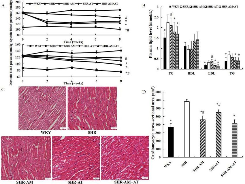
6                                     HUANG et al: CONNEXIN 43 PHOSPHORYLATION IN SH MODEL RATS

Figure 4. Comparison of T‑Cx43 and P‑Cx43 protein expression in the thoracic aorta using immunofluorescence double labeling. (A) Results of immunofluo‑
rescence double labeling analysis in thoracic aortic tissue. Green fluorescence, T‑Cx43; red fluorescence, P‑Cx43. Magnification, x400. Fluorescence intensity
for (B) T‑Cx43 and (C) P‑Cx43 of thoracic aorta. Data are expressed as the mean ± SD; n=8; *P
EXPERIMENTAL AND THERAPEUTIC MEDICINE 20: 80, 2020                                                     7

Ethics approval and consent to participate                                17. Martins‑Marques T, Catarino S, Zuzarte M, Marques C,
                                                                              Matafome P, Pereira P and Girão H: Ischaemia‑induced autophagy
                                                                              leads to degradation of gap junction protein connexin43 in
All procedures and ethics associated with animals were                        cardiomyocytes. Biochem J 467: 231‑245, 2015.
reviewed and approved by Medical School of Xi'an Jiaotong                 18. Xue J, Yan X, Yang Y, Chen M, Wu L, Gou Z, Sun Z,
                                                                              Talabieke S, Zheng Y and Luo D: Connexin 43 dephosphoryla‑
University (approval no. 2018‑898).                                           tion contributes to arrhythmias and cardiomyocyte apoptosis
                                                                              in ischemia/reperfusion hearts. Basic Res Cardiol 114: 40,
Patient consent for publication                                               2019.
                                                                          19. Hradec J, Zamorano J and Sutradhar S: Post hoc analysis of
                                                                              the cluster randomized usual care versus caduet investiga‑
Not applicable.                                                               tion assessing long‑term risk (CRUCIAL) trial. Curr Med Res
                                                                              Opin 29: 589‑596, 2013.
                                                                          20. Lu J, Liu F, Chen F, Jin Y, Chen H, Liu D and Cui W: Amlodipine
Competing interests                                                           and atorvastatin improve ventricular hypertrophy and diastolic
                                                                              function via inhibiting TNF‑α, IL‑1β and NF‑κ B inflammatory
The authors declared that they have no competing interests.                   cytokine networks in elderly spontaneously hypertensive rats.
                                                                              Biomed Pharmacother 83: 330‑339, 2016.
                                                                          21. Zhang ZY, Li Y, Li R, Zhang AA, Shang B, Yu J and Xie XD:
References                                                                    Tetrahydrobiopterin protects against radiation‑induced growth
                                                                              inhibition in H9c2 cardiomyocytes. Chin Med J (Engl) 129:
 1. Olsen MH, Angell SY, Asma S, Boutouyrie P, Burger D,                      2733‑2740, 2016.
    Chirinos JA, Damasceno A, Delles C, Gimenez‑Roqueplo AP,              22. Jin Y, Jing M, Zhang L, Song S and Ma X: Internet access and
    Hering D, et al: A call to action and a lifecourse strategy to            hypertension management among the elderly population: A
    address the global burden of raised blood pressure on current             nationally representative cross‑sectional survey in China. J Med
    and future generations: The lancet commission on hypertension.            Internet Res 31: e11280, 2019.
    Lancet 388: 2665‑2712, 2016.                                          23. Kearney PM, Whelton M, Reynolds K, Muntner P, Whelton PK
 2. Ettehad D, Emdin CA, Kiran A, Anderson SG, Callender T,                   and He J: Global burden of hypertension: Analysis of worldwide
    Emberson J, Chalmers J, Rodgers A and Rahimi K: Blood pres‑               data. Lancet 365: 217‑223, 2005.
    sure lowering for prevention of cardiovascular disease and death:     24. Delvaeye T, Vandenabeele P, Bultynck G, Leybaert L and
    A systematic review and meta‑analysis. Lancet 387: 957‑967, 2016.         Krysko DV: Therapeutic targeting of connexin channels: New
 3. Vriz O, Magne J, Jarosh J, Bossone E, Aboyans V and Palatini P:           views and challenges. Trends Mol Med 24: 1036‑1053, 2018.
    Local carotid arterial stiffness is an independent determinant        25. Nagasawa K, Takahashi K, Matsuura N, Takatsu M, Hattori T,
    of left ventricular remodeling in never‑treated hypertensive              Watanabe S, Harada E, Niinuma K, Murohara T and Nagata K:
    patients. Blood Press 28: 23‑33, 2019.                                    Comparative effects of valsartan combination with cilnidipine or
 4. Johnson RD and Camelliti P: Role of non‑myocyte gap junctions             amlodipine on cardiac remodeling and diastolic dysfunction in
    and connexin hemichannels in cardiovascular health and disease:           Dahl salt‑sensitive rats. Hypertens Res 38: 39‑47, 2015.
    Novel therapeutic targets? Int J Mol Sci 19: 866, 2018.               26. Chen Y, Chang Y, Zhang N, Guo X, Sun G and Sun Y:
 5. Desai CS, Ning H and Lloyd‑Jones DM: Competing cardio‑                    Atorvastatin attenuates myocardial hypertrophy in spontane‑
    vascular outcomes associated with electrocardiographic left               ously hypertensive rats via the C/EBPβ/PGC‑1α/UCP pathway.
    ventricular hypertrophy: The atherosclerosis risk in communities          Cell Physiol Biochem 46: 1009‑1018, 2018.
    study. Heart 98: 330‑334, 2012.                                       27. Naydenov Naydenov S, Margaritov Runev N, Ivanov Manov E
 6. Opie LH, Commerford PJ, Gersh BJ and Pfeffer MA: Controversies            and Georgieva Torbova‑Gigova S: Efficacy and safety of a
    in ventricular remodeling. Lancet 367: 356‑367, 2006.                     single‑pill combination of atorvastatin/amlodipine in patients
 7. Camargo LL, Harvey AP, Rios FJ, Tsiropoulou S, Da Silva RNO,              with arterial hypertension and dyslipidemia. Acta Clin Croat 57:
    Cao Z, Graham D, McMaster C, Burchmore RJ, Hartley RC, et al:             464‑472, 2018.
    Vascular Nox (NADPH oxidase) compartmentalization, protein            28. Song D, Liu X, Liu R, Yang L, Zuo J and Liu W: Connexin 43
    hyperoxidation, and endoplasmic reticulum stress response in              hemichannel regulates H9c2 cell proliferation by modulating
    hypertension. Hypertension 72: 235‑246, 2018.                             intracellular ATP and [Ca2+]. Acta Biochim Biophys Sin
 8. Schaffer AL, Buckley NA and Pearson SA: Who benefits from                 (Shanghai) 42: 472‑482, 2010.
    fixed‑dose combinations? Two‑year statin adherence trajecto‑          29. Seki A, Nishii K and Hagiwara N: Gap junctional regulation
    ries in initiators of combined amlodipine/atorvastatin therapy.           of pressure, fluid force, and electrical fields in the epigenetics
    Pharmacoepidemiol Drug Saf 26: 1465‑1473, 2017.                           of cardiac morphogenesis and remodeling. Life Sci 129: 27‑34,
 9. Chen HJ, Yao L, Chen TG, Yu M, Wang LH and Chen JZ:                       2015.
    Atorvastatin prevents connexin43 remodeling in hypertrophied          30. Zhai H, Dai W and Wang Y: Metoprolol protects cardiomyocytes
    left ventricular myocardium of spontaneously hypertensive rats.           in rabbit model of heart failure by regulating Cx43. Exp Ther
    Chin Med J (Engl) 120: 1902‑1907, 2007.                                   Med 15: 1902‑1905, 2018.
10. Waters D, Higginson L, Gladstone P, Kimball B, Le May M,              31. Nao T, Ohkusa T, Hisamatsu Y, Inoue N, Matsumoto T, Yamada J,
    Boccuzzi SJ and Lespérance J: Effects of monotherapy with                 Shimizu A, Yoshiga Y, Yamagata T, Kobayashi S, et al:
    an HMG‑CoA reductase inhibitor on the progression of                      Comparison of expression of connexin in right atrial myocar‑
    coronary atherosclerosis as assessed by serial quantitative arteri‑       dium in patients with chronic atrial fibrillation versus those in
    ography. The Canadian coronary atherosclerosis treatment trial.           sinus rhythm. Am J Cardiol 91: 678‑683, 2003.
    Circulation 3: 959‑968, 1994.                                         32. Xing D, Kjølbye AL, Nielsen MS, Petersen JS, Harlow KW,
11. Salameh A: Life cycle of connexins: Regulation of connexin                Holstein‑Rathlou NH and Martins JB: ZP123 increases gap
    synthesis and degradation. Adv Cardiol 42: 57‑70, 2006.                   junctional conductance and prevents reentrant ventricular
12. Söhl G and Willecke K: Gap junctions and the connexin protein             tachycardia during myocardial ischemia in open chest dogs.
    family. Cardiovasc Res 62: 228‑232, 2004.                                 J Cardiovasc Electrophysiol 14: 510‑520, 2003.
13. Rodriguez‑Sinovas A: Cx43 phosphorylation and cardioprotec‑           33. Wang LJ, Ma KT, Shi WY, Wang YZ, Zhao L, Chen XY, Li XZ,
    tion. Cardiovasc Res 83: 613‑614, 2009.                                   Jiang XW, Zhang ZS and Li L and Si JQ: Enhance gap junc‑
14. Egan Benova T, Szeiffova Bacova B, Viczenczova C, Diez E,                 tion channel activity between vascular smooth muscle cells in
    Barancik M and Tribulova N: Protection of cardiac cell‑to‑cell            cerebral artery of spontaneously hypertensive rats. Clin Exp
    coupling attenuate myocardial remodeling and proarrhythmia                Hypertens 39: 295‑305, 2017.
    induced by hypertension. Physiol Res 65 (Suppl 1): S29‑S42, 2016.
15. Cheng G, Chen CY, Shou XL and Han XY: Effects of captopril                               This work is licensed under a Creative Commons
    on the expression of connexin 43 in thoracic aorta from spon‑                            Attribution-NonCommercial-NoDerivatives 4.0
    taneous hypertensive rats. J Shaanxi Med 44: 1571‑1573, 2015.                            International (CC BY-NC-ND 4.0) License.
16. Martins‑Marques T, Catarino S, Marques C, Matafome P,
    Ribeiro‑Rodrigues T, Baptista R, Pereira P and Girão H: Heart
    ischemia results in connexin43 ubiquitination localized at the
    intercalated discs. Biochimie 112: 196‑201, 2015.
You can also read