Comparative transcriptome profiles of large and small bodied large scale loaches cultivated in paddy fields
←
→
Page content transcription
If your browser does not render page correctly, please read the page content below
www.nature.com/scientificreports
OPEN Comparative transcriptome
profiles of large and small bodied
large‑scale loaches cultivated
in paddy fields
Liulan Zhao1,4, Kuo He1,4, Qing Xiao1,4, Qiao Liu1, Wei Luo1, Jie Luo1, Hongmei Fu1, Jiayao Li2,
Xugan Wu2, Jun Du3, Quan Gong3, Xun Wang1* & Song Yang1*
Fish culture in paddy fields is a traditional aquaculture mode, which has a long history in East Asia.
Large-scale loach (Paramisgurnus dabryanus) fast growth is suitable for paddy fields aquaculture
in China. The objective of this study was to identify differential expression genes (DEGs) in the
brain, liver and muscle tissues between large (LG, top 5% of maximum total length) and small
(SG, top 5% of minimum total length) groups using RNA-seq. In total, 150 fish were collected each
week and 450 fish were collected at twelfth week from three paddy fields for all the experimental.
Histological observation found that the muscle fibre diameter of LG loaches was greater than that of
SG loaches. Transcriptome results revealed that the high expression genes (HEGs) in LG loaches (fold
change ≥ 2, p < 0.05) were mainly concentrated in metabolic pathways, such as “Thyroid hormone
signalling pathway”, “Citrate cycle (TCA cycle)”, “Carbon metabolism”, “Fatty acid metabolism”,
and “Cholesterol metabolism”, and the HEGs in SG loaches were enriched in the pathways related
to environmental information processing such as “Cell adhesion molecules (CAMs)”, “ECM− receptor
interaction” and “Rap1 signalling pathway”; cellular processes such as “Tight junction”, “Focal
adhesion”, “Phagosome” and “Adherens junction”. Furthermore, IGFs gene family may play an
important role in loach growth for their different expression pattern between the two groups. These
findings can enhance our understanding about the molecular mechanism of different growth and
development levels of loaches in paddy fields.
Fish culture in paddy fields is an ancient and widely practiced farming model in Southeast Asia for its great
advantages over traditional fish farming, such as resource conservation, environmental friendliness and economic
benefits1,2. In most southern China areas, there are sufficient water in the paddy fields from April to October,
and the water temperature during this season is suitable for the rapid growth of fish3. The appropriately modified
paddy fields are suitable for the cultivation of some omnivorous fish such as loach and carp with large popularity.
The large-scale loach (Paramisgurnus dabryanus) is one of the fastest growing fish species that has been widely
cultivated in Southeast A sia4. The large-scale loach is a omnivorous species living in mild water environment,
distributed in the middle and lower reaches of the Yangtze River system5. It is known among consumers for its
richness in vitamins, minerals, essential amino acids and other trace elements meanwhile low in cholesterol6.
The large-scale loach had been widely raised cross China as one of the most important species in paddy field
aquaculture7. However, compared with intensive farming, the fish raised in paddy field farming has some dis-
advantages in terms of poor body uniformity, which will affect the market of farmed fish.
Body size is a prominent and important feature of fish in aquaculture, which is driven by muscle differentia-
tion and growth, and it is also one of the most important factors affecting p roduction8. The growth and devel-
opment rate of fish varies from species to individual, and is greatly affected by environmental factors such as
temperature and diet9. As we all know, progeny often inherit the epigenetic traits of their parents including body
size and growth rate. In addition to being affected by the environmental factors, these genetic traits will also be
regulated by gene f unction10. Several candidate genes that could be critical for fish growth and development have
1
College of Animal Science and Technology, Sichuan Agricultural University, Chengdu 611130, China. 2Engineering
Research Center of Aquaculture, Shanghai Ocean University, Shanghai 200090, China. 3Fisheries Institute, Sichuan
Academy of Agricultural Science, Chengdu 611731, China. 4These authors contributed equally: Liulan Zhao, Kuo
He and Qing Xiao. *email: xunwang@sicau.edu.cn; ysys210@hotmail.com
Scientific Reports | (2021) 11:4936 | https://doi.org/10.1038/s41598-021-84519-9 1
Vol.:(0123456789)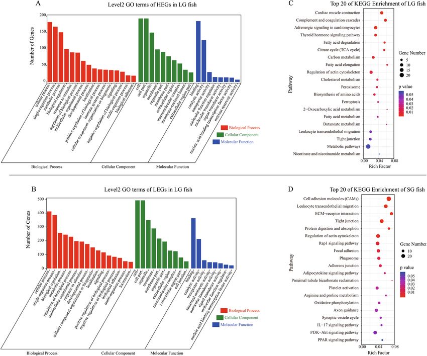
www.nature.com/scientificreports/
Figure 1. The weekly morphological characteristics (total length) of large-scale loach from large group (LG)
and small group (SG). (A) The weekly total length of LG and SG loach (n = 150 fish individuals). (B) The normal
distribution of loach total length at 12th week (n = 450 fish individuals). Vertical bars represent mean ± SD; *
show the significant difference in total length between LG and SG; *p < 0.05, **p < 0.01.
been identified from previous studies, such as neuropeptide Y (NPY) and Insulin-like growth factors (IGFs),
etc.11,12. As the neural body center, the brain plays an important role in regulating various life activities. It can
secrete a variety of growth factors (such as GH) to promote the growth of the tissues13, or regulate the secretion
of growth factors from other tissues (such as liver)14. The interaction of these hormones is critical to the regula-
tion of muscle growth, which directly determine the body size of the fish.
In this study, RNA-Seq was used to identify differentially expressed genes of three tissues (brain, liver, and
muscle) between large (LG, top 5% of maximum total length) and small (SG, top 5% of minimum total length)
large-scale loaches cultivated in paddy fields with the same genetic background. The purpose of this research is
to improve our understanding the biological mechanism behind the loach growth as well as providing basis for
future breeding at a molecular level.
Results
Morphological characteristics and histological observation. Weekly morphological characteristics
(total length) of LG fish were significantly higher than those of SG fish (p < 0.05), and the gap between two
groups was getting more enlarge during the 12 weeks (Fig. 1A). Specifically, the total length of LG fish reached
twice that of SG fish in the twelfth week (p < 0.05). The normal distribution of total length of large-scale loaches
at week 12 was shown in Fig. 1B, and the total length was mainly concentrated from 60 to 80 cm.
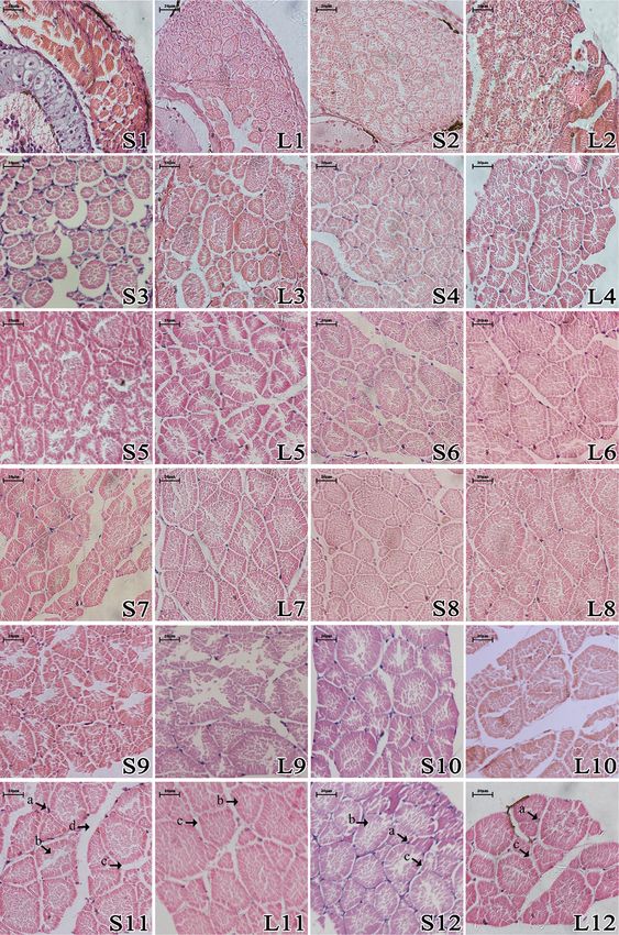
The comparisons of muscle histology between the LG and SG fish were presented in Fig. 2, in which we could
find that the muscle fibers diameter and density were different between the two groups. Then, the muscle fibre
diameter and density of two groups were measured and the statistical results were demonstrated in Fig. 3. The
muscle fibers diameter was greater in the LG fish, while the density was lower in the LG (Fig. 3).
RNA‑seq data and annotation of unigenes. To identify the differentially expressed genes associated
with differential body size in large-scale loach, a total of 18 cDNA libraries were constructed from LG and SG and
generated about 123.63 Gb sequencing data in total. After trimming and quality control, we obtained an average
of 45.79 M clean reads from the 18 cDNA libraries, and all clean Q30 base rates were over 94% (Table S1). After
assembly and remove redundancy, there were 28,620 (22.52%) unigenes were over 1000 bp in length among
127,062 unigenes (mean length: 820 bp, N50: 1546 bp, percent GC: 40.90%). More detailed information of the
filtered reads was presented in Table S2. Finally, 55,933 unigenes (44.02%) were functionally aligned by BLASTx
against databases totally, including GO, KEGG, NCBI-nr, and UniProt.
GO and KEGG enrichment analysis of the DEGs. After DEGs analysis, 189, 175 and 320 high expres-
sion genes (HEGs) were identified in the LG fish brain, liver and muscle, and 241, 481 and 668 HEGs were
identified in the SG fish brain, liver and muscle, respectively (Fig. 4A–C). Excluded the redundancy DEGs in two
or three tissues, a total of 1861 DEGs were identified among the three samples (Supplementary excel). For the
next GO and KEGG analysis, the DEGs were divided into two parts, which were composed of 684 HEGs in LG
fish and 1631 HEGs in SG fish. According to Go Term and previous studies, we finally selected growth-related
genes for heatmap analysis, which showed the IGFs gene family may play an important role in loach growth as
their expression levels were different between the two groups (Fig. 4D). These growth-related genes were fibro-
blast growth factor (FGF1, 6, 7 and 8), fibroblast growth factor binding protein 1 (FGFBP1), myogenic factor
5 (MYF5), myocyte enhancer factor 2 (MEF2), myogenic differentiation (MYOD), insulin like growth factor
(IGF1 and 2), insulin like growth factor binding protein (IGFBP1, 2, 3 and 7, IGF2BP1).
Scientific Reports | (2021) 11:4936 | https://doi.org/10.1038/s41598-021-84519-9 2
Vol:.(1234567890)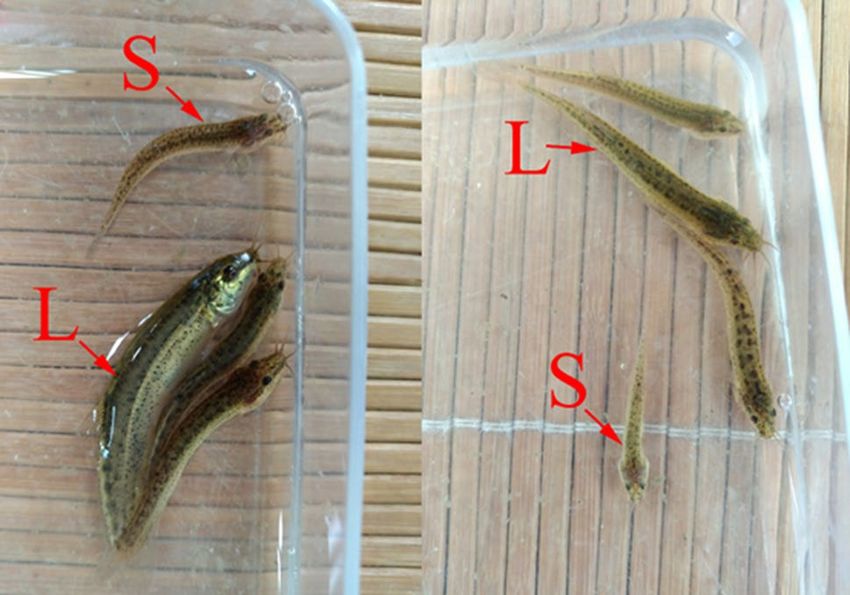
www.nature.com/scientificreports/

                                  Figure 2.  The weekly comparison of muscle histology between LG and SG loach. Note L large group, S small
                                  group; the number represents weeks. (a) Cell nucleus; (b) muscle fiber; (c) endomysium; (d) perimysium.

Scientific Reports |   (2021) 11:4936 |               https://doi.org/10.1038/s41598-021-84519-9                                              3

                                                                                                                                        Vol.:(0123456789)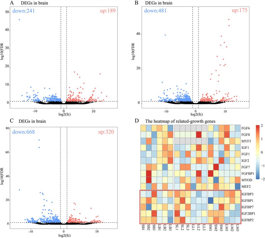
www.nature.com/scientificreports/
Figure 3. The weekly comparison of muscle fiber diameter (A) and density (B) between LG and SG loach. Note
The different lowercase letters above the bar represent the comparison within the SG loach and the different
uppercase letters represent the comparison within the LG loach. Vertical bars represent mean ± SD (n = 6 fish
individuals); * show the significant difference in total length between LG and SG; *p < 0.05, **p < 0.01.
GO enrichment analysis was performed to investigate the putative roles of these DEGs, which were classified
into biological process (BPs), cellular component (CCs) and molecular function (MFs) categories (Fig. 5A,C).
For the BPs, the major categories were cellular processes, single-organism process, metabolic process, regulation
of biological processes, and cellular responses to stimulus. The major categories of the CCs were cell, cell parts,
membrane parts, organelle parts and extracellular parts. The DEGs involved in binding and catalytic activity
were also most represented among the MFs.
In order to identify the functional biochemical pathways of predicted proteins encoded by DEGs, KEGG
pathway enrichment analysis was performed for both the HEGs in two groups (three tissues together). The top
20 of KEGG pathway enrichment analysis of two groups HEGs were showed in Fig. 5B,D, respectively. KEGG
pathway enrichment analysis of HEGs in LG fish (p < 0.05) showed a significant enrichment of metabolic path-
ways, such as “Thyroid hormone signalling pathway”, “Citrate cycle (TCA cycle)”, “Carbon metabolism”, “Fatty
acid metabolism”, and “Cholesterol metabolism”. The HEGs in SG fish were enriched in the pathways related to
environmental information processing such as “Cell adhesion molecules (CAMs)”, “ECM–receptor interaction”
and “Rap1 signalling pathway”; cellular processes such as “Tight junction”, “Focal adhesion”, “Phagosome” and
“Adherens junction”.
Validation of RNA‑Seq analysis. To confirm the RNA-seq data, four genes of each tissue were selected
for qRT-PCR validation. These genes included IGFBP1 (insulin-like growth factor binding protein-1), ID1
(inhibitor of DNA binding 1), MDKa (midkine a), and PPA1b (pyrophosphatase 1b). To adjust for variations in
starting template, gene expression was be normalized against β-actin for each tissue, then target genes mRNA
were quantified using the 2−ΔΔCT method, and the level of significance was determined by one-way analysis of
variance (ANOVA) with SPSS Statistics 22.0. While the relative expression levels were not perfectly consistent,
the qRT-PCR can provide additional basis for the RNA-seq results (Fig. S1).
Discussion
Intra-specific differences were first proposed by Darwin, who argued that individual differences and overpro-
duction within a species or variety were the basis of natural selection15. The individual growth varies a lot in
different species. The coefficient of individual growth variation in most mammals is 7%-10%, while in most fish
is approximate 20%-35%16. Fish body size differences are mainly formed by differences in trunk skeletal muscle
growth, which depends on the proliferation and hypertrophy of muscle fibers (muscle cells)17. During the growth
process, mammalian muscle growth mainly depends on the increase of muscle fibers diameter, but less on the
increase of muscle fibers q uantity18,19. Unlike mammals and birds, fish muscle fibers maintain the ability to pro-
liferate and hypertrophy for life20. For example, in larval sea bass (Dicentrarchus labrax), white muscle growth
is mainly through differentiation to form new muscle fibers, while in larval guppy (Poecilia reticulata), white
muscle growth is mainly achieved by increasing the size of muscle fi bers21,22. Considering the development in
other fish species, we hypothesize that loach muscles grow predominantly by means of increasing the diameter
of muscle fibers in this study (Figs. 2 and 3).
The results of transcriptome sequencing revealed that the metabolism of LG loaches was more vigorous com-
pared with that of SG loaches. The HEGs of the large loach in this study were significantly enriched in “Thyroid
hormone signalling pathway”, “Citrate cycle (TCA cycle)”, “Carbon metabolism” and “Fatty acid metabolism”,
and “Cholesterol metabolism”, which are all important regulators of growth, development and metabolism23–25.
Scientific Reports | (2021) 11:4936 | https://doi.org/10.1038/s41598-021-84519-9 4
Vol:.(1234567890)www.nature.com/scientificreports/
Figure 4. The high expressed genes (HEGs) of LG or SG loach. (A–C) the volcano map of HEGs in the brain,
liver and muscle, respectively. (D) the heatmap of related-growth genes between LG and SG loach. LB, LL, LM:
the brain, liver, and muscle of large group, SB, SL, SM: the brain, liver, and muscle of small group. FDR false
discovery rate; fc fold change.
It was also found in rainbow trout that the expression of lipid-metabolism-related genes was up-regulated in
fast growth fish compared with slow growth fish, which was consistent with our findings26. In addition, the
genes related to the growth were mainly involved in energy metabolism, carbohydrate and lipid metabolism,
and cytoskeletal composition, which the similar expression pattern were also observed in the study of salmonid
liver and muscle gene e xpression27–29. Generally, insulin-like growth factors (IGFs) were generally thought to
be up-regulated in tissues of rapidly growing individuals, such as Nile tilapia (Oreochromis Niloticus)30, channel
catfish (Ictalurus punctatus)31, and Arctic charr (Salvelinus alpinus)32. It was found in this study that the gene IGF1
and IGF2 were different in two groups, but the difference was not significant (FDR > 0.05), which was similar
to previous studies11,33,34. However, IGFBP1 (insulin-like growth factor binding protein-1) of brain and liver
and IGFBP7 of liver expressed in SG group were both higher than that in LG loaches in this study (FDR < 0.05;
Fig. 4D). In a recent study of rainbow trout, it was also found that small fish had higher expression of IGFBP1 in
sh26. Previous studies demonstrated that IGFBP1 could inhibit IGF binding to cell surface
the liver than in large fi
receptors and thereby inhibit IGF-mediated mitogenic and cell metabolic actions35. Likewise, overexpression of
IGFBP1a/b in zebrafish would retard embryonic development and g rowth36,37. Therefore, in addition to having
stronger metabolic activity, one of the internal factors for the fast growth of large fish may be related to the lower
expression of IGFBP1 and IGFBP7.
The binding protein of IGFs genes (IGFBPs) are known to inhibit cell growth and differentiation by binding
specifically IGFs38,39, which can be influenced by many factors. Previous studies have shown that fasting increases
hepatic IGFBPs levels, and IGFBPs drops back to normal after refeeding11,40. Whether in large-scale farming
or in the natural environment, the smaller individuals have lower social status, as well as mating and feeding
rights41,42. We could speculate that it might be more difficult for small individual loach to obtain food in paddy
fields culture environment, which led to its more delayed growth and development. Furthermore, the HEGs in
SG fish were enriched in the pathways related to environmental information processing such as cell adhesion
molecules (CAMs), ECM–receptor interaction and Rap1 signalling pathway; cellular processes such as tight
Scientific Reports | (2021) 11:4936 | https://doi.org/10.1038/s41598-021-84519-9 5
Vol.:(0123456789)www.nature.com/scientificreports/
Figure 5. The GO annotation and KEGG pathways enrichment with HEGs of LG or SG loach. Note The HEGs
in both groups include all the HEGs in the three tissues (brain, liver, and muscle).
junction, focal adhesion, phagosome and adherens junction. These functional pathways consist of a complex
mixture of structural and functional macromolecules and have their important roles in tissue and organ morpho-
genesis in maintaining cell and tissue structure and functions43–46. Thus, our results provide additional evidence
there was a lag in the development of the small fish compared to the large fish in the paddy cultivation system.
Conclusion
In this study, RNA-seq successfully identified that the differences in transcription levels of the loaches with
differential body sizes in integrated paddy field aquaculture. Compared with the slow-growing loach, the fast-
growing loach haver higher expression of metabolic genes. Furthermore, the transcription level of IGFBPs was
relatively low in the fast-growing loach, which were known to inhibit cell growth and differentiation. In addition,
large-scale loaches growth may be through the enlargement of muscle fibers.
Materials and methods
Ethics statement. All experiments were approved by the Institutional Animal Care and Use Committee
(IACUC) of the College of Animal Science and Technology of Sichuan Agricultural University, Sichuan, China,
under permit No. DKY-S20166409. All experimental methods were performed according to relevant guidelines
and regulations and the study was carried out in compliance with the ARRIVE guidelines.
Fish culture in paddy fields. The three paddy fields used in this experiment were performed disinfection
operation to kill wild aquatic animals with quicklime before the experiment. In addition, the border of the paddy
fields surrounded with a net were used to block other aquatic animals from entering the pond, such as frogs,
ensured that there were only experimental fish in the paddy fields.
Scientific Reports | (2021) 11:4936 | https://doi.org/10.1038/s41598-021-84519-9 6
Vol:.(1234567890)www.nature.com/scientificreports/
Figure 6. The selection of large-scale loach for LG and SG. Note L selected in LG; S selected in SG.
All experimental fish with the same genetic background came from a professional aquaculture farm of Neiji-
ang, China (N: 104°56′27.16″, E: 29°27′32.52″). The experimental fish were cultivated in three paddy fields with
an approximate area of 866.67 m2, when their total length was nearly 4 cm in an indoor pond. The experiment was
performed from July to September, and the water quality parameters were as follows: temperature was 12–24 °C;
dissolved oxygen was 5.3–6.7 mg/ml; pH was 7.5–8.1. Culture density was approximately twenty thousand fish
per paddy field. The fish were fed twice a day at 09:00 and 17:00 (2 ~ 4% body weight feeding rate), and were
subjected to the same daily management.
Partition of large and small size fish. For weekly samples, the total length of fish was measured from 50
random individuals in each paddy field, and normal distributions were made based on the total length of 150
individuals. Then, top 5% of maximum total length was defined as the large group (LG); similarly, top 5% of min-
imum total length was defined as the small group (SG). At the twelfth week, 150 individuals were measured for
the total length from each paddy field and used to divide LG and SG fish (LG and SG fish were showed in Fig. 6).
Sample collection. Based on the definition of LG and SG fish, 3 large fish and 3 small fish were sampled
weekly and fixed in fresh Bouin’s solution after being anaesthetized with MS-222 (Tricaine methane sulfonate,
100 ppm) for histological observation. For the first four weeks, the entire body of the fish was preserved from
dorsal fin to caudal fin, while a piece of muscle (0.5 cm × 0.5 cm × 0.5 cm) was obtained from fish during subse-
quent weeks.
At the twelfth week, 3 large fish and 3 small fish from each paddy field were rapidly sampled brain (B), liver (L)
and muscle (M) for RNA-Seq, after being anaesthetized using MS-222 (100 ppm). The samples were immediately
frozen in liquid nitrogen, and then stored in -80 °C freezer for subsequent analysis.
Histological characteristics and observation. Fixed samples were wrapped in gauze and dehydrated
in an ethanol series, then infiltrated in xylene, and embedded in paraffin wax finally. Tissues each were cut
into 6 μm sections serially using a rotary microtome according to routine procedures. Muscle sections were
stained with haematoxylin and eosin (HE) for histological analysis and examined on microscope slides. Digital
images were captured using a Nikon Eclipse Ti-S (Nikon Instruments Inc, Japan) and measured with Image Pro
Plus software (Media Cybernetics, USA). The muscle fiber density was the number of muscle fibers per square
millimetre. In detail, the muscle fibers numbers were counted within a 100 μm × 100 μm square under micro-
scope after HE staining (200 × digital images). Only muscle fibers more than half its size within the square were
counted. The muscle fiber diameter was the geometric mean of the √ long and short diameter of the 100 muscle
fibers per fish (400 × digital images; geometric mean formula: G = ab ; G: geometric mean; a: long diameter; b:
short diameter).
Total RNA extraction and cDNA library construction. After three large fish or three small fish from
each paddy filed were pooled into one sequencing sample, three LG samples and three SG samples were used
sequencing, respectively. Three tissues (B: brain, L: liver and M: muscle) were used to sequence in each sam-
ple groups. Total RNA was extracted using the total RNA extraction reagent RNAiso Plus (Takara Bio Inc,
Japan) according to the manufacturer’s protocol. After confirming the quality of RNA with agarose gel electro-
phoresis and Nanodrop 2000 (Thermo Scientific, USA), RNA that integrity number (RIN) greater than 8 and
OD260/280 > 1.80 was used for mRNA library construction47. The qualified RNA was sent to the Annoroad
Gene Technology Corporation (Beijing, China) for library preparation and sequenced using Illumina HiSeq
2000 by sequencing strategy: paired-end sequencing and raw reads length 150 bp.
Scientific Reports | (2021) 11:4936 | https://doi.org/10.1038/s41598-021-84519-9 7
Vol.:(0123456789)www.nature.com/scientificreports/
Sequencing data analysis. Low-quality, adaptor-polluted and high content of unknown bases (N)-con-
taining sequencing reads were filtered (the adaptor sequences; unknown bases more than 10%; the percentage
of no more than Q 5 bases is over 50% in a read). The Q30 of the clean data was calculated, and all downstream
analyses were performed using the clean, high-quality data. Trinity (http://trinityrnaseq.sourceforge.net/, ver-
sion: v2.0.6)48 was used to perform de novo assembly with clean reads, and Tgicl (http://trinityrnaseq.sourceforg
e.net/, version: v2.0.6)49 then used to cluster transcripts to unigenes.
After assembly, unigenes were used for functional annotation against the NT (Non-redundant protein
sequences Database), NR (Nucleotide Sequence Database), Uniprot (Universal Protein), COG (Cluster of
Orthologous Groups of proteins), GO (Gene Ontology) and KEGG (Kyoto Encyclopedia of Genes and Genomes)
databases. Differential expression analysis was performed using edgeR50 in the OmicShare tools, an online plat-
form for data analysis (www.omicshare.com/tools). The default parameters of edgeR were used, and differential
expression genes (DEGs) were selected according to log2 (fold change) ≥ 1 and p value < 0.05. The GO and KEGG
pathway analyses were then carried out with the differentially expressed genes of all the three t issues50.
Validation of RNA‑seq analysis by real‑time PCR. Four related genes were randomly selected for
quantitative real-time PCR (qRT—PCR) to identify the accuracy of RNA-seq results. 3 fish (three tissues: brain,
liver, and muscle) were randomly selected from large and small group fish, respectively. β-actin was used as
the reference gene. Total RNA was used to synthesize mRNA cDNA by using TianGen FastKing RT Kit (With
gDNase). The emission intensity was detected by Step One real-time PCR system (Applied Biosystems) under
the following steps: initial denaturation step at 95 °C for 20 s, 40 thermal cycling steps consisted of 3 s at 95 °C,
30 s at 60 °C. The target gene qRT-PCR primers were designed with reference to the sequences data of this
study by Primer 5.0 (The reference gene primers are shown in Table S3). All reactions were run in triplicate
and included no template controls for each gene and the quantitative results were quantified using the 2−ΔΔCT
method51.
Statistical analysis. Statistical analysis was performed using SPSS 22.0 software (SPSS, Chicago, IL, USA).
Data were presented as the mean ± SEM, and significant differences (p < 0.05) were identified using one-way
analysis of variance (ANOVA)52.
Data availability
Data has been deposited in the SRA under the study accession code PRJNA623189.
Received: 18 February 2020; Accepted: 3 February 2021
References
1. Liu, J. & Cai, Q. Integrated aquaculture in Chinese lakes and paddy fields. Ecol. Eng. 11(1–4), 49–59 (1998).
2. Lu, S. G. & Huang, C. P. Economic and ecological effects of fish culture in paddy fields. Chin. J. Ecol. 10, 26 (1988).
3. Weimin, M. Recent developments in rice-fish culture in China: A holistic approach for livelihood improvement in rural areas. In
Success stories in Asian aquaculture 15–40 (Springer, Berlin, 2010).
4. Li, C. et al. Transcriptome characterization and SSR discovery in large-scale loach Paramisgurnus dabryanus (Cobitidae, Cyprini-
formes). Gene 557(2), 201–208 (2015).
5. You, C., Yu, X. & Tong, J. Detection of hybridization between two loach species (Paramisgurnus dabryanus and Misgurnus anguil-
licaudatus) in wild populations. Environ. Biol. Fishes 86(1), 65 (2009).
6. Gao, Y. et al. Effects of fulvic acid on growth performance and intestinal health of juvenile loach Paramisgurnus dabryanus (Sau-
vage). Fish Shellfish Immunol. 62, 47–56 (2017).
7. You, C.-H., Tong, J.-G. & Yu, X.-M. Microsatellite DNA analysis on genetic diversity of seven populations of Paramisgurnus dab-
ryanus. J. Hydroecol. 33(1), 84–91 (2012).
8. Talesara, C. & Urfi, A. A histophysiological study of muscle differentiation and growth in the common carp Cyprinus carpio Var.
communis. J. Fish Biol. 31(1), 45–54 (1987).
9. Stoiber, W. et al. Cellularity changes in developing red and white fish muscle at different temperatures: Simulating natural envi-
ronmental conditions for a temperate freshwater cyprinid. J. Exp. Biol. 205(16), 2349–2364 (2002).
10. Brogiolo, W. et al. An evolutionarily conserved function of the Drosophila insulin receptor and insulin-like peptides in growth
control. Curr. Biol. 11(4), 213–221 (2001).
11. Amaral, I. P. & Johnston, I. A. Experimental selection for body size at age modifies early life-history traits and muscle gene expres-
sion in adult zebrafish. J. Exp. Biol. 215(22), 3895–3904 (2012).
12. Van der Merwe, M., Franchini, P. & Roodt-Wilding, R. Differential growth-related gene expression in abalone (Haliotis midae).
Mar. Biotechnol. 13(6), 1125–1139 (2011).
13. Devlin, R. H., Sundström, L. F. & Muir, W. M. Interface of biotechnology and ecology for environmental risk assessments of
transgenic fish. Trends Biotechnol. 24(2), 89–97 (2006).
14. Seiliez, I., Sabin, N. & Gabillard, J.-C. FoxO1 is not a key transcription factor in the regulation of myostatin (mstn-1a and mstn-1b)
gene expression in trout myotubes. Am. J. Physiol. 301(1), R97–R104 (2011).
15. Darwin, C. On the origin of species by means of natural selection. Am. Midland Nat. 49(3), 395–405 (1859).
16. Gjedrem, T. Contribution from selective breeding to future aquaculture development. J. World Aquacult. Soc. 3, 33–45 (1997).
17. Stoiber, W. et al. Tonic fibres in axial muscle of cyprinid fish larvae: their definition, possible origins and functional importance.
Anat. Embryol. 205(2), 113–124 (2002).
18. Picard, B., Lefaucheur, L., Berri, C. C. & Duclos, M. J. Muscle fibre ontogenesis in farm animal species. Reprod. Nutr. Dev. 42(5),
415–431 (2002).
19. Braun, T. & Gautel, M. Transcriptional mechanisms regulating skeletal muscle differentiation, growth and homeostasis. Nat. Rev.
Mol. Cell Biol. 12(6), 349 (2011).
20. Alami-Durante, H. et al. Growth and multiplication of white skeletal muscle fibres in carp larvae in relation to somatic growth
rate. J. Fish Biol. 50(6), 1285–1302 (1997).
Scientific Reports | (2021) 11:4936 | https://doi.org/10.1038/s41598-021-84519-9 8
Vol:.(1234567890)www.nature.com/scientificreports/
21. Veggetti, A., Mascarello, F., Scapolo, P. A. & Rowlerson, A. Hyperplastic and hypertrophic growth of lateral muscle in Dicentrarchus
labrax (L.). Anat. Embryol. 182(1), 1–10 (1990).
22. Veggetti, A. et al. Muscle growth and myosin isoform transitions during development of a small teleost fish, Poecilia reticulata
(Peters) (Atheriniformes, Poeciliidae): A histochemical, immunohistochemical, ultrastructural and morphometric study. Anat.
Embryol. 187(4), 353–361 (1993).
23. Zhang, Y. F. et al. Tetrabromobisphenol A disrupts vertebrate development via thyroid hormone signaling pathway in a develop-
mental stage-dependent manner. Environ. Ence Technol. 48(14), 8227–8234 (2014).
24. Kelley, D. E., Goodpaster, B., Wing, R. R. & Simoneau, J. A. Skeletal muscle fatty acid metabolism in association with insulin
resistance, obesity, and weight loss. Am. J. Physiol. 277(6 Pt 1), E1130–E1141 (1999).
25. Dietschy, J. M. & Turley, S. D. Thematic review series: brain Lipids. Cholesterol metabolism in the central nervous system during
early development and in the mature animal. J. Lipid Res. 45(8), 1375–1397 (2004).
26. Kocmarek, A. L., Ferguson, M. M. & Danzmann, R. G. Differential gene expression in small and large rainbow trout derived from
two seasonal spawning groups. BMC Genom. 15(1), 57 (2014).
27. Devlin, R. H., Sakhrani, D., Tymchuk, W. E., Rise, M. L. & Goh, B. Domestication and growth hormone transgenesis cause similar
changes in gene expression in coho salmon (Oncorhynchus kisutch). Proc. Natl. Acad. Sci. USA 106(9), 3047–3052 (2009).
28. Rise, M. L. et al. Multiple microarray platforms utilized for hepatic gene expression profiling of GH transgenic coho salmon with
and without ration restriction. J. Mol. Endocrinol. 37(2), 259–282 (2006).
29. White, S. L., Sakhrani, D., Danzmann, R. G. & Devlin, R. H. Influence of developmental stage and genotype on liver mRNA levels
among wild, domesticated, and hybrid rainbow trout (Oncorhynchus mykiss). Bmc Genom. 14, 673 (2013).
30. Huang, C. W. et al. Differential expression patterns of growth-related microRNAs in the skeletal muscle of Nile tilapia. J. Anim.
Sci. 90(12), 4266–4279 (2012).
31. Small, B. C., Davis, K. B. & Peterson, B. C. Elucidating the effects of cortisol and stress on economically important traits in channel
catfish. North Am. J. Aquacult. 70(2), 223–235 (2008).
32. Macqueen, D. J. et al. The parallel evolution of dwarfism in Arctic charr is accompanied by adaptive divergence in mTOR-pathway
gene expression. Mol. Ecol. 20(15), 3167–3184 (2011).
33. Amaral, I. P. G. & Johnston, I. A. Insulin-like growth factor (IGF) signalling and genome-wide transcriptional regulation in fast
muscle of zebrafish following a single-satiating meal. J. Exp. Biol. 214(13), 2125–2139 (2011).
34. Bower, N. I. & Johnston, I. A. Discovery and characterization of nutritionally regulated genes associated with muscle growth in
Atlantic salmon. Physiol. Genom. 42A(2), 114–130 (2010).
35. Lee, P. D., Conover, C. A. & Powell, D. R. Regulation and function of insulin-like growth factor-binding protein-1. Proc. Soc. Exp.
Biol. Med. 204(1), 4–29 (1993).
36. Kajimura, S., Aida, K. & Duan, C. Insulin-like growth factor-binding protein-1 (IGFBP-1) mediates hypoxia-induced embryonic
growth and developmental retardation. Proc. Natl. Acad. Sci. USA 102(4), 1240–1245 (2005).
37. Kamei, H. et al. Duplication and diversification of the hypoxia-inducible IGFBP-1 gene in zebrafish. PLoS ONE 3, e3091 (2008).
38. Murphy, N. et al. Insulin-like growth factor-1, insulin-like growth factor-binding protein-3, and breast cancer risk: Observational
and Mendelian randomization analyses with 430 000 women. Ann. Oncol. 31(5), 641–649 (2020).
39. Lu, Z. et al. Reprogramming of human fibroblasts into osteoblasts by insulin-like growth factor-binding protein 7. Stem Cells
Transl. Med. 9(3), 403–415 (2020).
40. Murphy, L. J., Seneviratne, C., Moreira, P. & Reid, R. E. Enhanced expression of insulin-like growth factor binding protein4 in the
fasted rat: The effects of insulin and growth hormone administration. Endocrinology 128, 689–696 (1991).
41. Leips, J., Helen Rodd, F. & Travis, J. The adaptive significance of population differentiation in offspring size of the least killifish,
Heterandria formosa. Ecol. Evol. 3(4), 948–960 (2013).
42. Pottinger, T. G. & Carrick, T. R. Stress responsiveness affects dominant-subordinate relationships in rainbow trout. Horm. Behav.
40(3), 419–427 (2001).
43. Petra, B. et al. Identification of central regulators of calcium signaling and ECM–receptor interaction genetically associated with
the progression and recurrence of atrial fibrillation. Front. Genet. 9, 162 (2018).
44. Erbaba, B. et al. Zebrafish brain RNA sequencing reveals that cell adhesion molecules are critical in brain aging. Neurobiol. Aging
94, 164–175 (2020).
45. Ramos, C. J., Lin, C., Liu, X. & Antonetti, D. A. The EPAC-Rap1 pathway prevents and reverses cytokine-induced retinal vascular
permeability. J. Biol. Chem. 293(2), 717–730 (2018).
46. Petit, V. & Thiery, J. P. Focal adhesions: Structure and dynamics. Biol. Cell 92(7), 477–494 (2000).
47. Sun, J. L. et al. Analysis of miRNA-seq in the liver of common carp (Cyprinus carpio L.) in response to different environmental
temperatures. Funct. Integr. Genom. 19(2), 265–280 (2019).
48. Grabherr, M. G. et al. Full-length transcriptome assembly from RNA-Seq data without a reference genome. Nat. Biotechnol. 29(7),
644 (2011).
49. Pertea, G. et al. TIGR Gene Indices clustering tools (TGICL): A software system for fast clustering of large EST datasets. Bioinfor-
matics 19(5), 651–652 (2003).
50. Robinson, M. D., McCarthy, D. J. & Smyth, G. K. edgeR: A Bioconductor package for differential expression analysis of digital
gene expression data. Bioinformatics 26(1), 139–140 (2010).
51. Livak, K. J. & Schmittgen, T. D. Analysis of relative gene expression data using real-time quantitative PCR and the 2 −ΔΔCT Method.
Methods 25(4), 402–408 (2001).
52. Yang, S. et al. Mixed diets reduce the oxidative stress of common carp (Cyprinus carpio): Based on microRNA sequencing. Front.
Physiol. 10, 631 (2019).
Acknowledgements
The authors thank Dr. Li Mingzhou and Dr. Ma Jideng for their valuable input into the project design, and the
authors are grateful to Feng Siyuan for assistance in data analysis of the RNA-seq results. In addition, the authors
thank Mr. Liang from the professional aquaculture farms of Neijiang, China for providing the experimental site
and experimental fish.
Author contributions
S.Y. managed the grants, supervised the laboratory work, and led the design and coordination of this study.
L.L.Z., K.H. and W.X. designed the experiments. Q.X., W.L., H.M.F., J.D., J.L. (Jie Luo) and Q.L. performed
the fish cultivation and sample collection. Q.X., H.M.F. and Q.G. performed the experiments. Q.X. wrote the
manuscript draft. K.H., J.Y.L. (Jiayao Li) and X.G.W. edited and revised the manuscript. All authors read and
approved the final manuscript.
Scientific Reports | (2021) 11:4936 | https://doi.org/10.1038/s41598-021-84519-9 9
Vol.:(0123456789)www.nature.com/scientificreports/
Funding
This research was supported by National Key Research and Development Project (2019YFD0900302) and
the Science and Technology Research Program of Chongqing Municipal Education Commission (Grant No.
KJZD-K201803501).
Competing interests
The authors declare no competing interests.
Additional information
Supplementary Information The online version contains supplementary material available at https://doi.
org/10.1038/s41598-021-84519-9.
Correspondence and requests for materials should be addressed to X.W. or S.Y.
Reprints and permissions information is available at www.nature.com/reprints.
Publisher’s note Springer Nature remains neutral with regard to jurisdictional claims in published maps and
institutional affiliations.
Open Access This article is licensed under a Creative Commons Attribution 4.0 International
License, which permits use, sharing, adaptation, distribution and reproduction in any medium or
format, as long as you give appropriate credit to the original author(s) and the source, provide a link to the
Creative Commons licence, and indicate if changes were made. The images or other third party material in this
article are included in the article’s Creative Commons licence, unless indicated otherwise in a credit line to the
material. If material is not included in the article’s Creative Commons licence and your intended use is not
permitted by statutory regulation or exceeds the permitted use, you will need to obtain permission directly from
the copyright holder. To view a copy of this licence, visit http://creativecommons.org/licenses/by/4.0/.
© The Author(s) 2021
Scientific Reports | (2021) 11:4936 | https://doi.org/10.1038/s41598-021-84519-9 10
Vol:.(1234567890)You can also read