Case Report A Successful Esthetic Approach of Gingival Depigmentation Using Microneedling Technique and Ascorbic Acid (Vitamin C) - Hindawi.com
←
→
Page content transcription
If your browser does not render page correctly, please read the page content below
Hindawi
Case Reports in Dentistry
Volume 2022, Article ID 3655543, 7 pages
https://doi.org/10.1155/2022/3655543

Case Report
A Successful Esthetic Approach of Gingival Depigmentation Using
Microneedling Technique and Ascorbic Acid (Vitamin C)

                                    1                                      2
          Diana Mostafa                 and Shaden M. Alotaibi
          1
              Clinical Periodontology Department, Faculty of Dentistry, Alexandria University, Alexandria, Egypt
          2
              Preventive Dental Sciences, Vision colleges for dentistry and Nursing, Riyadh, Saudi Arabia

          Correspondence should be addressed to Diana Mostafa; dr.dianamostafa@gmail.com

          Received 22 September 2021; Revised 23 February 2022; Accepted 5 April 2022; Published 25 April 2022

          Academic Editor: Giuseppe Alessandro Scardina

          Copyright © 2022 Diana Mostafa and Shaden M. Alotaibi. This is an open access article distributed under the Creative Commons
          Attribution License, which permits unrestricted use, distribution, and reproduction in any medium, provided the original work is
          properly cited.

          A gingival depigmentation is a periodontal plastic procedure that is performed in order to remove melanocytic pigmentation. A
          variety of different modalities have been proposed for removing hyperpigmentation involving surgical scraping, gingival autograft,
          cryotherapy, electrosurgery, and lasers. However, the microneedling technique is a nonsurgical procedure that creates microholes
          to facilitate the penetration of topical medications across the connective tissues. Case Description. A healthy female patient aged 25
          years with a pigmented gingiva seeking gingival depigmentation. On examination, a dark brown ribbon of hyperpigmentation was
          observed within the mandibular attached gingiva. The case was diagnosed as physiological moderate gingival pigmentation
          (pigmentation index score = 3). The patient was interested in achieving aesthetic results with minimally invasive, nonexpensive
          procedures. Based on the patient’s concerns, the microneedling technique using vitamin C was suggested and consented. We
          used a dermapen device to microneedle the gingiva until bleeding pinpoints were observed; then, topical ascorbic acid was
          applied. After 3 days, our outcomes revealed an excellent aesthetic pink gingival appearance. Conclusions and Practical
          Implications. Compared to other minimally invasive techniques, our technique is less expensive and more risk-free. Our novel
          technique of using dermapen and topical ascorbic acid has shown promising results to our case which gives new perspectives
          for its application in gingival depigmentation.

1. Introduction                                                           adverse psychological impact on patients, especially those
                                                                          with short lips and high smile lines. However, gingival pig-
Since perioaesthetics have become a major demand in den-                  mentation (GP) tends to be more prominent among dark-
tistry, treatment protocols should not only address func-                 skinned individuals. It is more likely to be observed in the
tional and biological problems but also establish harmony                 anterior area than the posterior, and the mandible is more
between white and pink (teeth and gingiva). A typical                     commonly affected than the maxilla. The degree of GP is
macroanatomical feature of healthy gingiva is its pink colour             affected by melanocyte activity, melanosomes number, dis-
which diverges into various shades depending on the degree                persion degree, and degradation of the pigment [1, 2].
of keratinization, the thickness of the gingiva, the degree of                Gingival hyperpigmentation was classified according to
vascularization, reduction of haemoglobin, and the presence               melanin index by Hanioka et al. [3] into three classes; class
of melanocytic cells [1].                                                 0 indicates no pigmentation, class 1 represents solitary units
     Hyperpigmented gingiva is caused by a wide range of                  of pigmentation in papillae only, and class 2 displays a con-
external or internal influences. It is maybe due to physiolog-             tinuous ribbon of gingival pigmentation (GP). In addition,
ical or pathological factors, which cause the excessive depo-             the oral pigmentation index was introduced by Dummett
sition of melanin granules in melanocytes in the basal and                and Gupta in 1971 [4] as scores for GP according to its col-
suprabasal layers of the epithelium [2]. Gingival hyperpig-               our degree; score 1 is given to pink gingiva (no pigmenta-
mentation can cause a cosmetic problem that may have an                   tion), score 2 indicates light brown pigmentation (mild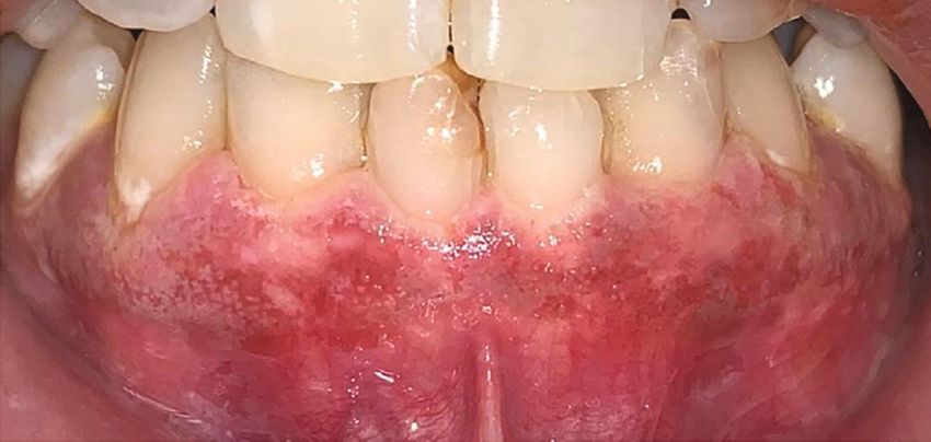
2                                                                                                     Case Reports in Dentistry

                                 Figure 1: Preoperative picture of moderate gingival pigmentation.

pigmentation), score 3 represents medium brown or mixed            dermapen to enhance the absorption of topical AA (vitamin
brown pink and brown pigmentation (moderate pigmenta-              C) into pigmented gingival mucosa.
tion), and score 4 indicates deep brown or bluish-black pig-
mentation (heavy pigmentation). In 2017, the American
Academy of Periodontology and the European Federation              2. Aim of the Study
of Periodontology proposed a new classification of peri-            The purpose of the study was to assess the performance of
odontal and peri-implant diseases [5] in which gingival col-       the microneedling technique using dermapen with topical
our alterations are diagnosed as “gingival pigmentations”          ascorbic acid paste on gingival depigmentation.
and it was classified into melanoplakia, smoker’s melanosis,
drug-induced pigmentations, and amalgam tattoo.
    Gingival depigmentation (GD) is considered a periodon-         3. Case Description
tal plastic procedure whereby melanocytic pigmentation is
removed. A variety of different modalities have been pro-           A healthy 25-year-old female patient complained of aesthi-
posed for removing hyperpigmentation involving bur abra-           cally unappealing dark colour in the lower front gum area
sion, surgical scraping, gingival autograft, cryotherapy,          (Figure 1). According to her detailed history, hyperpigmen-
electrosurgery, and laser [6]. In addition, some studies sug-      tation was evident from birth, which implied physiological
gest that ascorbic acid (vitamin C) could be used to treat gin-    hyperpigmentation. In addition, it was reported that the
gival pigmentation [7].                                            patient had never taken any medications or been diagnosed
    However, the microneedling (MN) technique is a non-            with any systemic conditions which would affect the gingival
surgical procedure that is known as collagen induction ther-       colour. Besides, her family and social histories were insignif-
apy involving repetitive punctures on the skin. In                 icant. According to her, she had never smoked or been
dermatology, MN has been utilized considerably in recent           exposed to second-hand smoke. Furthermore, her complaint
years as it is an effective, simple, economical, well-tolerated,    had not been addressed in an oral setting previously.
and cosmetically and therapeutically beneficial procedure
[8]. The MN serves to separate the cells instead of cutting        4. Clinical Findings and Diagnostic Assessment
through forming microconduits which increases the skin’s
permeability and blood flow into the epidermis. This process        On clinical examination, no abnormalities were identified
facilitates the penetration of topical medications across the      extraorally. Intraoral examination revealed a minimal accu-
stratum corneum layer. Besides, growth factors are produced        mulation of plaque and supragingival calculus, indicating
promoting the regeneration of collagen and elastin [8, 9].         an acceptable level of oral hygiene. She had an average pla-
    However, ascorbic acid (AA) has been demonstrated as a         que index of 0.8 and an average gingival index of 1 with
water-soluble antioxidant and an essential nutrient for colla-     no bleeding upon probing. Neither clinical attachment loss
gen biosynthesis [10]. It plays a role in immunomodulation         nor radiographic bone loss was determined. There was only
as well as the elimination of the hyperpigmented spots [11]        mild marginal gingival inflammation accompanied by rolled
by interacting with the copper ions at the tyrosinase active       margins and blunted interdental papillae.
site and inhibiting the activity of the enzyme tyrosinase,             A dark brown ribbon of hyperpigmentation was noticed
thereby diminishing melanin production [10].                       within the gingival mucosa of the mandibular arch confined
    While many clinical investigations have been docu-             to the attached gingiva region from right to left 2nd premo-
mented, on the effectiveness of the MN technique in treating        lars (Figure 1). The case was diagnosed as physiological
scars and wrinkles, promoting skin rejuvenation, and man-          moderate                 gingival               pigmentation
aging pigmentation disorders [8, 12–14], scarce dental stud-       (pigmentation index score = 3) according to the Dummett-
ies have been conducted on its application in the oral cavity.     Gupta Oral Pigmentation Index [4] with extended ribbon-
For the current case, we utilized the MN technique using           like melanin pigmentation (melanin index class 2).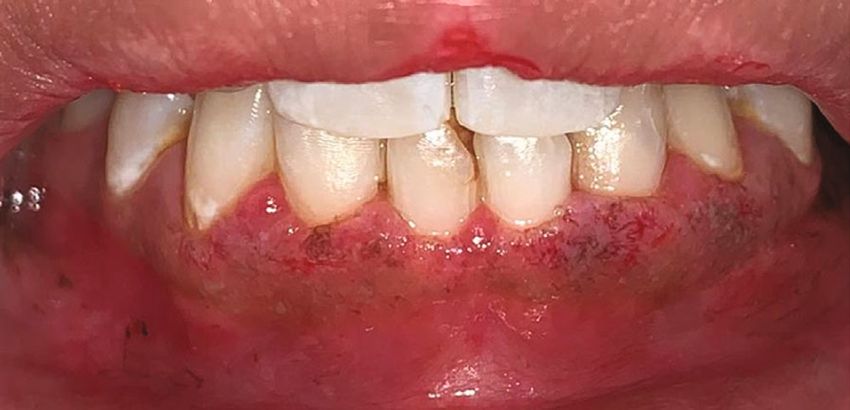
Case Reports in Dentistry 3
Figure 2: Microneedling technique using dermapen and ascorbic acid (vitamin C) application.
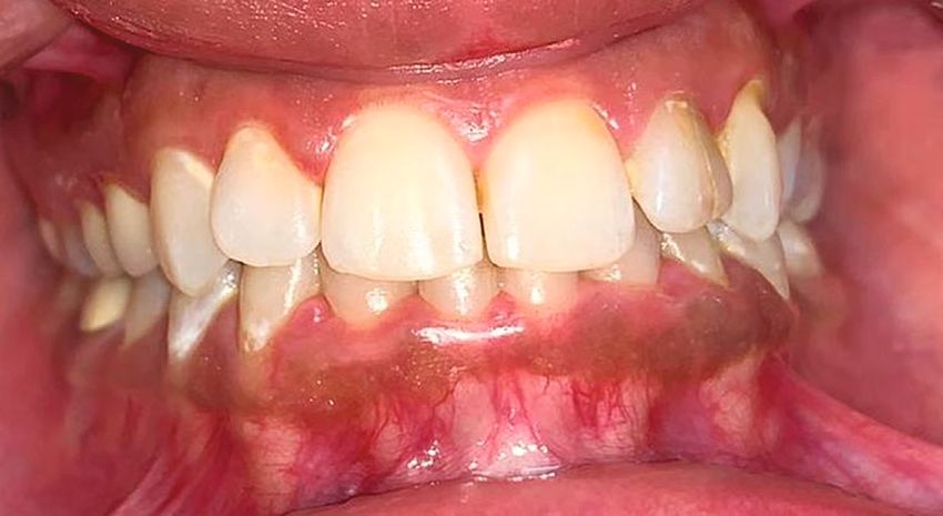
5. Therapeutic Intervention tion. Clinical outcomes showed complete disappearance of
gingival pigmentation. Generally, after the first and second
The patient was most concerned about achieving aesthetic application, healing was normal and satisfactory providing
results with minimally invasive, nonexpensive procedures. excellent aesthetic results where the pigmentation index
In light of the patient’s concerns, the MN technique was sug- score decreased to zero with reduction of melanin index
gested. The patient was provided with a detailed explanation (class 0) as presented in Figures 3(c) and 3(d). After the
of this off-label procedure, its instructions, and potential 6th month of the follow-up period, repigmentation of the
complications. Informed consent was established as well as gingiva was detected showing light brown solitary pigmen-
permission for this off-label procedure, photos, and ted areas as depicted in Figure 4.
publication.
As a preliminary routine, supragingival scaling was per-
formed and oral hygiene instructions were provided by the
7. Discussion
dental hygienist. We subsequently scheduled the procedure Although the GP does not exhibit any medical concerns,
and performed it aseptically. Local anaesthesia 2% ligno- patients frequently complain of “black gums” seeking cos-
caine (1 : 80,000 adrenaline) was administered by infiltration metic solutions. In this case presentation, the patient was
technique in the mandibular region. To microneedle the gin- diagnosed with physiological pigmentation based on the
gival mucosa, we used a dermapen device model M8 with 24 negative history of any pathological cause, since the physio-
microneedles arranged in rows, which was adjusted with logic pigmentation is probably genetic in nature.
1.5 mm depth at the 6th mode speed of 700 cycles/min. GD is considered a periodontal plastic procedure
The dermapen was used in intermittent motion on the sex- whereby the gingival hyperpigmentation is removed or
tant gingival area for 30-40 seconds/tooth. When bleeding reduced through various techniques such as scalpel scraping,
pinpoints were observed on all areas of pigmented gingiva, bur abrasion, free gingival graft, cryosurgery, electrosurgery,
the gingival mucosa was irrigated with a saline solution and laser [6]. In the present case, we achieved GD by a novel
and sterile gauze was applied to dry the area. Then, topical technique using microneedling and topical AA. It is a mini-
AA powder (1000 mg/ml) was mixed with saline in a small mally invasive, well-tolerated, safe, not expensive, and less
glass dish forming a paste. The mixed slurry paste was time-consuming procedure.
applied to the gingival mucosa using for 10 minutes as To our knowledge, this is the first study to introduce NM
shown in Figure 2. The treated area was left without dress- with AA in gingival depigmentation. Nevertheless, there was
ing. Our patient was instructed to refrain from drinking a study done by Ozsagir et al. [15] who compared the use of
acidic or hot beverages for 24 hours and to not brush her i-PRF alone or in combination with microneedling for gingi-
lower teeth for one day to avoid any mechanical trauma to val augmentation. They concluded that after 6 months, the
the gingiva. Following the procedure, neither mouthwashes group using i-PRF combined with microneedling showed a
nor medications were prescribed for her. The photos were statistically significant increase in gingival thickness com-
taken before, during, and at follow-up appointments until pared to the group using only i-PRF.
6 months. Although, the FDA has legally authorized microneedling
devices to improve the appearance of facial acne scars, facial
6. Clinical Follow-Up and Outcomes wrinkles, and abdominal scars in patients aged 22 years or
older, these devices are still considered as off-label proce-
One day after the procedure, the attached gingiva exhibited dures for oral use. However, there are various types of MN
an increase in volume and an alteration in texture as well devices including dermaroller, dermastamp, and dermapen.
as the appearance of white and red-coloured areas, indicat- Unlike other MN devices, dermapen is the only device that
ing tissue inflammation (Figure 3(a)). Neither pain nor ten- can be applied intraorally due to its small size, changeable
derness was reported, and only discomfort sensation was head. This increases the accessibility inside the mouth and
experienced. On the third day, all of these findings dimin- makes it easy to be used for a larger number of patients. This
ished resulting in the pink appearance of the gingiva device is a wireless electric device with automated features
(Figure 3(b)). The same procedure was performed after 2 that allow the operator to modify the speeds, pressures,
weeks, resulting in same the postoperative tissue inflamma- and depth of penetration, thus reducing the probability of
tion which diminished on the 3rd day of the second applica- operator-related side effects.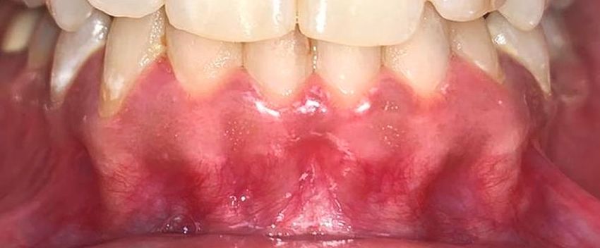
4                                                                                                              Case Reports in Dentistry

    (a) Immediately after microneedling and ascorbic acid application                   (b) After one day of the procedure

                    (c) After 3 days of the procedure                                          (d) After 3 months

                                     Figure 3: (a–d) Postoperative pictures of gingival depigmentation.

                                   Figure 4: Recurrence of gingival pigmentation started after 6 months.

    In the present study, the patient agreed and signed                 reduced pigmentation incidence scores and area of pigmen-
explicit written consent for this off-label procedure being              tation. As well as comparing injectable AA to conventional
used as a part of research. During the procedure, it was nec-           scalpel depigmentation by Yussif et al. [19] in 2019 who con-
essary to generate precise bleeding pinpoints by MN to                  cluded that injecting AA into pigmented cells presented
achieve optimal results [16]. These microholes allow the                comparable results to conventional surgery.
therapeutic medication to penetrate easily into the connec-                 Furthermore, our findings were consistent with the
tive tissues, stimulating new collagen production and affect-            results of Shimada et al. [20] who applied topical vitamin
ing melanocytes [8].                                                    C gel to the gingiva and concluded that AA inhibited mela-
    Then, we used topical AA as a therapeutic medication                nin pigmentation. However, a study made by El-Mofty et al.
that was reported to be effective in reducing melanogenesis              [21] stated that intramucosal AA injections were better and
directly and thus promoting depigmentation, because mela-               more efficacious than topical AA gels for GD.
nin acts as a reservoir for reactive oxygen species (ROS), Cu,              Following the first day of the procedure, the gingiva
and calcium within the cells. Following AA entry into the               showed an increase in volume and a change in texture with
target tissue, it binds to melanin causing a deficiency of               white and red-coloured areas representing gingival inflam-
ROS, Cu, and Ca, resulting in the reduction in melanin pro-             mation, oedema, and irritation. These results were corre-
duction [17]. As to say that AA affects melanocyte function              sponding with numerous clinical studies in dermatology
rather than the number, as opposed to other approaches                  which revealed that the MN technique can cause postopera-
relying on the destruction of melanocytes.                              tive transit oedema and erythema that resolves usually after
    Our outcomes revealed excellent aesthetic results, and              1-3 days [22–25].
these were in agreement with Yussif et al. [18] who used                    In contrast to any other depigmentation procedure, the
intraepithelial injections of 1-1.5 ml AA (200-300 mg) in               patient was able to see these excellent aesthetic results after
the gingival mucosa and reported that its direct delivery               3 days (Figure 3). However, healing after scalpel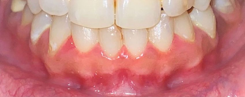
Case Reports in Dentistry                                                                                                           5

depigmentation takes 7-10 days, and other procedures take        new perspectives for its application. Although our technique
more than 2 weeks to heal [26, 27]. In addition, a dressing      is a simple and safe GD method, it should be performed cau-
is not necessary after the procedure since microholes are cre-   tiously; otherwise, tissue destruction and gingival recession
ated without perfused bleeding and heal quickly, unlike scal-    may result. Our results were limited to this particular case.
pel depigmentation where the gingiva should be covered           Therefore, the present findings should be confirmed in
with dressing as it leaves denuded connective tissue and         future randomized controlled clinical trials, including histo-
heals by secondary intention [5]. Moreover, a free gingival      logical analysis.
graft can be used for GD, but it involves a second surgical
site (donor site) and it has unaesthetic colour matching [28].   Consent
     Compared to other minimally invasive techniques, our
technique poses less risk of complications and is less expen-    Patient consent for publication was obtained.
sive. However, lasers are considered easy, fast, and efficient
modalities that have hemostasis and decontamination              Conflicts of Interest
effects, but they are expensive and tissues heal within 1-2
weeks [29, 30]. Additionally, electrosurgery has been            The authors report no conflicts of interest.
reported to cause heat accumulation and undesired tissue
destruction, and it takes 5-14 days for complete healing         Authors’ Contributions
[31]. While cryosurgery may cause postoperative swelling
and increase soft tissue destruction as well as difficulty in      DM contributed to conceptualization, manuscript writing
controlling the depth and freezing duration, also it takes 3-    and editing, and clinical work. SA contributed to conceptu-
4 weeks for complete keratinization [26].                        alization, manuscript preparation, and writing.
     Pigment recurrence is described as a spontaneous and
recurrent process that occurs within 24 hours up to 8 years      References
following the depigmentation procedure which poses a chal-
                                                                  [1] C.-R. Yuri, “Gingival melanosis:, a review of diagnosis and
lenge to a dentist [28]. Methods of treatment, number of
                                                                      treatment criteria,” Odontoestomatología, vol. 21, no. 33,
recall periods, genetic and ethnic factors, tobacco consump-          pp. 54–61, 2019.
tion, and hormonal factors influence the gingival repigmen-        [2] Y. Ciçek and U. Ertaş, “The normal and pathological pigmen-
tation [32]. It has been hypothesized that the rate of melanin        tation of oral mucous membrane: a review,” The Journal of
formation is higher in darker individuals [33]. Also, gingival        Contemporary Dental Practice, vol. 4, no. 3, pp. 76–86, 2003.
pigmentation is more in the anterior gingiva than posterior       [3] T. Hanioka, K. Tanaka, M. Ojima, and K. Yuuki, “Association
ones which have been attributed to sunlight exposure [34].            of melanin pigmentation in the gingiva of children with par-
Many authors [35–37] who used surgical technique showed               ents who smoke,” Pediatrics, vol. 116, no. 2, pp. e186–e190,
early recurrence of pigmentation after 15-56 days of follow-          2005.
up periods. However, the majority of the available literature     [4] C. O. Dummet and G. Barens, “Oromucosal pigmentation: an
has shown a lower recurrence rate for cryosurgery and lasers          updated literary review,” Journal of Periodontology, vol. 42,
[38]. Nakamura et al. [39] described repigmentation after 24          no. 11, pp. 726–736, 1971.
months after laser depigmentation. Additionally, previous         [5] P. Corteillini and N. F. Bissada, “Mucogingival conditions in
studies [40–43] have reported gingival pigmentation recur-            the natural dentition: narrative review, case definitions, and
rence after 12-48 months following cryosurgery                        diagnostic considerations,” Journal of Clinical Periodontology,
depigmentation.                                                       vol. 45, pp. S190–S198, 2018.
     In our case report, repigmentation was observed after 6      [6] A. Thangavelu, S. Elavarasu, and P. Jayapalan, “Pink esthetics
months (Figure 4). This might be due to the proliferation             in periodontics - gingival depigmentation: a case series,” Jour-
of melanocytes and their migration to the depigmented area            nal of Pharmacy & Bioallied Sciences, vol. 4, Suppl 2, pp. S186–
[27]. These recurrence findings vary from those of Sheel               S190, 2012.
et al. [44] who used scalpel depigmentation along with the        [7] R. M. Sanadi and R. S. Deshmukh, “The effect of vitamin C on
                                                                      melanin pigmentation - a systematic review,” J Oral Maxillofac
monthly local application of AA, which gave positive aes-
                                                                      Pathol., vol. 24, no. 2, pp. 374–382, 2020.
thetic results, but recurrence was observed after 9 months.
                                                                  [8] C. Iriarte, O. Awosika, M. Rengifo-Pardo, and A. Ehrlich,
This may be related to the amount of AA that gingival tis-
                                                                      “Review of applications of microneedling in dermatology,”
sues are exposed to.                                                  Clinical, Cosmetic and Investigational Dermatology, vol. -
     However, our procedure is well tolerated by patients and         Volume 10, pp. 289–298, 2017.
can be repeated from time to time for more aesthetic results.     [9] A. Singh and S. Yadav, “Microneedling: advances and widen-
                                                                      ing horizons,” Indian Dermatology Online Journal, vol. 7,
8. Conclusions                                                        no. 4, pp. 244–254, 2016.
                                                                 [10] J. Velisek and K. Cejpek, “Biosynthesis of food constituents:
Clinical experience, affordability, and personal preferences           vitamins, water-soluble vitamins, part 2 – a review,” Czech
should be considered when selecting a technique for treating          Journal of Food Sciences, vol. 25, pp. 49–64, 2008.
gingival hyperpigmentation. Our novel technique of using         [11] S. J. Padayatty and M. Levine, “Vitamin C: the known and the
dermapen and topical ascorbic acid in the treatment of gin-           unknown and Goldilocks,” Oral Diseases, vol. 22, no. 6,
gival pigmentation showed promising results, which gives              pp. 463–493, 2016.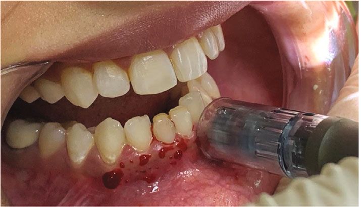
6                                                                                                                Case Reports in Dentistry

[12] M. T. McCrudden, E. McAlister, A. J. Courtenay, P. González-              ment of plantar warts,” Journal of Cosmetic Dermatology,
     Vázquez, T. R. Raj Singh, and R. F. Donnelly, “Microneedle                vol. 18, no. 1, pp. 124–128, 2019.
     applications in improving skin appearance,” Experimental           [26]   K. Almas and W. Sadiq, “Surgical treatment of melanin-
     Dermatology, vol. 24, no. 8, pp. 561–566, 2015.                           pigmented gingiva: an esthetic approach,” Indian Journal of
[13] F. Al Qarqaz and A. Al-Yousef, “Skin microneedling for acne               Dental Research, vol. 13, no. 2, pp. 70–73, 2002.
     scars associated with pigmentation in patients with dark skin,”    [27]   R. Kathariya and A. R. Pradeep, “Split mouth de-epithelization
     Journal of Cosmetic Dermatology, vol. 17, no. 3, pp. 390–395,             techniques for gingival depigmentation: a case series and
     2018.                                                                     review of literature,” J Indian Soc Periodontol., vol. 15, no. 2,
[14] J. W. Jung, W. O. Kim, H. R. Jung, S. A. Kim, and Y. W. Ryoo,             pp. 161–168, 2011.
     “A face-split study to evaluate the effects of microneedle radio-   [28]   S. A. Mokeem, “Management of gingival hyperpigmentation
     frequency with Q-switched Nd: YAG laser for the treatment of              by surgical abrasion: report of three cases,” Saudi Dental Jour-
     melasma,” Annals of Dermatology, vol. 31, no. 2, pp. 133–138,             nal, vol. 18, pp. 162–166, 2006.
     2019.
                                                                        [29]   P. Atsawasuwan, K. Greethong, and V. Nimmanon, “Treat-
[15] Z. B. Ozsagir, E. Saglam, B. Sen Yilmaz, J. Choukroun, and                ment of gingival hyperpigmentation for esthetic purposes by
     M. Tunali, “Injectable platelet-rich fibrin and microneedling              Nd: YAG laser: report of 4 cases,” Journal of Periodontology,
     for gingival augmentation in thin periodontal phenotype: a                vol. 71, no. 2, pp. 315–321, 2000.
     randomized controlled clinical trial,” Journal of Clinical Peri-
                                                                        [30]   H. Tal, D. Oegiesser, and M. Tal, “Gingival depigmentation by
     odontology, vol. 47, no. 4, pp. 489–499, 2020.
                                                                               erbium: YAG laser: clinical observations and patient
[16] A. Gowda, B. Healey, H. Ezaldein, and M. Merati, “A system-               responses,” Journal of Periodontology, vol. 74, no. 11,
     atic review examining the potential adverse effects of micro-              pp. 1660–1667, 2003.
     needling,” The Journal of Clinical and Aesthetic Dermatology,
                                                                        [31]   M. J. Oringer, Ed., Electrosurgery in Dentistry, W.B. Saunders
     vol. 14, no. 1, pp. 45–54, 2021.
                                                                               Co, Philadelphia, 2nd ed. edition, 1975.
[17] T. H. Tsai, C. J. Huang, W. H. Wu, W. C. Huang, J. H. Chyuan,
                                                                        [32]   R. Hegde, A. Padhye, S. Sumanth, A. Jain, and N. Thukral,
     and P. J. Tsai, “Antioxidant, cell-protective, and anti-
                                                                               “Comparison of surgical stripping; erbium-doped: yttrium,
     melanogenic activities of leaf extracts from wild bitter melon
                                                                               aluminum, and garnet laser; and carbon dioxide laser tech-
     (Momordica charantia Linn var abbreviata Ser) cultivars,”
                                                                               niques for gingival depigmentation: a clinical and histologic
     Botanical Studies, vol. 55, no. 1, p. 78, 2014.
                                                                               study,” Journal of Periodontology, vol. 84, no. 6, pp. 738–748,
[18] N. M. Yussif, S. O. Zayed, S. A. Hasan, and S. S. Sadek, “Eval-           2013.
     uation of injectable vitamin C as a depigmenting agent in
     physiologic gingival melanin hyperpigmentation: a clinical         [33]   R. E. Billingham, “Dendritic cells in pigmented human skin,”
     trial,” Rep Opinion, vol. 8, pp. 113–120, 2016.                           Journal of Anatomy, vol. 83, no. 2, pp. 109–115, 1949.
[19] N. M. Yussif, A. R. Abdel Rahman, and E. Elbarbary, “Mini-         [34]   R. B. Raut, M. A. Baretto, F. S. Mehta, M. K. Sanjana, and K. L.
     mally invasive non-surgical locally injected vitamin C versus             Shourie, “Gingival pigmentation: its incidence amongst the
     the conventional surgical depigmentation in treatment of gin-             Indian adults,” JAIDA, vol. 26, pp. 9-10, 1954.
     gival hyperpigmentation of the anterior esthetic zone: a pro-      [35]   T. M. Ginwalla, B. C. Gomes, and B. R. R. Verma, “Surgical
     spective comparative study,” Clinical Nutrition Experimental,             removal of gingival pigmentation (a preliminary study),” Jour-
     vol. 24, pp. 54–65, 2019.                                                 nal of the Indian Dental Association, vol. 38, no. 6, pp. 147–50
[20] Y. Shimada, H. Tai, A. Tanaka et al., “Effects of ascorbic acid            passim, 1966.
     on gingival melanin pigmentation in vitro and in vivo,” Jour-      [36]   T. K. Pal, K. K. Kapoor, C. C. Parel, and K. Mukherjee, “Gingi-
     nal of Periodontology, vol. 80, no. 2, pp. 317–323, 2009.                 val melanin pigmentation – a study on its removal for
[21] M. El-Mofty, S. Elkot, A. Ghoneim, D. Yossri, and O. M.                   esthetic,” J Indian Soc of Periodontology, vol. 3, pp. 52–54,
     Ezzatt, “Vitamin C mesotherapy versus topical application                 1994.
     for gingival hyperpigmentation: a clinical and histopatholo-       [37]   C. O. Dummett and T. E. Bolden, “Post surgical clinical repig-
     gical study,” Clinical Oral Investigations, vol. 25, no. 12,              mentation of the gingiva,” Oral Surgery, Oral Medicine, Oral
     pp. 6881–6889, 2021.                                                      Pathology, Oral Radiology, and Endodontics, vol. 16, no. 3,
[22] S. Yadav and S. Dogra, “A cutaneous reaction to microneed-                pp. 353–365, 1963.
     ling for postacne scarring caused by nickel hypersensitivity,”     [38]   D. S. Alasmari, “An insight into gingival depigmentation tech-
     Aesthetic Surgery Journal, vol. 36, no. 4, p. NP168–170, 2016.            niques: the pros and cons,” Int J Health Sci (Qassim)., vol. 12,
[23] S. Dogra, S. Yadav, and R. Sarangal, “Microneedling for acne              no. 5, pp. 84–89, 2018.
     scars in Asian skin type: an effective low cost treatment modal-    [39]   Y. Nakamura, M. Hossain, K. Hirayama, and K. Matsumoto,
     ity,” Journal of Cosmetic Dermatology, vol. 13, no. 3, pp. 180–           “A clinical study on the removal of gingival melanin pigmen-
     187, 2014.                                                                tation with the CO2 laser,” Lasers in Surgery and Medicine.,
[24] T. M. Leheta, R. M. Abdel Hay, R. A. Hegazy, and Y. F. El                 vol. 25, no. 2, pp. 140–147, 1999.
     Garem, “Do combined alternating sessions of 1540 nm nona-          [40]   L. Jokar, M. Bayani, H. Hamidi, M. Keivan, and S. Azari-Mar-
     blative fractional laser and percutaneous collagen induction              habi, “A comparison of 940 nm diode laser and cryosurgery
     with trichloroacetic acid 20% show better results than each               with liquid nitrogen in the treatment of gingival physiologic
     individual modality in the treatment of atrophic acne scars?              hyperpigmentation using split mouth technique: 12 months
     A randomized controlled trial,” The Journal of Dermatological             follow up,” J Lasers Med Sci., vol. 10, no. 2, pp. 131–138, 2019.
     Treatment, vol. 25, no. 2, pp. 137–141, 2014.                      [41]   A. R. S. Shirazi, A. M. Taghavi, and F. Khorakian, “Treatment
[25] M. R. Al‐Naggar, A. S. Al‐Adl, A. R. Rabie, M. R. Abdelkhalk,             of gingival physiologic pigmentation in adolescent using cryo-
     and M. L. Elsaie, “Intralesional bleomycin injection vs                   surgery technique with liquid nitrogen: one year follow up,”
     microneedling-assisted topical bleomycin spraying in treat-               Journal of Cosmetic Dermatology, vol. 33, pp. 331–342, 2010.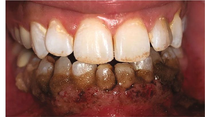
Case Reports in Dentistry                                                  7

[42] C.-J. Yeh, “Cryosurgical treatment of melanin-pigmented gin-
     giva,” Oral Surgery, Oral Medicine, Oral Pathology, Oral Radi-
     ology, and Endodontics, vol. 86, no. 6, pp. 660–663, 1998.
[43] T. Haim, L. Jacob, and K. Avital, “Cryosurgical depigmenta-
     tion of the gingiva - a case report,” Journal of Clinical Peri-
     odontology, vol. 14, no. 10, pp. 614–617, 1987.
[44] V. Sheel, P. Purwar, J. Dixit, and P. Rai, “Ancillary role of vita-
     min C in pink aesthetics,” BML Case Reports, vol. 2015, no. -
     jun08 1, p. bcr2014208559, 2015.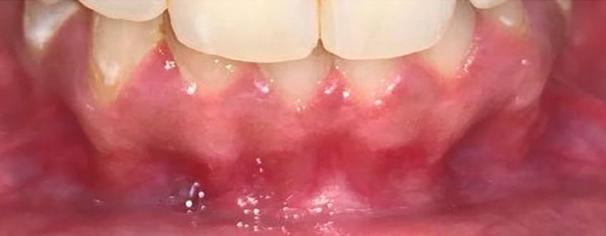
You can also read