Case Report: Solitary Extramedullary Plasmacytoma in the Cervix Misdiagnosed as Cervical Cancer - Frontiers
←
→
Page content transcription
If your browser does not render page correctly, please read the page content below
CASE REPORT
                                                                                                                                                  published: 04 June 2021
                                                                                                                                           doi: 10.3389/fonc.2021.685070

                                              Case Report: Solitary Extramedullary
                                              Plasmacytoma in the Cervix
                                              Misdiagnosed as Cervical Cancer
                                              Ji Wang 1†, Lin Jiang 1†, Xuejin Ma 1, Tingchao Li 2, Heng Liu 3, Xiaoxi Chen 3 and Shiguang Li 1*
                                              1Department of Radiology, The First People’s Hospital of Zunyi, The Third Affiliated Hospital of Zunyi Medical University,
                                              Zunyi, China, 2 Department of Pathology, The First People’s Hospital of Zunyi, The Third Affiliated Hospital of Zunyi Medical
                                              University, Zunyi, China, 3 Department of Radiology, Medical Imaging Center of Guizhou Province, Affiliated Hospital of Zunyi
                                              Medical University, Zunyi, China

                                              Solitary plasmacytoma (SP) is a malignant tumor caused by the monoclonal proliferation of
                                              plasma cells, representing less than 5% of plasma cell tumors. SP can be categorized into
                                              two groups: solitary bone plasmacytoma (SBP) and solitary extramedullary plasmacytoma
                                              (SEP). SEP most commonly occurs in the head and neck and is rarely located in the
                                              reproductive system. Here, we report a case of a 77-year-old woman with SEP in the cervix
                           Edited by:
                          Wenyin Shi,         who had a 7-day history of vaginal bleeding. Ultrasonography and magnetic resonance
           Thomas Jefferson University,       imaging (MRI) showed an oval mass in the cervix, which was initially considered as
                         United States
                                              neoplastic lesions and highly suspected to be cervical cancer, but cervical leiomyoma
                         Reviewed by:
                               Fiori Alite,
                                              and other benign tumors cannot be completely excluded. Subsequently, cervical biopsy
    Geisinger Commonwealth School of          showed that the tumor was SEP, and then the patient underwent surgery. The
                Medicine, United States
                                              postoperative pathological diagnosis was also SEP, which confirmed the radiologist’s
                         Anuj Mahindra,
           Scripps Clinic, United States      misjudgment. In conclusion, SEP that occurs in the cervix is remarkably rare, and only nine
                     *Correspondence:         cases have been reported in the cervix. No case reports to date have described in detail the
                            Shiguang Li       imaging findings of cervical SEP. This study demonstrates the MRI imaging characteristics
                  imaging_sgli@163.com
†
                                              of a patient with SEP of the cervix and reviews the imaging findings of SEP reported in the
 The authors have contributed equally
to this work and share first authorship
                                              previous literature, in order to provide more extensive insights for radiologists to consider the
                                              differential diagnosis of cervical lesions.
                     Specialty section:
                                              Keywords: solitary extramedullary plasmacytoma, solitary plasmacytoma, cervical cancer, MRI, case report
           This article was submitted to
                     Radiation Oncology,
                 a section of the journal
                   Frontiers in Oncology
                                              CASE PRESENTATION
               Received: 01 April 2021
               Accepted: 18 May 2021          A 77-year-old female patient was admitted to hospital because of a 7-day history of abnormal vaginal
              Published: 04 June 2021         bleeding, with no abdominal pain or distention. Gynecological examination revealed cervical atrophy,
                               Citation:      and the normal shape of the cervix disappeared. There was no contact bleeding of the cervix and no
     Wang J, Jiang L, Ma X, Li T, Liu H,      tenderness of the posterior fornix. She had no other symptoms, and the results of the laboratory
                Chen X and Li S (2021)
                                              examinations were normal. Ultrasonography showed hypoechoic nodule in the cervix. The pelvic
    Case Report: Solitary Extramedullary
            Plasmacytoma in the Cervix
                                              magnetic resonance imaging (MRI) scan found a well-defined oval mass of size 2.3×1.8×2.8 cm in the
      Misdiagnosed as Cervical Cancer.        cervix, which destroyed the continuity of the cervical stroma (Figure 1A). On MRI, the mass was
              Front. Oncol. 11:685070.        homogeneous, slightly hyperintense on the T2-weighted images (Figures 1A, D), and isointense on the
       doi: 10.3389/fonc.2021.685070          T1-weighted images (Figures 1B, E). Decreased apparent diffusion coefficient (ADC) value were

Frontiers in Oncology | www.frontiersin.org                                           1                                          June 2021 | Volume 11 | Article 685070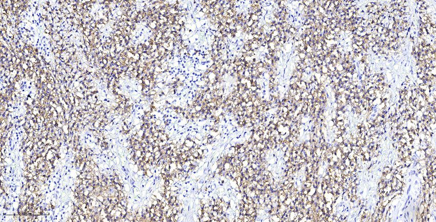
Wang et al.                                                                                                                       Case Report: SEP in the Cervix

                    A                                      B                                         C

                    D                                      E                                         F

 FIGURE 1 | (A, B, D, E) MRI scans showed a well-defined oval mass located in cervix, which destroyed the stroma (A). The mass showed slight hyperintensity on
 T2-weighted MR images (A, D) and isointensity on T1-weighted MR images (B, E). (C) The ADC map showed low ADCs. (F) Sagittal contrast-enhanced MR image
 showed mild to moderate heterogeneous enhancement of the lesion, presenting rim-enhancement.

detected in the cervical lesion (Figure 1C). Post-contrast                         cavity, sinuses, nasopharynx, and larynx (1). Other common sites
enhancement images showed mild to moderate heterogeneous                           are gastrointestinal tract, urogenital tract, skin, lung and breast
enhancement and pronounced peripheral rim enhancement                              (2). These areas are rich in lymphoid tissues and plasmacyte cells
(Figure 1F). According to the patient’s age, clinical manifestations               (3, 4). SP is more often found in male patients over 40 years of
and imaging features, the radiologist’s initial consideration was                  age (5). According to 9 previously reported cervical SEP cases (6,
neoplastic lesions and the possibility of cervical cancer was high.                7) and our case, the patients’ age ranges from 21 to 77 years old,
The possibility of cervical leiomyoma cannot be fully ruled out,                   and the mean age is 41 years old. The clinical manifestations are
however. The patient accepted colposcopic examination and                          diverse, such as coital bleeding, vaginal discharge, vaginal
underwent cervical biopsy and endocervical curettage under                         bleeding, pelvic pain, etc. However, some patients are
colposcopic guidance. The biopsy specimens revealed SEP on                         asymptomatic (6). Our patient presented with vaginal bleeding.
microscopic examination. Additionally, bone scan revealed no                       Postmenopausal women with vaginal bleeding should first be
abnormal skeletal uptake, and no abnormal plasma cells were                        excluded from common malignancies such as endometrial
found in the bone marrow, which excluded the diagnosis of                          cancer and cervical cancer. In addition, other diseases such as
multiple myeloma (MM). Following discussion, the clinicians                        endometrial polyps, submucous uterine fibroids and cervical
decided that the best treatment strategy was surgery with                          polyps should also be given consideration, but SEP should also
postoperative radiotherapy. Then, the patient underwent a                          be considered, despite its rare occurrence. The computed
laparoscopic extensive hysterectomy, bilateral salpingo-                           tomography (CT) imaging finding of SEP is a low-attenuation
oophorectomy, and pelvic lymph node dissection. The resected                       mass with slight heterogeneity. On MRI, SEP usually manifests as
mass size was about 3.0×2.0 cm. The result of histopathological                    a well-defined or ill-defined soft-tissue tumor with mass effect,
examination revealed a SEP of cervix. Immunohistochemical                          showing iso/hypo-intense on the T1-weighted images and iso/
staining was positive with CD38, CD138, Kappa (Figure 2) and                       hyper-intense on the T2-weighted images compared with
negative with Lambda. The Ki-67 proliferation index was low (5%).                  muscles. The contrast-enhanced imaging is described as mild
The patient received radiotherapy after surgery and remains well                   to marked heterogeneous enhancement on both CT and MRI (2,
through follow-up.                                                                 8, 9). On diffusion weight imaging (DWI), SEP can show
                                                                                   restricted diffusion, suggesting high cellularity in the lesion
                                                                                   (10). Generally, the tumor rarely metastases to other sites but
DISCUSSION                                                                         is locally invasive and can destroy, infiltrate or encroach adjacent
                                                                                   structures (2, 8). Besides, necrosis may occur in larger tumors
SEP can occur in any part of the extramedullary sites but                          (2). By reviewing the literatures of SEP, we have not found the
predominantly in the head and neck, particularly in the nasal                      occurrence of hemorrhage, calcification, and fat (10). CT has

Frontiers in Oncology | www.frontiersin.org                                    2                                        June 2021 | Volume 11 | Article 685070
Wang et al. Case Report: SEP in the Cervix
A B
C D
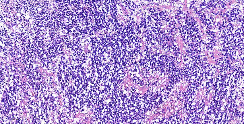
FIGURE 2 | Histological and immunohistochemical features of SEP. (A) Hematoxylin-eosin staining shows that the monoclonal well-differentiated plasma cells are
diffusely distributed and consistent in size. (B–D) Immunohistochemical staining presented CD38(+), CD138(+), Kappa (+). [Original magnifications: (A, B) 200×;
(C, D) 400×].
greater advantages in evaluating bone destruction comparing to SEP in the cervix needed to be distinguished from other
MRI (2). lesions such as abscesses, lymphomas and rectal stromal tumors.
In our case, the tumor showed slight hyperintensity on the Cervical abscess is not a common pelvic inflammatory disease
T2-weighted images and isointensety on the T1-weighted (PID). The abscess cavity contains a fluid component and is
images. The lesion was hyperintense on DWI and hypointense surrounded by edema (13). The abscess typically presents as
on ADC may indicate tumor cells are closely arranged. Following ring-like and/or septal enhancement (14). Lymphoma makes up
contrast enhancement, the tumor exhibited a mild to moderate 3.5% of all malignant tumors in women (15). Primary cervical
inhomogeneous enhancement and the edge appeared more lymphomas originate from the cervical stroma are characterized
obviously, which may suggest that the blood supply was more by rapid growth and rare necrosis (16, 17). Structure
abundant at the edge of the tumor. The tumor was oval with clear preservation and intact squamous epithelium are other features
borders. It destroyed the cervical stroma, which indicated of them (16). The typical imaging appearance of cervical
a g g r es s i v e tu m or b eh a vi o r s . S o m e o f t h e i m a g i n g lymphoma has been described as a mass with homogeneous
manifestations of our case are similar to cervical leiomyoma signal, unclear edge, and moderately homogeneous enhancement
and early cervical cancer. Uterine leiomyoma is benign tumor (16). Gastrointestinal stromal tumors (GISTs) are found in the
with well-defined borders and occurs primarily from the uterine gastrointestinal tract and need to be differentiated from cervical
body or less commonly from the cervix. The most specific tumors when they occur in the rectum (18). The occurrence of
manifestation of cervical leiomyoma is obviously low intensity rectal GISTs is more common in men aging from 50 to 60 years
on T2-weighted images (11). In this case, the signal intensity on old (19). Rectal GISIs are characterized by an exophytic growth
T2-weighted images was evidently not low enough and the lesion pattern, but anatomical continuity with the rectum can be
was invasive, not consistent with the manifestations of typical observed on the imaging (19). Small GITIs (≤5cm) are usually
leiomyoma. Cervical cancer often manifests as a round, lobulated round or oval, with homogeneously obvious and persistent
or irregular soft tissue mass on MRI, showing iso/slight hypo- enhancement, whereas large GITIs (>5cm) are lobulated with
intensity on the T1-weighted images, medium hyper/hyper- mild, inhomogeneous gradual enhancement, and cystic changes
intensity on the T2-weighted images, and obvious high signal are often seen in the tumor (18). It has been reported that GISIs
on DWI with a corresponding low signal on ADC maps. The can appear calcification, mostly focal and patchy (19). However,
malignant progression of cervical cancer can spread to the definitive diagnosis requires histological examination, and
vagina, parauterine tissues, adjacent organs, and distant imaging is of limited utility in differential diagnosis.
metastasis. Of note, cervical cancer is a hypervascular tumor, SEP is usually a localized low-grade malignant tumor, and the
in which the most significant imaging feature is early and therapeutic purpose is mainly local control (20). Because SP is
obvious enhancement on the dynamic contrast enhanced MRI highly sensitive to radiation (1), most authors generally agree
(DCE-MRI), but the late enhancement is lower than the normal that radiotherapy as the standard treatment of SP and excellent
cervical stroma (12). local tumor control rates can be achieved. A radiotherapy dose of
Frontiers in Oncology | www.frontiersin.org 3 June 2021 | Volume 11 | Article 685070
Wang et al.                                                                                                                                  Case Report: SEP in the Cervix

40–50 Gy is recommended by most of the authors (the daily dose is                           necessary to observe the recurrence or metastasis of patients
1.8-2 Gy), and the total treatment course is about 4 weeks (20). At                         through follow-up.
present, the treatment of SP includes radiotherapy, surgery or a                                In conclusion, we present an extremely rare case of SEP in the
combination of both (4). The efficacy of chemotherapy in the                                 cervix and its characterization on MRI. SEP should be considered
treatment of SEP is controversial and further research is needed.                           in the differential diagnosis of cervical lesions without a history
Surgical resection may as an option for the treatment of SEP with                           of multiple myeloma.
clear surgical margins or those that occur in rare area with small size
(20). However, some scholars consider that single surgery is not
recommended even in the case of complete resection. The
combination of surgery and radiotherapy can achieve better
                                                                                            DATA AVAILABILITY STATEMENT
therapeutic effects and survival results (21). The lesion presented in                      The raw data supporting the conclusions of this article will be
this case is located in a rare part of the cervix, with a clear boundary                    made available by the authors, without undue reservation.
and small size, so it is more appropriate to use surgery
plus postoperative radiotherapy. While current clinical treatments
for cervical cancer are mainly surgery, radiotherapy and
chemotherapy. The major treatment strategy for early cervical                               ETHICS STATEMENT
cancer is surgery, and the treatment of middle and advanced
                                                                                            Written informed consent was obtained from the patient for the
cervical cancer is primarily radiotherapy supplemented with
                                                                                            publication of this case report.
chemotherapy. However, patients with recurrent or metastatic
cervix cancer have dismal prognosis. Therefore, early detection of
cervix lesion and make a correct diagnosis would be extremely helpful
to choose optimal therapy and improve the survival rate of patients.                        AUTHOR CONTRIBUTIONS
    The progression of SP includes the appearance of new
plasmacytoma or the progression to MM. SBP is more likely to                                JW and LJ: manuscript writing. TL: pathological review. XM, XC,
progress to MM than SEP (1). Risk factors for progression to                                and HL: manuscript revision. SL: conception and critical review.
MM include persistence of a serum monoclonal protein after                                  All authors contributed to the article and approved the
treatment and abnormal serum SFLC ratio (< 0.26 or > 1.65).                                 submitted version.
Furthermore, minimal bone marrow plasmacytosis is a
contributing factor to decreased progression-free survival.
Furthermore, some authors identified age (≤60 years), tumor                                  ACKNOWLEDGMENTS
size (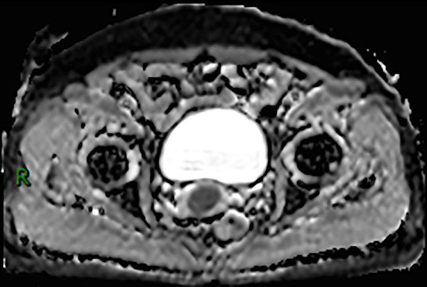
Wang et al.                                                                                                                               Case Report: SEP in the Cervix

17. Chan JK, Loizzi V, Magistris A, Hunter MI, Rutgers J, DiSaia PJ, et al.          21. Ozsahin M, Tsang RW, Poortmans P, Belkacé mi Y, Bolla M, Dinçbas FO, et al.
    Clinicopathologic Features of Six Cases of Primary Cervical Lymphoma.                Outcomes and Patterns of Failure in Solitary Plasmacytoma: A Multicenter
    Am J Obstet Gynecol (2005) 193(3 Pt 1):866–72. doi: 10.1016/j.ajog.                  Rare Cancer Network Study of 258 Patients. Int J Radiat Oncol Biol Phys
    2005.04.044                                                                          (2006) 64(1):210–7. doi: 10.1016/j.ijrobp.2005.06.039
18. Yu MH, Lee JM, Baek JH, Han JK, Choi BI. MRI Features of Gastrointestinal
    Stromal Tumors. AJR Am J Roentgenol (2014) 203(5):980–91. doi: 10.2214/          Conflict of Interest: The authors declare that the research was conducted in the
    AJR.13.11667                                                                     absence of any commercial or financial relationships that could be construed as a
19. Jiang Z-X, Zhang S-J, Peng W-J, Yu B-H. Rectal Gastrointestinal Stromal          potential conflict of interest.
    Tumors: Imaging Features With Clinical and Pathological Correlation. World
    J Gastroenterol (2013) 19(20):3108–16. doi: 10.3748/wjg.v19.i20.3108             Copyright © 2021 Wang, Jiang, Ma, Li, Liu, Chen and Li. This is an open-access
20. Tsang RW, Campbell BA, Goda JS, Kelsey CR, Kirova YM, Parikh RR, et al.          article distributed under the terms of the Creative Commons Attribution License
    Radiation Therapy for Solitary Plasmacytoma and Multiple Myeloma:                (CC BY). The use, distribution or reproduction in other forums is permitted, provided
    Guidelines From the International Lymphoma Radiation Oncology Group.             the original author(s) and the copyright owner(s) are credited and that the original
    Int J Radiat Oncol Biol Phys (2018) 101(4):794–808. doi: 10.1016/j.ijrobp.       publication in this journal is cited, in accordance with accepted academic practice. No
    2018.05.009                                                                      use, distribution or reproduction is permitted which does not comply with these terms.

Frontiers in Oncology | www.frontiersin.org                                      5                                             June 2021 | Volume 11 | Article 685070
You can also read