Challenging diagnosis of cervical vagal nerve schwannoma
←
→
Page content transcription
If your browser does not render page correctly, please read the page content below
Journal of Surgical Case Reports, 2022, 4, 1–4
                                                                                                                      https://doi.org/10.1093/jscr/rjac084
                                                                                                                                             Case Report

case report

Challenging diagnosis of cervical vagal
nerve schwannoma
Lina Pankratjevaite1 , 2 ,† , Niloofar Sherazi Dreyer3 , *,† , Albertas Dauksa4 and Valdas Sarauskas5

                                                                                                                                                               Downloaded from https://academic.oup.com/jscr/article/2022/4/rjac084/6562927 by guest on 10 June 2022
1 Department   of Breast Surgery, Rigshospitalet, Copenhagen University Hospital, Copenhagen, Denmark
2 Department   of Breast Surgery, Herlev and Gentofte Hospital, Copenhagen University Hospital, Herlev, Gentofte, Denmark
3 Department of Otorhinolaryngology, Head and Neck Surgery, Rigshospitalet, Copenhagen University Hospital, Copenhagen, Denmark
4 Department of Surgery, Medical Academy, Hospital of Lithuanian University of Health Sciences, Kaunas, Lithuania
5 Department of Pathology, Medical Academy, Hospital of Lithuanian University of Health Sciences, Kaunas, Lithuania

*Correspondence address. Tel: 0045 27437041; E-mail: nilou.sherazi@gmail.com
† L. Pankratjevaite and N. S. Dreyer have contributed equally to the paper.

Abstract
Schwannoma arising from vagal nerve is a rare tumour. It is a slow-growing, benign mass, but rarely it might undergo malignant
transformation. We report a case of a 55-year-old woman with asymptomatic Xth cranial nerve schwannoma in the left side of
the neck. Initially, during the ultrasound examination, the tumour was misconceived to be a malignant lymph node. The patient
underwent complete surgical excision of it. Histopathological examination revealed typical features of schwannoma. Clinical diagnose
of cervical vagal nerve schwannoma is difficult. Magnetic resonance imaging is as an accurate diagnostic tool for these tumours.
Surgical excision is the treatment of choice.

INTRODUCTION                                                                    the left side of the neck. Her past medical history was
Schwannoma (syn. neurilemoma, neuroschwannoma)                                  significant for thyroid nodules diagnosed 4 years ago,
is a benign, encapsulated neoplasm originating from                             arterial hypertension and vertebrobasilar insufficiency
  Schwann cells of peripheral, cranial or autonomic                             lasting few years. There was no family history of malig-
nerves [1]. Parapharyngeal space is the most common                             nancy. Her social history was non-significant.
place of the head and neck region where extracranial                               US revealed normal size, with hypoechogenic nodules
schwannomas arise [2]. Vagal nerve (Xth cranial nerve)                          thyroid gland and a heterogenic 2.6 × 1.4-cm size ovoid
schwannoma is a rare, slow-growing tumour [1, 3, 4].                            shape mass in the left side of the neck (Fig. 1). The
However, rarely it might become malignant [4]. Vagal                            tumour was misunderstood to be a lymph node and
nerve schwannoma most often affects people between                              malignant process was suspected. Fine needle aspiration
the third and fifth decades [1, 5, 6]. There is no sex-related                  cytology (FNAC) of the mass was performed, but it was
predisposition [1, 5, 6]. It is usually asymptomatic [6] but                    inconclusive. The patient was referred to the Department
sometimes hoarseness, pain, or cough may be present                             of Surgery for suspected malignant lymph node excision.
[4]. In advanced cases, symptoms, such as dysphonia,                            During physical examination, her blood pressure was
dysphagia, dyspnoea, pharyngeal or airway obstruction,                          150/90 mmHg and heart rate was 88 times per min. Her
may develop and are suggested to be correlated with                             electrocardiography showed sinus rhythm. Blood labora-
schwannoma size. Magnetic resonance imaging (MRI) is                            tory findings were within normal limits. Clinical exami-
a gold standard used to assess vagal nerve schwannomas                          nation revealed firm, non-tender, ∼3 × 2-cm size mass in
and to evaluate their extent [1, 3, 6]. Surgical excision is                    the left side of the neck.
the treatment of choice for these tumours [3, 6].                                  The patient underwent surgery. Under general anaes-
                                                                                thesia, a cervical incision was done. An encapsulated,
                                                                                well-circumscribed, yellowish, ovoid-shaped, 3 × 1.5-cm
CASE REPORT                                                                     size mass was found between the internal jugular vein
A 55-year-old woman was admitted to the ambulatory                              and the carotid artery. It originated from the vagal nerve.
for elective ultrasound (US) to assess the thyroid gland                        During the excision of the mass, the patient’s pulse
and neck. The patient complained of a palpable lump in                          decreased from 90 beats per min to 20 beats per min.

Received: January 25, 2022. Accepted: February 20, 2022
Published by Oxford University Press and JSCR Publishing Ltd. © The Author(s) 2022.
This is an Open Access article distributed under the terms of the Creative Commons Attribution License (https://creativecommons.org/licenses/by/4.0/), which
permits unrestricted reuse, distribution, and reproduction in any medium, provided the original work is properly cited.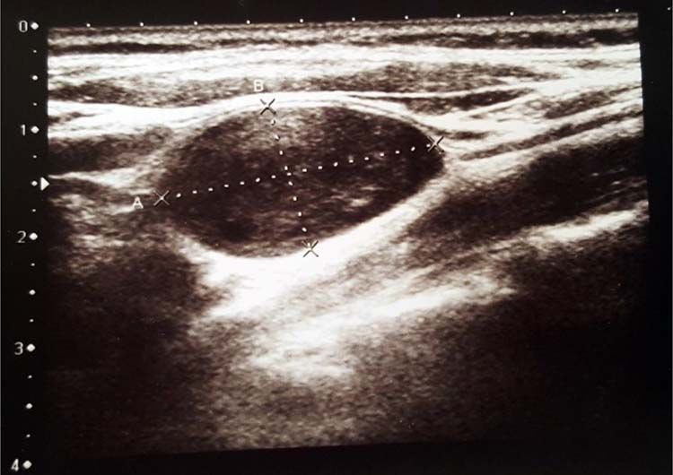
2 | L. Pankratjevaite et al.
Figure 1. US showed ovoid-shaped mass.
Figure 3. Nuclear palisading around fibrillary process (Verocay bodies)
Downloaded from https://academic.oup.com/jscr/article/2022/4/rjac084/6562927 by guest on 10 June 2022
is seen in cellular area; cells are narrow, elongated and wavy with
tapered ends interspersed with collagen fibres.
only complained about asymptomatic neck lump. Some
authors say that FNAC should be used as a routine
procedure for diagnosing the origin of all neck masses
[3]. However, the usefulness of preoperative diagnosis
of vagal nerve schwannoma by FNAC is controversial
[1, 7, 8]. The quality of the specimen and the experience
of cytopathologist influence the preoperative diagnostic
accuracy of FNAC [1]. Moreover, sometimes FNAC can be
dangerous and if the cervical mass clinically and radio-
Figure 2. Biphasic tumour: compact hypercellular Antoni A area (right) logically is suspected to be benign, it is not recommended
and myxoid hypocellular Antoni B area (left).
to do needle or open biopsy because the treatment is still
surgical [8].
After the mass was removed, the patient’s pulse got nor- Radiologic imaging plays an important role in diagnos-
malized. The tumour was removed with a nerve-sparing ing vagal nerve schwannoma. On US images, it appears
technique. as a round or elliptical cross-section with a clear bor-
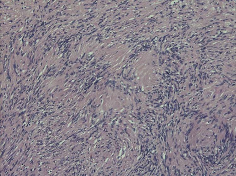
Histopathological examination revealed typical fea- der tumour [1]. Computed tomography (CT) shows vagal
tures of schwannoma (Figs 2 and 3). Tumour was well nerve schwannoma as a well-covered, well-defined mass
demarcated, encapsulated, composed of spindle cells, [1, 4], which is usually of higher attenuation than muscle
organized in a palisading fashion and had hypocellular on contrast-enhanced images [4].
myxoid component with large vessels. Tumour cells had However, MRI is a gold standard to assess vagal nerve
an ill-defined cytoplasm and elongated nucleus. There schwannomas and to evaluate their extent [1, 3, 6].
was no mitotic activity. MRI evaluation typically shows well-circumscribed mass
Post-operatively, the patient complained of coughing lying between the internal jugular vein and the carotid
which disappeared after 2 months. At follow-up, 2 years artery [6]: isotense or hypointense signal on T1-weighted
after the surgery, there was no recurrence of the tumour. images and hyperintense signal on T2-weighted images
are seen [12]. Moreover, MRI is important for differential
diagnosis and treatment planning. It is important to
DISCUSSION notice, that our patient did not have nervus vagus
Schwannoma is a benign tumour originating from cra- schwannoma’s symptoms and complained only of a
nial, peripheral or autonomic nerves except optic and palpable lump in a neck. A US was performed and the
olfactory nerves [1, 7]. Usually, it is solitary but some- mass in the neck was misunderstood to be a malignant
times it might be multiple [5, 7]. About 25% of the cases lymph node. Due to inconclusive FNAC, the patient was
of schwannoma occur in the head and neck area [8]. sent directly to surgical lymph node biopsy and during
Vagal nerve schwannoma is a rare tumour [1, 3, 4, 6– the surgery it was discovered that it was not a lymph
9], which rarely undergoes malignant transformation [4]. node. Our case shows that preoperative MRI or CT scan
It most often affects people between the third and fifth of the neck can be very helpful for diagnosis and for
decades [1, 5, 6]. Both sexes are affected equally [5, 6]. planning the treatment.
Vagal nerve schwannoma usually is an asymptomatic, Macroscopically schwannoma looks like yellowish-
slow-growing tumour [1, 4]. Sometimes symptoms, such white, well-circumscribed mass [1, 6]. Microscopically,
as hoarseness or paradoxical coughing during palpation the baseline features of schwannoma are Antoni type
of the mass, may be present [1, 4, 6, 7, 10]. This sign is A tissue and Antoni type B tissue [1, 4]. Necrosis,
unique to vagal nerve schwannoma [1, 6, 10]. None of haemorrhage and cystic degeneration are other specific
these symptoms were present in our case. The patient features [1, 4].Cervical vagal nerve schwannoma | 3
Anamnesis, imaging studies play an important role in cervical vagal schwannoma. FNAC should be performed
the differential diagnosis of vagal nerve schwannoma. with precaution. Complete excision of the tumour is the
The differential diagnosis of vagal nerve schwannoma treatment of choice. We recommend to perform surgery
includes neurofibromas, metastatic lymph nodes, lym- with intraoperative nerve monitoring.
phoma, paragangliomas, glomus vagale tumours/carotid
body tumours, schwannomas of cervical sympathetic CONFLICT OF INTEREST STATEMENT
origin, branchial cleft anomalies, cysts of the structures
None declared.
of the neck (e.g. thyroid and parathyroid) and vascular
malformations of the neck [4]. Blood samples and some-
times even genetic assessment may be necessary for FUNDING
diagnosis. It is very important to distinguish vagal nerve None.
schwannoma from other diseases as it can influence
further examination and the treatment. For example, if
CONSENT
Downloaded from https://academic.oup.com/jscr/article/2022/4/rjac084/6562927 by guest on 10 June 2022
on radiologic imaging schwannoma could not be dis-
tinguished from neck paraganglioma, FNAC is not rec- Written informed consent for patient information to be
ommended and it is even dangerous to be performed published was provided by the patient.
due to the risk of hypertensive crisis after FNAC. More-
over, it is important to differentiate schwannoma from REFERENCES
hyperfunctional neck paraganglioma, as the latter may
1. Lahoti BK, Kaushal M, Garge S, Aggarwal G. Extra vestibular
be associated with intraoperative hemodynamic insta-
schwannoma: a two year experience. Indian J Otolaryngol Head
bility and hypertensive crisis due to acute release of Neck Surg 2011;63:305–9.
catecholamines during surgery. 2. Shrikrishna BH, Jyothi AC, Kulkarni NH, Mazhar MS. Extracranial
The treatment of choice of vagal nerve schwannoma head and neck schwannomas: our experience. Indian J Otolaryn-
is a complete surgical excision [1, 3, 4, 8]. However, reg- gol Head Neck Surg 2016;68:241–7.
ular observation with imaging could be offered to poor 3. Chiun KC, Tang IP, Prepageran N, Jayalakshmi P. An extensive
surgical candidates and asymptomatic, older patients. cervical vagal nerve schwannoma: a case report. Med J Malaysia
Nerve sparing dissection should be performed for 2012;67:342–4.
benign schwannoma [1, 6, 8]. ‘Subtotal resection’ 4. Sreevatsa MR, Srinivasarao RV. Three cases of vagal nerve
schwannoma and review of literature. Indian J Otolaryngol Head
with or without intracapsular enucleation may be
Neck Surg 2011;63:310–2.
performed [13]. This technique allows higher rate of
5. Langner E, Del Negro A, Akashi HK, Araújo PP, Tincani AJ, Martins
nerve preservation and lower rates of post-operative
AS. Schwannomas in the head and neck: retrospectives analysis
vocal fold palsy [14]. If it is not possible to spare the of 21 patients and review of the literature. Sao Paulo Med J
nerve, end-to-end anastomosis or interposition of nerve 2007;125:220–2.
graft might be done [15]. For malignant schwannomas, 6. Chiofalo MG, Longo F, Marone U, Franco R , Petrillo A, Pezzullo
the best treatment option is wide excision where possible L. Cervical vagal schwannoma. A case report Acta Otorhinolaryngol
[15]. Moreover, vagal nerve schwannoma might be Ital 2009;29:33–5.
removed by an endoscopic gasless unilateral axillo- 7. Vijendra SS, Rao RA, Prasad V, Haseena S, Niprupama M. A giant
breast approach [16]. However, excision of vagal nerve vagal schwannoma with unusual extension from skull base to
schwannoma has a risk of vocal cord paralysis [6, the mediastinum. J Can Res Ther 2015;11:970–3.
8. Imperatori A, Dionigi G, De Monte L, Conti V, Rotolo N.
15]. The intraoperative neurostimulation techniques
Cervico-mediastinal schwannoma of the vagus nerve: resec-
as intermittent intraoperative nerve monitoring and
tion with intraoperative nerve monitoring. Updates Surg 2011;63:
continuous monitoring may reduce the risk of injury to
59–61.
the nerves and prevent from post-operative neurological 9. Shinohara Y, Matsumoto T, Kiga N, Tojyo I, Fujita S. Neurilem-
complications, such as true vocal cord paralysis/palsy, moma of the vagus nerve in the poststyloid parapharyngeal
hoarseness and dysphagia [8, 17]. In present case, vagal space. J Clin Diagn Res 2016;10:ZD17-19.
nerve schwannoma was completely excised; however, 10. Simsek G, Sahan M, Gunsoy B, Arikok A, Akin I. Schwannoma
the patient post-operatively complained of cough that of the cervical vagus nerve: a rare benign neurogenic tumor.
spontaneously disappeared within 2 months. Otolaryngology Online J 2013;3:95–103.
Recurrence risk following vagal schwannoma surgery 11. Mehta KS, Gupta D, Koul N, et al. Vagal nerve schwannoma -
is extremely low and there are no data that present a rare neoplasm with a rare presentation and newer surgical
management technique. IOSR J Dent Sci Med Sci 2013;9:60–5.
recurrence following the specific choice of surgery [14].
12. Jiten N, Puspakishore M, Sudhiranjan T, Upasana R, Sobita P,
Kalpana Th. Cervical schwannoma of the vagus nerve: diag-
nostic and therapeutic challenge. Otolaryngology Online J 2016;6:
745–8.
CONCLUSIONS
13. Obholzer R, Borsetto D, Sandison A. Function preservation for
Vagal nerve schwannoma is a benign, usually asymp- resection of vagal schwannoma of the head and neck: Are
tomatic tumour. Rarely, it might become malignant. MRI we talking about the same technique? Head Neck 2020;42:
is a gold standard to assess preoperative diagnosis of 3469–70.4 | L. Pankratjevaite et al.
14. Sandler ML, Sims JR, Sinclair C, Sharif KF, Ho R, Yue LE. Vagal 16. Hwang KR, Kim JW, Kim HK, Lee SW. A cervical vagal schwan-
schwannomas of the head and neck: a comprehensive review noma mimicking a parathyroid cyst. Clin Exp Otorhinolaryngol
and a novel approach to preserving vocal cord innervation and 2014;7:153–6.
function. Head Neck 2019;41:2450–66. 17. Ijichi K, Kawakita D, Maseki S, Beppu S, Takano G, Murakami
15. Colreavy MP, Lacy PD, Hughes J, Bouchier-Hayes D, Brennan P, S. Functional nerve preservation in extracranial head and neck
O’Dwyer AJ. Head and neck schwannomas—a 10 year review. schwannoma surgery. JAMA Otolaryngol Head Neck Surg 2016;142:
Laryngol Otol 2000;114:119–24. 479–83.
Downloaded from https://academic.oup.com/jscr/article/2022/4/rjac084/6562927 by guest on 10 June 2022You can also read