Leptospirosis as Unusual Trigger of Systemic Lupus Erythematosus
←
→
Page content transcription
If your browser does not render page correctly, please read the page content below
pISSN: 2093-940X, eISSN: 2233-4718
                              Journal of Rheumatic Diseases Vol. 26, No. 1, January, 2019
                              https://doi.org/10.4078/jrd.2019.26.1.79                                                               Case Report

Leptospirosis as Unusual Trigger of Systemic Lupus
Erythematosus
Jinhee Lee, Yang Ree Kim, Chong-Hyeon Yoon
Department of Internal Medicine, Uijeongbu St. Mary's Hospital, College of Medicine, The Catholic University of Korea, Seoul, Korea

Systemic lupus erythematosus (SLE) is a chronic autoimmune disorder of an unknown origin. The role of leptospirosis as a trig-
gering factor for SLE is unknown. This paper reports an uncommon case of SLE following a leptospira infection. A 29-year-old
female was referred due to fevers, myalgia, and facial edema with rash. Laboratory investigations revealed a hepatic dysfunc-
tion, significantly raised lactate dehydrogenase with marked leukopenia and thrombocytopenia. A diagnosis of leptospirosis
was confirmed. The patient was treated with antibiotic therapy for leptospirosis. She developed dyspnea after one week. The
echocardiogram revealed global hypokinesia with a decreased ejection fraction. A positivity of antinuclear, anti-DNA, and an-
ti-Smith antibodies, together with clinical and laboratory improvement by steroid therapy, led to the diagnosis of SLE. This case
highlights the presence of concurrent SLE and leptospirosis. As the symptoms of SLE are similar to leptospirosis, accurate diag-
nosis through high suspicion is essential for appropriate treatment. (J Rheum Dis 2019;26:79-82)

Key Words. Systemic lupus erythematosus, Leptospirosis, Myocarditis

                       INTRODUCTION                                                     es from self-limited febrile episodes to severe organ
                                                                                        dysfunction. This paper reports an uncommon case of
 Systemic lupus erythematosus (SLE) is a chronic auto-                                  newly diagnosed SLE during the treatment of leptospirosis.
immune disorder with an unknown origin [1]. Many re-
searchers have focused on the role of infection in the                                                             CASE REPORT
pathogenesis of SLE, particularly infections caused by vi-
ruses, bacteria, and parasites [2]. The mechanisms, how-                                 A 29-year-old woman visited the emergency department
ever, remain poorly understood. Some studies have                                       with fever, myalgia, and facial edema with a rash since the
shown that autoimmune diseases, such as Guillain-Barré                                  previous 7 days during late summer. She had no sig-
syndrome [3], autoimmune hepatitis [4], and auto-                                       nificant prior medical history. The facial rash was a non-
immune epilepsy [5], were a result of leptospirosis. On                                 pruritic, erythematous patch with multiple crust on the
the other hand, the role of leptospirosis as a triggering                               nose and cheeks, and diffuse swelling was observed on
factor for SLE is unknown.                                                              the whole face. Two weeks before the onset of illness, the
 Leptospirosis is a zoonosis caused by the spirochetes of                               patient traveled to the riverside. Her temperature was
                                                                                             o
the genus Leptospira. Transmission may follow direct con-                               38.9 C, pulse 107 beats/min, blood pressure 95/70
tact with soil or water contaminated with the urine of in-                              mmHg, and respiratory rate 25 breaths/min. The labo-
fected rodents [6]. Leptospirosis is a biphasic illness                                 ratory data revealed the following: white blood cell
                                                                                                           9
characterized by an early septicemic phase and a delayed                                (WBC) 1.000×10 /L, hemoglobin 8.7 g/dL, platelet
                                                                                               9
immune phase. The clinical spectrum of the disease rang-                                86×10 /L, erythrocyte sedimentation rate 19 mm/h

Received:May 8, 2018, Revised:June 1, 2018, Accepted:June 9, 2018
Corresponding to:Chong-Hyeon Yoon        http://orcid.org/0000-0003-2305-4637
                 Division of Rheumatology, Department of Internal Medicine, Uijeongbu St. Mary’s Hospital, College of Medicine, The Catholic
                 University of Korea, 271 Cheonbo-ro, Uijeongbu 11765, Korea. E-mail:chyoon@catholic.ac.kr

Copyright ⓒ 2019 by The Korean College of Rheumatology. All rights reserved.
This is a Open Access article, which permits unrestricted non-commerical use, distribution, and reproduction in any medium, provided the original work is properly cited.

                                                                                  79
Jinhee Lee et al.
(normal 0∼10), C-reactive protein (CRP) 0.23 mg/dL cutaneous fat layer. The patient was immediately com-
(normal <0.3), procalcitonin 0.385 ng/mL (normal menced on empirical intravenous antibiotics with piper-
<0.046), aspartate aminotransferase (AST) 570 U/L, acillin/tazobactam for presumed facial cellulitis. On the
alanine aminotransferase (ALT) 516 U/L, lactate de- 4th day, her fever improved but the leukopenia, thrombo-
hydrogenase (LDH) 1,213 U/L, and creatine phosphoki- cytopenia and elevated liver enzymes persisted. A viral
nase (CPK) 246 U/L. Urinalysis showed protein 1+ and examination revealed negative serology for hepatitis A, B,
few bacteria. The chest X-ray showed a normal heart size C, herpes simplex virus, human immunodeficiency virus,
and lung parenchyma. The electrocardiography (ECG) cytomegalovirus, Epstein–Barr virus, and parvovirus. The
was normal. The chest and abdominal computed tomog- antibodies to leptospira were positive in 1:160 by the mi-
raphy (CT) scan revealed no significant abnormal croscopic agglutination test (MAT) method. The patient
findings. The paranasal sinus CT scan showed diffuse was suspected of leptospirosis and doxycycline was
swelling of the face with a streaky infiltration in the sub- added. One week later, the measured level of antibodies
to leptospira was 1:1,280, which was more than four
times higher than the first, confirming leptospirosis.
Despite the combinations of antibiotic therapy, the labo-
ratory abnormalities were not improved. On the 13th day,
she suddenly complained of shortness of breath and chest
tightness. The facial rash was persisted. The laboratory
9
data showed WBC 2,120×10 /L, hemoglobin 10.2 g/dL,
9
platelet 45×10 /L, CRP 0.21 mg/dL, AST 675 U/L, ALT
167 U/L, LDH 1,626 U/L, and CPK 2,503 U/L. Urinalysis
was normal. The chest X-ray showed cardiomegaly with
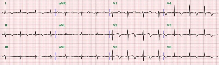
left-sided pleural effusion (Figure 1). ECG showed a si-
nus rhythm with a T wave inversion in leads II, III, and V2∼
V6 (Figure 2A). The cardiac enzyme showed CK-MB
10.28 ng/mL (normal 0.2∼2.88), troponin-I 0.063
ng/mL (normal <0.78), and pro-BNP 6,268 pg/mL
(normal 5∼125). Echocardiography revealed global hy-
pokinesia with a left ventricular ejection fraction of 42%
Figure 1. Chest X-ray showed cardiomegaly with left-sided and a moderate amount of pericardial effusion. The anti-
pleural effusion on the 13th day of illness. nuclear antibody was positive in 1:1,600 titers. The an-
Figure 2. (A) Electrocardiogram
showing a normal sinus rhythm
with T wave inversion in leads
II, III and V2 to V6. (B) After the
prednisolone treatment, the elec-
trocardiogram showed a nor-
mal sinus rhythm with normally
positive T wave.
80 J Rheum Dis Vol. 26, No. 1, January, 2019
Leptospirosis as Unusual Trigger of Systemic Lupus Erythematosus

ti-dsDNA, anti-Ro/La, anti-Smith, and anti-ribosomal P           timing of symptoms and response to antibiotics, fever
antibodies were positive. The complement levels were             and proteinuria are more likely to be related to
low (C3 13 mg/dL [normal 76∼139]; C4, 2.7 mg/dL                  leptospirosis.
[normal 12∼37]) and the direct Coomb’s test was                    The patient had an erythematous rash with multiple
positive. She fulfilled the Systemic Lupus International         crust on the nose and both cheeks and diffuse swelling on
Collaborating Clinics criteria for a diagnosis of SLE [7].       the whole face. In leptospirosis, the nonspecific macular
Therefore, she was diagnosed with SLE concomitant with           rash can be seen and is usually not severe, transient and
leptospirosis. The left ventricular dysfunction was be-          not itchy [9,10]. The patient’s rash was not consistent
lieved to be caused by the SLE flare, and treatment with 1       with the typical patterns observed in leptospirosis. A lu-
mg/kg/day of prednisolone was begun. The shortness of            pus rash is a butterfly-shaped rash that appears over the
breath, liver dysfunction, and pancytopenia improved af-         bridge of the nose and cheeks. Facial edema with crust ac-
ter 1 week of prednisolone treatment. The abnormal find-         companied by erythematous rash has rarely been re-
ings on ECG and chest X-ray were returned to normal              ported in SLE [11]. In the present case, the patient’s rash
(Figure 2B). The echocardiography conducted 2 weeks af-          improved drastically after prednisolone therapy. Therefore,
ter prednisolone therapy showed a normal left ventricular        the patient’s facial rash is considered to be a presentation
ejection fraction without pericardial effusion. The patient      of SLE.
was discharged with 60 mg of prednisolone. After dis-              At the time of admission, her symptoms were consid-
charge, the dose of prednisolone was reduced gradually to        ered to be infectious disease, but CRP was normal.
5 mg for 5 months. After one month, leukopenia, hair loss        According to Crouzet et al. [12], high CRP levels are ob-
and facial rash occurred. The dose of prednisolone was in-       served in most patients of leptospirosis. On the other
creased to 20 mg and her symptoms improved. The dose             hand, elevated CRP was 67%∼75% in a retrospective
of steroid was then tapered. The patient was on medi-            study of leptospirosis [13]. Therefore, a normal or only
cation with prednisolone 5 mg and hydroxychloroquine             slightly elevated CRP does not exclude a leptospirosis
in the outpatient clinic.                                        infection.
                                                                   The most severe clinical manifestation in this case was
                    DISCUSSION                                   left ventricular dysfunction due to myocarditis. Myocarditis
                                                                 is an immune-inflammatory state of the myocardium
  Leptospirosis has been reported worldwide, particularly        [14]. In many studies, myocarditis associated with lep-
during the rainy season in late summer or early autumn           tospirosis is rare and is improved by antibiotics therapy
[6]. The incubation period is usually 1∼2 weeks. A con-          [15]. In the present case, the clinical and laboratory find-
firmatory diagnosis is based on isolation of the organism,       ings deteriorated despite the antibiotics therapy but im-
on a positive result in the polymerase chain reaction, or        proved rapidly after prednisolone therapy. Therefore, my-
on seroconversion or fourfold or greater between the             ocarditis is considered to have developed by SLE rather
acute and convalescent specimens in the MAT method               than leptospirosis.
[8]. In cases with strong clinical evidence of leptospirosis,      In conclusion, this paper reported a case of newly diag-
a single antibody titer of >1:800 is evidence of a current       nosed SLE during the treatment of leptospirosis. As SLE
or recent infection.                                             and leptospirosis share similar clinical features, accurate
  In the present case, the early symptoms were fever,            diagnosis through high suspicion is essential for appro-
myalgia, and facial edema with rash. Flu-like illness, such      priate treatment. Leptospirosis may be considered a rare
as fever and myalgia, might be considered the pre-               cause of SLE, but further studies will be needed to de-
sentation of the septicemic phase of leptospirosis. The          termine the role of leptospirosis as a trigger factor for
patient’s fever and myalgia improved during antibiotics          SLE.
therapy but facial rash was not changed. In addition, pro-
teinuria, which was observed at admission, was not de-                                 SUMMARY
tected after the antibiotic treatment. Renal involvement
in leptospirosis is common and proteinuria, pyuria, and            This case highlights the presence of concurrent SLE and
microscopic hematuria are observed frequently in uri-            leptospirosis. As the symptoms of SLE are similar to lep-
nalysis [8]. According to the patient's travel history, the      tospirosis, accurate diagnosis through high suspicion is

www.jrd.or.kr                                                                                                            81
Jinhee Lee et al.
essential for appropriate treatment. ternational collaborating clinics classification criteria for
systemic lupus erythematosus. Arthritis Rheum 2012;64:
2677-86.
CONFLICT OF INTEREST 8. Budihal SV, Perwez K. Leptospirosis diagnosis: competancy
of various laboratory tests. J Clin Diagn Res 2014;8:
No potential conflict of interest relevant to this article 199-202.
9. Puca E, Pilaca A, Kalo T, Pipero P, Bino S, Hysenaj Z, et al.
was reported.
Ocular and cutaneous manifestation of leptospirosis ac-
quired in Albania: a retrospective analysis with implications
REFERENCES for travel medicine. Travel Med Infect Dis 2016;14:143-7.
10. Barnabe C, Fahlman N. Overlapping clinical features of lu-
1. Laxminarayana D. Molecular insights into systemic lupus pus and leptospirosis. Clin Rheumatol 2008;27 Suppl
erythematosus pathogenesis. Clin Med Insights Pathol 1:S23-5.
2014;20;7:7-9. 11. Castro LA, Davis DM, Davis MD, Bruce AJ, Pittelkow MR.
2. Esposito S, Bosis S, Semino M, Rigante D. Infections and Facial edema and crusted patches: a precursor to life-threat-
systemic lupus erythematosus. Eur J Clin Microbiol Infect ening acute systemic lupus erythematosus. J Am Acad
Dis 2014;33:1467-75. Dermatol 2007;56(5 Suppl):S126-7.
3. Bal AM, Bharadwaj RS, Gita N, Joshi SA, Thakare JP. 12. Crouzet J, Faucher JF, Toubin M, Hoen B, Estavoyer JM.
Guillain-Barre syndrome in a pediatric patient following in- Serum C-reactive protein (CRP) and procalcitonin (PCT)
fection due to Leptospira. Jpn J Infect Dis 2003;56:29-31. levels and kinetics in patients with leptospirosis. Eur J Clin
4. Urganci N, Kalyoncu D, Cayonu N, Erdem E, Yildirmak Y, Microbiol Infect Dis 2011;30:299-302.
Yilmaz B. Acute liver failure, autoimmune hepatitis, and lep- 13. Mori M, VAN Esbroeck M, Depoorter S, Decaluwe W,
tospirosis: a case report. Pediatr Emerg Care 2011;27:963-5. Vandecasteele SJ, Fretin D, et al. Outbreak of leptospirosis
5. Makhija P, Gopinath S, Kannoth S, Radhakrishnan K. A case during a scout camp in the Luxembourg Belgian province,
of post-leptospirosis autoimmune epilepsy presenting with Belgium, summer 2012. Epidemiol Infect 2015;143:1761-6.
sleep-related hypermotor seizures. Epileptic Disord 2017; 14. Navinan MR, Rajapakse S. Cardiac involvement in
19:456-60. leptospirosis. Trans R Soc Trop Med Hyg 2012;106:515-20.
6. Levett PN. Leptospirosis. Clin Microbiol Rev 2001;14: 15. Trivedi SV, Bhattacharya A, Amichandwala K, Jakkamsetti
296-326. V. Evaluation of cardiovascular status in severe leptospirosis.
7. Petri M, Orbai AM, Alarcón GS, Gordon C, Merrill JT, Fortin J Assoc Physicians India 2003;51:951-3.
PR, et al. Derivation and validation of the systemic lupus in-
82 J Rheum Dis Vol. 26, No. 1, January, 2019You can also read