Classical and computed tomographic anatomical analyses in a not-so-cryptic Alviniconcha species complex from hydrothermal vents in the SW Pacific ...
←
→
Page content transcription
If your browser does not render page correctly, please read the page content below
Laming et al. Frontiers in Zoology (2020) 17:12
https://doi.org/10.1186/s12983-020-00357-x
RESEARCH Open Access
Classical and computed tomographic
anatomical analyses in a not-so-cryptic
Alviniconcha species complex from
hydrothermal vents in the SW Pacific
Sven R. Laming1,2,3*, Stéphane Hourdez4, Marie-Anne Cambon-Bonavita2 and Florence Pradillon1
Abstract
The chemosymbiotic gastropod Alviniconcha (Provannidae), first described in 1988, is one of the most emblematic
hydrothermal-vent taxa described from the Central Indian Ridge and the Southwest (SW) Pacific. Symbiotic bacteria
found in the gill of Alviniconcha are thought to be their principal source of nutrition. In the SW Pacific, species
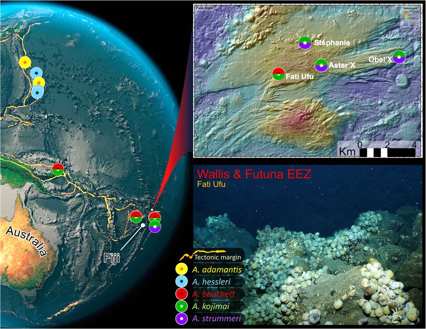
distributions for A. kojimai, A. boucheti – and to a lesser extent A. strummeri – overlap. While Alviniconcha species do
not appear to truly co-exist in these highly energetic but spatially limited habitats, certain species regularly co-occur
within a single vent field and in rare instances, the same edifice. Past research suggests that SW-Pacific Alviniconcha
species might aggregate around fluids with distinct geothermal profiles. These small-scale distribution patterns have
been attributed to differences in their symbiont assemblages or host physiologies. However, little is known about
the anatomy of most Alviniconcha species, beyond that detailed for the type species Alviniconcha hessleri, whose
geographic range does not overlap with other congeners. In fact, species within this genus are currently described
as cryptic, despite the absence of any comparative morphological studies to assess this. To test whether the genus
is genuinely cryptic and identify any functional differences in host anatomy that might also mediate habitat partitioning
in SW Pacific species, the current study examined the morphoanatomy of A. kojimai, A. boucheti and A. strummeri from
the Fatu Kapa vent field, an area of hydrothermal activity recently discovered north of the Lau Basin near the Wallis and
Futuna Islands and the only known example where all three species occur within adjacent vent fields. A combination of
detailed dissections, histology and X-ray computed tomography demonstrate that A. kojimai, A. strummeri and A. boucheti
are readily identifiable based on shell morphology and ornamentation alone, and therefore not truly cryptic. These traits
provide a rapid and reliable means for species identification. However, aside from some subtle differences in radular
morphology, these species of Alviniconcha exhibit conserved anatomical features, providing no evidence that functional
host anatomy is implicated in habitat partitioning. This provides support for the current belief that host-
species distributions are probably governed by symbiont-mediated physiological factors.
Keywords: Deep sea, Chemosymbiotic, Periostracum, Gastropod, Habitat partitioning, Computed tomography,
Histology, Taxonomy, 3D model
* Correspondence: svenlaming@ua.pt
1
Ifremer, Laboratoire Environnement Profond (REM/EEP/LEP), Plouzané,
France
2
Ifremer, Univ Brest, CNRS, UMR6197, Laboratoire de Microbiologie des
Environnements Extrêmes (REM/EEP/LM2E), Plouzané, France
Full list of author information is available at the end of the article
© The Author(s). 2020 Open Access This article is licensed under a Creative Commons Attribution 4.0 International License,
which permits use, sharing, adaptation, distribution and reproduction in any medium or format, as long as you give
appropriate credit to the original author(s) and the source, provide a link to the Creative Commons licence, and indicate if
changes were made. The images or other third party material in this article are included in the article's Creative Commons
licence, unless indicated otherwise in a credit line to the material. If material is not included in the article's Creative Commons
licence and your intended use is not permitted by statutory regulation or exceeds the permitted use, you will need to obtain
permission directly from the copyright holder. To view a copy of this licence, visit http://creativecommons.org/licenses/by/4.0/.
The Creative Commons Public Domain Dedication waiver (http://creativecommons.org/publicdomain/zero/1.0/) applies to the
data made available in this article, unless otherwise stated in a credit line to the data.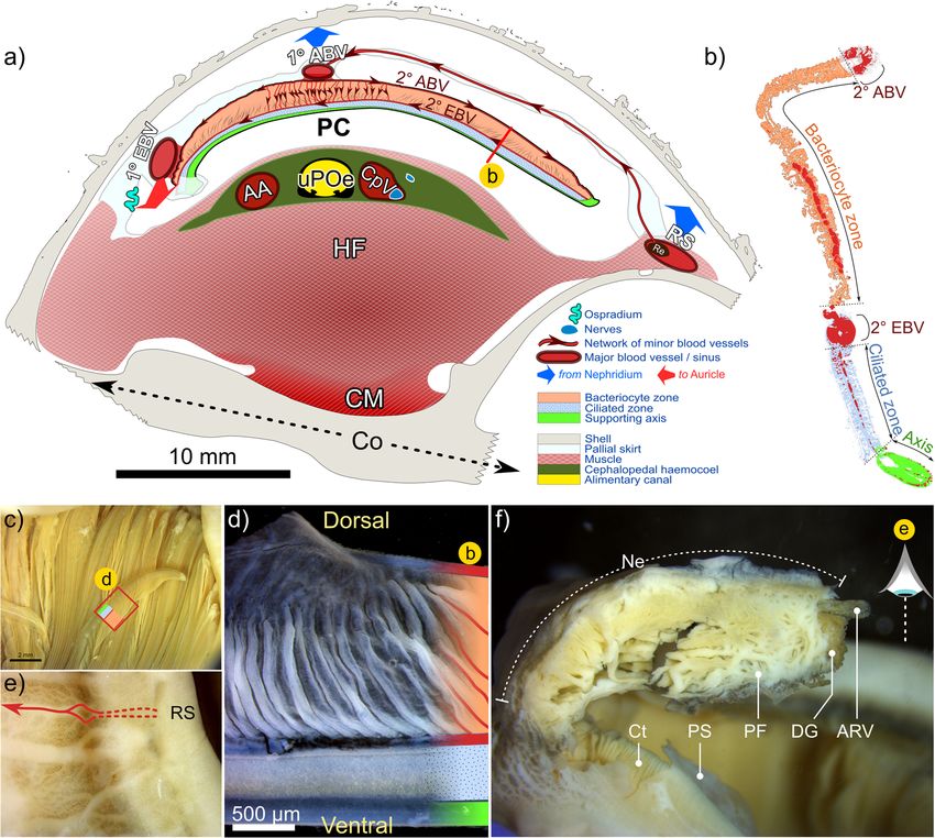
Laming et al. Frontiers in Zoology   (2020) 17:12                                                                Page 2 of 27

Background                                                     could be confidently related to a given clade ([1], but
The chemosymbiotic gastropod genus Alviniconcha                also in [8]). Few studies have presented species mor-
(Provannidae) is one of the most abundant, emblematic          phoanatomy in much detail to support either argument
taxa of hydrothermal-vent communities described from           ([9] presents gross anatomy, but in the context of a
the Central Indian Ridge and the SW Pacific, including         symbiosis-based paper) and none have employed an ex-
the Mariana volcanic arc and the Mariana, Manus,               haustive, comparative approach.
North-Fiji and Lau back-arc basins [1]. This genus was            The molecular assignments of Johnson et al. [1] have
erected in 1988, with a preliminary description of Alvini-     nonetheless helped to clarify the distribution of the six
concha hessleri Okutani & Ohta, 1988, based on speci-          species now described, all of which are obligate
mens from the Mariana Back-arc Basin [2]. Soon after in        hydrothermal-vent fauna. Geographic ranges of the five
1993, an emended diagnosis of this species provided a          described species from the Pacific abut one another at
detailed account of adult morphoanatomy and larval-            an oceanic-basin scale. Records of more than one Alvini-
shell characteristics for the first time [3], based on new     concha species over smaller spatial scales (< 100-km
Alviniconcha specimens collected from the vent sites           separation) are restricted to select regions in the SW
“White Lady” (North-Fiji Basin) and “Vai LiIi” (Lau            Pacific [1]. Current understanding indicates that separate
Basin, with additional juveniles from “Hine Hina”). In         species do not form mixed colonies, though they have
that paper, Warén and Ponder identified a high degree          been recorded in different places on a single edifice
of intraspecific plasticity in larval-shell morphology and     [11]. Alviniconcha kojimai and A. boucheti regularly
stated more generally that the species’ shell was “of little   occur in proximity to one another throughout their
use for classification” ([3]: p. 56). The same year, another   broad species distributions in the Manus and North-Fiji
study analysing the genetic diversity of Alviniconcha          Basins, sometimes within a single vent field at sites only
specimens from the same sites presented strong evi-            10–100 m apart. In the northern Lau Basin, the boundary
dence for the existence of multiple species [4]; unfortu-      of A. boucheti’s geographic range, they are both recorded
nately, material from the original Mariana Back-arc            from shared edifices but in these instances, one species is
population was lacking, precluding any confirmation that       always overwhelmingly dominant (A. boucheti dominates
A. hessleri was one of the species at these sites. Evidence    at Kilo Moana KM-2, all Tow Cam vent sites, and ABE-1,
for the existence of an Alviniconcha species complex has       while A. kojimai dominates at ABE-2 and -4). Alvini-
grown in the decades that have followed [5–8], as has          concha kojimai records continue farther south in the Lau
our understanding of a genus-wide dependency upon              Basin where, at Tu’i Malila vent sites, it occurs on adjacent
chemosymbiosis for nutrition (e.g. [9–13]), expanding          or shared edifices in lower numbers with the dominant
on earlier, pioneering research in this field [14, 15].        species A. strummeri [11], the latter being an otherwise-
Robust molecular diagnoses based on concatenated               rare member of the genus with a geographic distribution
marker gene analyses have recently been published for          restricted to the southern Lau basin and predominantly at
the remaining species: A. adamantis, A. boucheti, A. koji-     Tu’i Malila (at time of writing, see Fig. 1).
mai, A. marisindica and A. strummeri Johnson, Warén,              Alviniconcha form dense aggregations around the bases
Tunnicliffe, Van Dover, Wheat, Schultz & Vrijenhoek            and walls of active chimneys [17] which issue chemically
2015, resulting in six distinct evolutionary lineages in-      reduced fluids that provide both energy and carbon for
cluding that of A. hessleri [1]. An inherent outcome,          chemosynthesis. High thermal tolerances [18] and bran-
however, is that species identification remains entirely       chial, chemosynthetic, bacterial symbioses [9–15] which
dependent upon molecular analyses. Conflicting ac-             facilitate high potential growth rates [18] and probably
counts may be found in the literature of the utility of        represent the host’s primary source of nutrition [19], en-
morphological features in distinguishing Alviniconcha          able Alviniconcha to take direct advantage of these re-
species. Denis and colleagues [4] commented on what            sources. Residing in the host’s hypertrophied ctenidium,
they believed to be species-specific shell features as         these intracellular – but occasionally intracytoplasmic or
further evidence to support their hypothesis for the pres-     intravacuolar [15] – symbionts may also provide a second-
ence of multiple species in Alviniconcha; this was also        ary fluid-detoxification role. Denis et al. [4] were the first
touched upon, albeit briefly as a side note to a prelimin-     to identify a potential correlation between the spatially
ary description of another provannid from the SW               discrete – but often proximate – distribution patterns ob-
Pacific. Yet, several studies have stated the opposite, be     served for SW Pacific Alviniconcha species and local-scale
it because of confounding phenotypic plasticity (inad-         geochemistry. Research since has suggested that these
vertently based on undescribed species rather than the         small-scale differences in host-species distributions across
species thought to be under investigation [3, 16]), or –       sites with marked differences in fluid composition, could
as exemplified by the molecular diagnoses for Alvini-          relate to the distinct bacterial symbiont assemblages that
concha – because no distinguishing morphological traits        each host species possesses [10, 11]. Most Alviniconcha
Laming et al. Frontiers in Zoology      (2020) 17:12                                                                                       Page 3 of 27

 Fig. 1 Map of SW-pacific region, Alviniconcha species records and Fatu Kapa vent-field study sites. Photo of habitat taken in northern area of Fati
 Ufu during cruise. The current distribution records for the five Pacific species indicate that only A. kojimai and A. boucheti are found together with
 any regularity. The distribution of the sixth species, A. marisindica, is restricted to sites on the Central Indian Ocean Ridge (not shown). Image
 credits: Globe Amrita Carroll; Fatu Kapa Map Futuna 3 cruise, AUV AsterX; Fati Ufu site photo Futuna 3 cruise, HOV Nautile

species, including A. kojimai and A. strummeri, host                          undermine our capacity to validate this hypothesis. It is
Gammaproteobacteria-dominated symbioses that are                              not yet known whether there are adaptive functional
closely related to known chemoautotrophic [9–11, 14, 15],                     traits specific to each host species that might also be im-
sulphur-oxidising bacteria that predominantly use the Cal-                    plicated in the distribution of SW Pacific Alviniconcha
vin–Benson–Bassham cycle for carbon fixation [20]. In                         species. More fundamentally, such gaps impede real-
the remaining two species, A. boucheti and A. marisindica,                    time species identification. Currently, the discrimination
symbioses are dominated by sulphur-oxidising bacteria                         of Alviniconcha to species level requires a molecular
from a separate phylum, the Campylobacterota [9, 10] –                        approach involving DNA extraction, amplification and
formerly known as the Epsilonproteobacteria [21, 22] –                        sequencing. This is typically performed after sample
that also utilise H2-oxidation [23–25] under high-H2 con-                     collection and processing and therefore cannot inform
centrations (several mM, [25]) and likely fix carbon                          experimental design and sampling strategies a priori or
through the Reverse Tricarboxylic Acid cycle [20]. Thus,                      alterations frequently necessary due to the logistics of
one hypothesis that might explain the small-scale habitat                     sampling deep-sea environments. Anatomical studies
partitioning observed in SW Pacific Alviniconcha species,                     can provide great insight regarding the evolutionary ad-
is that optimal conditions for chemosynthesis differ for                      aptations that organisms have developed to survive in
each host species, as a function of the unique metabolic                      their environment. For chemosymbiotic species, accom-
capabilities or physiological requirements of their sym-                      modating bacterial partners at high densities typically re-
biont assemblages. Interestingly, phylogenetic analyses of                    quires the alteration of an existing organ, such as tissue
recently published symbiont genomes from SW pacific                           hypertrophy and differentiation in gills (many examples
Alviniconcha species indicate that chemoautotrophic                           in [26]) or the oesophageal gland [27, 28], or the emer-
function in symbiotic bacteria is similar irrespective of                     gence of a novel specialised organ such as the siboglinid
host species [24], suggesting differences relating to gene                    trophosome [29], dedicated to this purpose. Diverse ad-
expression (e.g. [25]) and regulation and/or differences in                   aptations such as these are typically discovered through
host physiology may mediate habitat partitioning.                             detailed histological and anatomical analyses, offering
  However, knowledge gaps concerning Alviniconcha                             fascinating new ecological insights and evolutionary
anatomy – documented in the type species only –                               perspectives. However, due to a continued focus on
Laming et al. Frontiers in Zoology (2020) 17:12 Page 4 of 27
symbiotic tissues in most Alviniconcha species, accounts Additional file 1. The interactive model for A. strummeri
of remaining host anatomy are limited and sparse in is embedded in Additional file 2.
detail. To address this, the current study documents the
comparative morphoanatomy of members of this genus Shell morphology and ornamentation
from the Fatu Kapa vent field, an area of hydrothermal General observations
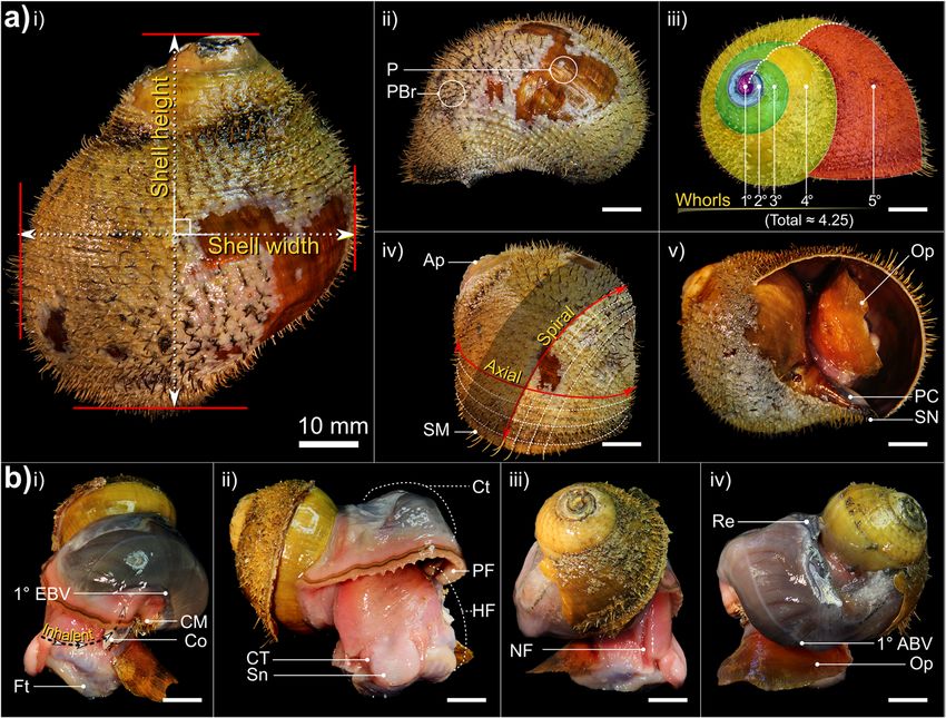
activity recently discovered north of the Lau Basin near Shells are dextral, hirsute and flexible with little minera-
the Wallis and Futuna Islands. This region is not only lised support, except the columella, which is calcareous
highly active, geothermally [30] and rich in metals and robust. Shell outlines are subglobular (e.g. Fig. 2,
[31–33] but also a rare instance where A. kojimai, A. Additional file 3). The number of whorls is dependent
boucheti and A. strummeri are documented at sites on shell height (SH) with maximum of 4.5–5 whorls at
only a few kilometres apart: an ideal scenario for examin- SH > 70 mm (Fig. 2a-iii). The larval protoconch is invari-
ing comparative host biology. The current study employs ably absent and spires are rounded (presumably due to
a comparative anatomical approach using morphology, dissolution and mechanical erosion), revealing a thick
microscopy and computed tomography (CT) to assess multi-layered periostracum and argenate ostracum. The
whether: 1) the three congeneric gastropod species A. koji- body whorl is greatly enlarged at around ~ 70–80% SH
mai, A. boucheti and A. strummeri, occurring in close (Fig. 2a, Additional file 3a). The aperture is large, sub-
proximity, are actually cryptic as documented, and; 2) oval with a slight parietal callus. The columella possesses
whether there are species-specific aspects of anatomy, out- a small siphonal notch only (Fig. 2a, Additional file 3a).
side of symbioses, that might be driving hypothesised All three study species possess a dense, regular, spiral ar-
habitat partitioning. rangement of non-calcareous periostracal bristles that
adorn the latter whorls, being absent on the apex. In all
Results species, bristle lengths (BL) within a single spiral row
Species distribution and identification were approximately congruent, with slight decreases in
Although species distribution data is limited for the length spirally, with decreasing whorl width. Within each
current samples collected in the Wallis and Futuna volcanic BL class (see next section), medial bristles were also
region, we know that at least some specimens of A. kojimai slightly longer than those nearest sutures, the junctures
and A. strummeri were taken from single gastropod between adjacent whorls (e.g. Fig. 2a, v). However, BL
patches, recorded for the first time (co-occurring less than displayed very striking, species-specific arrangements
a metre apart). Unfortunately, data on the proximity of A. when viewed axially (i.e. along co-marginal bristle lines,
boucheti and A. kojimai when co-occurring are unavailable, discussed below).
as specimens were a pooled collection from multiple sam-
pling locations during a dive where both species were found Species-specific shell characteristics
(though still only several metres apart). Mean SH for A. kojimai, A. strummeri and A. boucheti
Nucleotide sequence data from 106 specimens target- were SH 60.08 mm ± σ 14.2 (SHrange = 37.8–89.1 mm,
ing the 1228 bp fragment of the mitochondrial gene, n = 44), 55.33 mm ± σ 10.2 (SHrange = 46.1–53.7 mm, n =
cytochrome-c oxidase subunit I (COI), have been depos- 4) and 65.19 mm ± σ 5.2 (SHrange = 57.1–77.6 mm, n =
ited in GenBank (see Table 1). Anatomical descriptions 14) respectively, measured in a random subset of indi-
detailed below are listed by functional system. For each viduals. In specimens from the current study site, perios-
subsection, features applicable to all three study species traca were typically laurel-green to light brown in A.
(exemplified at times by figures featuring a single spe- kojimai and A. strummeri (due in part to a thin white
cies) are described first, followed by features identified layer of bacterial flocculent) and burnt-umber brown in
as specific to individual species, where applicable (sum- A. boucheti (Table 2). In A. kojimai and A. strummeri, a
marised in Table 2). Note that interactive anatomical columellar fold arises internally in the first (apical) whorl
models are available for CT-based visualisations per- and descends as an adapical spiral as far as 0.5 whorls
formed on A. kojimai and A. strummeri specimens. The back from the aperture opening (Fig. 3b, Add-
interactive model for A. kojimai is embedded in itional file 4). This fold is particularly pronounced in A.
Table 1 GenBank Accession numbers by species and site for the three Alviniconcha spp.
A. kojimai (66) A. strummeri (8) A. boucheti (32)
Vent sites Stéphanie (5) MT010417 – MT010420 (4) MT010483 (1) -
Obel’x (28) MT010421 – MT010444 (24) MT010484 – MT010487 (4) -
Aster’x (24) MT010445 – MT010465 (21) MT010488 – MT010490 (3) -
Fati Ufu (49) MT010466 – MT010482 (17) - MT010491 – MT010522 (32)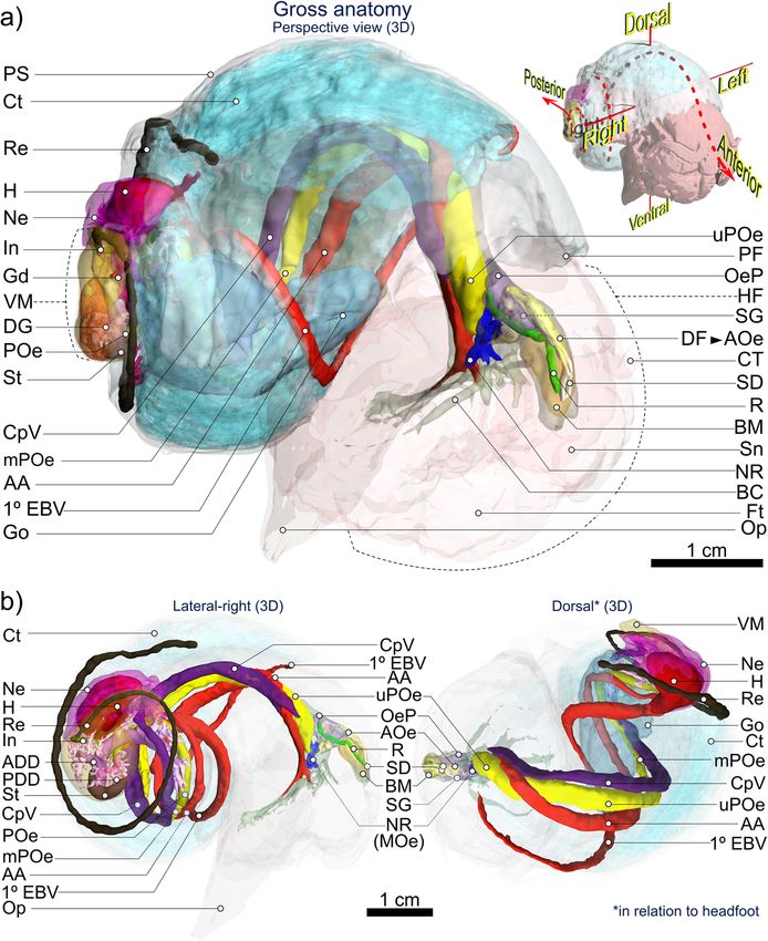
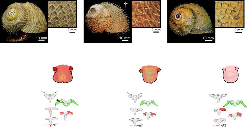
Laming et al. Frontiers in Zoology           (2020) 17:12                                                                                                  Page 5 of 27

Table 2 Summary of distinguishing characteristics among the three Alviniconcha spp.

Species were readily distinguishable based on periostraca. BL bristle length, RT rachidian, LT Lateral teeth. Mean BL calculated based on lengths from 10
individuals of A. kojimai and A. boucheti and 5 individuals of A. strummeri (based on three replicates ea.). *A fourth 2° bristle can occur in A. kojimai, very
occasionally and sporadically. † Photo is of an alcohol-fixed specimen (other photos from frozen specimens)

strummeri in which the columella is notably thicker (rela-                             ambiguous A. kojimai specimens were usually distinguish-
tive to shell width) than that of A. kojimai and A. boucheti                           able from A. boucheti. Contrary to the other species, bris-
(Additional file 4). A columellar fold is absent in A. bou-                            tles in A. strummeri were universally congruent with no
cheti (Fig. 3b, Additional file 4). The arrangement and                                obvious axial differences in BL (except general trends de-
lengths of periostracal bristles follow species-specific pat-                          scribed in 4.2.1) and comparatively longer than the longest
terns (observed in ninety specimens, Fig. 3a). In A. bou-                              (1°) BL of the other study species, with greater mean BL in
cheti and A. kojimai, BL alternated markedly in length                                 adults of equivalent SH (Table 1, Fig. 3).
axially (Table 2, Fig. 3a). Three discrete BL were identifi-
able in A. boucheti (− 80 °C, n = 29): long primary (1°),                              External morphoanatomy
shorter secondary (2°, at ~ 50% of primary BL) and much                                General observations
shorter tertiary bristles (3°, at < 10% of primary BL). Be-                            In a 4-whorl shelled specimen (~ 50–60 mm SH), soft-
tween each spiral row of 1° bristles were three rows of the                            body occupies the last (i.e. most recently formed) 2–2.5
shorter bristles, arranged in a − 3°-2°-3°- sequence (Fig. 3).                         whorls. The muscular head-foot occupies the last 0.1–0.25
In A. kojimai (− 80 °C, n = 52; 4%-formalin-fixed, n = 2),                             whorls, depending on the degree of extension beyond the
two to three markedly different BL were identifiable. Lon-                             aperture. When preserved, the snout is short with tight,
ger (1°) bristles were easily identifiable. Between each                               transverse epidermal folds both dorsally and ventrally,
spiral row of 1° bristles were three rows (occasionally four)                          suggesting some capacity for elongation. Two cephalic
of much shorter 2° bristles (5–10% of primary BL). The 2°                              tentacles, lacking basal eye spots, flank the snout (~ 2 x
bristles were generally congruent but in a minority of indi-                           snout length). The ventral underside of the snout is flush
viduals, medial 2° bristles were almost double the length                              with the anterior flank of the propodium and whitish-pink
of other 2°, thus superficially resembling the bristle ar-                             in colour, with a small distal mouth concealed from view
rangement of A. boucheti. However, a lattice-like network                              dorsally. In chemically preserved specimens, the mouth
of minute spiral and axial ridges connects bristles to one                             was tightly contracted and thus difficult to discern. The
another at their bases in A. kojimai (Additional file 3), a                            foot is large, wide and fleshy; the sole possesses translu-
feature not evident in the other two study species (Table                              cent pale blue-purple bulges within which are the pedal
1), though this feature can be obscured by bacterial floccu-                           blood sinuses (Additional file 3v, the anterior-most, med-
lent (as in Fig. 3a). By employing this characteristic,                                ial propodal bulge being considerably larger and ovoid:
Laming et al. Frontiers in Zoology      (2020) 17:12                                                                                        Page 6 of 27

 Fig. 2 Aspects of gross morphology and explanation of certain malacological terms. Example is A. boucheti: a i) abapertural view with shell
 dimensions, ii) comparison of bare (PBr) and adorned periostracum (P) in lateral-left view, iii) spiral distance from the apex, measured in whorls, iv)
 spiral and axial lines in relation to the apex (Ap) and shell margin (SM) and v) apertural view with only a slight parietal callus (PC) and siphonal
 notch (SN) present; b lateral-left, anterior, lateral-right and near-posterior views of a specimen with a whorl removed. An equivalent micrograph
 plate for A. kojimai may be found in Additional file 3. NB. the ctenidium (Ct) in A. boucheti is darker in appearance than that of A. kojimai and A.
 strummeri. Additional abbreviations: 1° ABV Primary afferent branchial vessel; 1° EBV Primary efferent branchial vessel; CM Columellar muscle; Co
 Columella; CT Cephalic tentacle; Ft Foot; HF Head-foot; NF Neck furrow; Op Operculum; PF pallial fringe; Re Rectum; Sn Snout

30% foot-width laterally). On both sides of the foot, a lat-                  head-foot laterally, posterior-ventral to the neck furrow’s
eral epipodial fold extends anteriorly from beneath the at-                   steepest point of descent. On the left, the pallial margin
tachment region for the operculum, terminating below                          is attached via a thickening, siphon-shaped fold of
the cephalic tentacles. A deep cephalopedal channel, or                       muscle (inhalant region, Fig. 2b, i), adhered to the shell
neck furrow (Fig. 2b-iii and Additional file 3, ‘NF’), demar-                 via the columellar muscle ~ 0.5 whorls back (itself con-
cated by parallel epidermal folds, descends from within                       tinuing ~ 0.75 whorls farther). The pallial cavity extends
the pallial cavity down the right flank of the head-foot, be-                 1–1.25 whorls back from the pallial margin (~ 0.1 whorls
coming wider and less defined as it terminates below the                      behind the outer apertural lip), decreasing rapidly in vol-
right cephalic tentacle on the anterior-most point of the                     ume posteriorly as the cavity narrows and loses height
epipodial fold. In some specimens, the neck furrow pos-                       (A. boucheti, Fig. 2b; A. kojimai, Fig. 4, Additional files 1
sesses another narrow, central fold (Additional file 5a),                     and 2; A. strummeri, Additional file 2). Viewed dorsally,
thus creating twinned channels, which ultimately diverge                      the ctenidium (extending left-to-right) and rectum (far-
below the right cephalic tentacle. The operculum is at-                       right) are visible through the pallial epithelium, which is
tached to the metapodium along the posterior-most edge                        delicate and translucent (e.g. Fig. 2). On the right side of
of the epipodial folds and is similar in all three species:                   the pallial cavity in areas where ctenidium is absent, the
ovo-quadrate, thickened proximally, with an irregular,                        pink (anterior) or purple-black (posterior) epithelial
curved distal margin. The nucleus is not evident.                             surface of the underlying cephalopedal haemocoel can
  The anterior pallial margin is muscular, possessing a                       be seen.
large dark-coloured co-marginal vessel and a distal an-                          The anterior-most cavity of the cephalopedal haemocoel
terior fold, from which project numerous, stout, tapering                     (i.e. the buccal cavity) is relatively narrow, laterally, and
papillae, each with a medial, dorsal fold. Papillae are                       tall, dorsoventrally (Figs. 4 and 5, Additional file 1 +
longest on the right of the pallial margin, small and                         model, Additional file 2 + model). In medial sagittal cross-
indistinct on the far left. On the right, the final few milli-                section it appears roughly inverse-triangular, comprising;
metres of pallial margin deflect inwards, attaching to the                    1) the ventrally directed region that houses the
Laming et al. Frontiers in Zoology      (2020) 17:12                                                                                       Page 7 of 27

 Fig. 3 Distinguishing morphoanatomical characteristics of the shell and head-foot . External, species-specific, morphological features include: a
 the lengths and arrangement of primary (1°), secondary (2°) and tertiary (3°) periostracal bristles; b the architecture and diameter of the columella
 where arrowheads indicate the columellar fold (absent in A. boucheti) and; c head-foot coloration (arrowheads indicate neck furrows).
 Abbreviations: CT Cephalic tentacles; Ft Foot; Mo Mouth; PF Pallial fringe; Sn Snout

circumoesophageal complex, comprising the oesophageal                         the buccal mass. Posterior to the buccal cavity, the cepha-
nerve ring and associated circulatory system branching                        lopedal haemocoel widens and flattens dorsoventrally,
out within the foot; 2) the anterior region – within the                      with irregular undulations visible externally on the pallial
snout – housing the buccal complex (i.e. mouth, buccal                        floor. When present, the anterior-most region of the
mass, anterior oesophageal opening and anterior                               gonad is visible as a bulge in the pallial floor on the far-left
oesophagus, salivary glands and buccal ganglia) and                           side of the pallial cavity level with the anterior end of colu-
nerves/blood-supply to the cephalic tentacles and; 3) the                     mellar muscle and continues posteriorly, until it meets
posteriorly directed (and dorsal-most) region, through                        with the pallial skirt and thus the cardio-renal complex
which the upper posterior oesophagus (posterior to the                        (Fig. 4, Additional file 1 + model, Additional file 2 +
oesophageal nerve ring), several nerves and two blood ves-                    model).
sels all pass (Figs. 4 and 5, Additional file 1 + model). The                    The visceral hump (and the visceral haemocoel within),
muscle wall from the right-to-dorsal side of the head-foot                    located posterior to the pallial skirt, occupies ~ 1 whorl,
is over twice as thick as the left, with the cephalopedal                     comprising the digestive glands, the cardio-renal complex,
haemocoel displaced left of the medial line posterior to                      lower posterior oesophagus, stomach and intestine (Fig. 4,
Laming et al. Frontiers in Zoology       (2020) 17:12                                                                                          Page 8 of 27

 Fig. 4 3D visualisation of Alviniconcha gross anatomy based on computed tomography (CT). Images are from the A. kojimai specimen scanned
 using computed tomography. A more detailed interactive model of this specimen is embedded in Supplementary figure 1 and a second interactive
 model for A. strummeri is included in Supplementary figure 2. Note that for clarity purposes, some tissues and features present in the interactive
 model, are not included in the above 3D visualisation (but see Figs. 6 and 8). Abbreviations: 1° EBV Primary efferent branchial vessel; AA Anterior aorta;
 ADD Anterior digestive duct; AOe Anterior oesophagus; BC Buccal cavity; BM Buccal mass; CpV Cephalopedal Vein; Ct Ctenidium; CT Cephalic
 tentacles; DF Dorsal fold; DG Digestive gland; Ft Foot; Gd Gonoduct; Go Gonad; H Heart; HF Head-foot; In Intestine; MOe Mid-oesophagus; mPOe Mid-
 posterior oesophagus; Ne Nephridium; NR Oesophageal nerve ring; OeP Oesophageal pouches; Op Operculum; PDD Posterior digestive duct; PF Pallial
 fringe; POe Lower posterior oesophagus; PS Pallial skirt; R Radula; Re Rectum; SD Salivary ducts; SG Salivary glands; Sn Snout; St Stomach; uPOe Upper
 posterior oesophagus; VM Visceral mass. Figure is of 3D renderings with exaggerated perspective, thus scales are approximate

Additional file 1 + model and Fig. 5b). The short intestine                     strummeri (Fig. 3). When contracted, the snout appears
soon merges into the rectum in the anterior-most region                         to be wider distally in A. kojimai, at times almost pear-
of the visceral hump, returning along the far-right side of                     shaped; in A. strummeri, near-square shaped and nar-
the pallial cavity for much of its length.                                      rower and in A. boucheti, the snout is very broad and
                                                                                round. However, these differences do not hold true for
Species-specific morphoanatomical characteristics                               live specimens (F. Pradillon, unpublished observations).
Muscle tissue in frozen specimens is pale pink in A. bou-                       The mouth is encircled by bright-red tissue in frozen
cheti but decidedly darker in A. kojimai and A.                                 specimens of A. kojimai (Additional file 3v).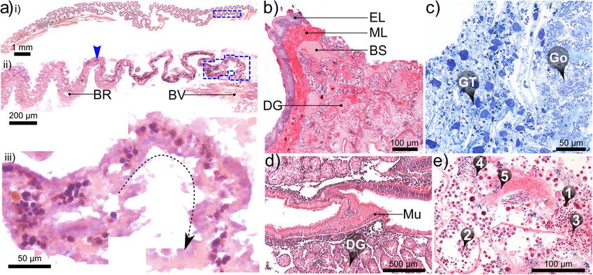
Laming et al. Frontiers in Zoology     (2020) 17:12                                                                                      Page 9 of 27

 Fig. 5 Elements of the alimentary and digestive system in Alviniconcha. Micrographs depict: a the anatomy of the buccal cavity; b internal and
 external morphology of the alimentary canal including: i) upper posterior oesophagus (uPOe) – posterior to the osophageal nerve ring encircling
 the mid-oesophagus (NR + MOe) – with ventral folds clearly visible and ii) transverse incision of uPOe immediately prior to mid-posterior
 oesophageal juction (in iii and iv) with T-profile of ventral folds highlighted. In iii) the mid-posterior oesophageal junction is marked by a
 decrease in diameter and (iv) the emergence of numerous dorsal folds, which rapidly become established (v). In vi) the lower posterior
 oesophagus (POe) enters the visceral haemocoel and communicates with the stomach posteriorly: arrow indicates the direction of travel. A
 schematic overview of stomach (St) anatomy in presented in c including micrographs of several anatomical features; in d scanning electron
 micrographs of pristine posterior regions of radulae from each species are provided. Pictures in a-c are from a formalin-fixed A. kojimai specimen.
 Additional abbreviations: AA Anterior aorta; ADD Anterior digestive duct; AOe Anterior oesophagus; BM Buccal mass; CpV Cephalopedal Vein; CT
 Connective tissue; DF Dorsal fold; DG Digestive gland; GS Gastric shield; In Intestine; MaT Major typhosole; MiT Minor typhosole; Mo Mouth; OeP
 Oesophageal pouches; PDD Posterior digestive duct; R Radula; Re Rectum; SA Sorting area; SS Style sac

Alimentary system overview                                                   and posterior to the nerve ring, between which is the mid-
As in all gastropods, the alimentary system is U-shaped.                     oesophagus, the short, constricted region that passes
In order of occurrence, the descending limb is composed                      through the oesophageal nerve ring. For additional clarity
of a mouth; buccal mass; relatively small salivary glands,                   however, the long posterior oesophagus is subdivided
and the long oesophagus, which extends from the buccal                       again herein into three regions, distinguishable by changes
cavity to the visceral hump, where it turns abruptly to                      in diameter and their internal morphology (Figs. 4, 5, 6,
enter the returning stomach posteriorly (Figs. 4, 5, 6,                      detailed later): the upper- (extending most of the cephalo-
Additional files 1 and 2 + models). Following conven-                        pedal haemocoel), mid- (posterior-most cephalopedal
tional nomenclature, the oesophagus is composed of                           haemocoel) and lower-posterior oesophagus (almost en-
three regions delineated by the location of the oesophageal                  tirely within visceral haemocoel). The returning limb of
nerve ring; the anterior and posterior regions are anterior                  the ‘U’ comprises the small stomach, which tapers
Laming et al. Frontiers in Zoology (2020) 17:12 Page 10 of 27
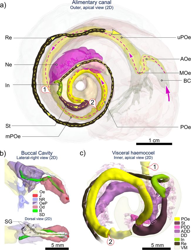
Fig. 6 2D (flattened) visualisation of the Alviniconcha alimentary system based on CT. Images are from the CT-scanned A. kojimai specimen.
Pictured: a overview of the alimentary system in Alviniconcha (excluding digestive glands), displaying the route of ingested material on its
passage between the mouth (large pink arrow) and the end of the rectum (Re); b enlarged view of buccal cavity with jaws (J) and radula (R),
nested in the curved odontophore (Od), with associated salivary ducts (SD) terminating in salivary glands (SG) left of the oesophagus (Oe) and
blind oesophageal pouches (OeP); c enlarged inner-side view of visceral mass (VM) showing the relative positions of the stomach (St) and
associated anterior and posterior digestive glands (ADD, PDD), lower posterior oesophagus (POe), intestine (In) and rectum. ① is upper-to-mid-
posterior oesophageal junction; ② is mid-to-lower posterior oesophageal junction. Figure is of flattened 2D renderings with exact scales.
Additional abbreviations: AOe Anterior oesophagus; BC Buccal cavity; DD digestive diverticula; MOe Mid-oesophagus; mPOe Mid-posterior
oesophagus; Ne Nephridium; NR Oesophageal nerve ring; uPOe Upper posterior oesophagus
anteriorly to form the style sac, the associated digestive anteriorly (Fig. 6). Salivary glands are found in close as-
glands, and ultimately the morphologically similar short sociation with the oesophagus, dorso-anterior to the
intestine and much longer rectum. oesophageal nerve ring but posterior to the buccal mass
(Figs. 4, 5, 6, Additional file 1 + model). They communi-
Buccal complex cate with the buccal cavity via separate, narrow dorsal
The buccal mass is small at 3–4 mm in length (SH ~ ducts, entering dorsally above the jaws (Fig. 6). Orien-
40–60 mm) and heart-shaped, narrowing ventrally and tated as an inverted “V” on the dorsal buccal wall, theLaming et al. Frontiers in Zoology (2020) 17:12 Page 11 of 27
two simple jaws are light brown, semi-circular, fragile, the limited number that were measured. In A. koji-
with minutely denticulate cutting edges (Fig. 6, Add- mai, SH of 50.4–78.8 mm equated to RL of 6.46–10.2
itional file 6a). The radular sac (and posterior end of mm (n = 3), in A. strummeri SH of 46.1 and 51.6 mm
radula) lies to the right of the oesophagus and is slightly equated to RL of 6.35 and 6.60 mm respectively and
coiled, dorsally (Figs. 5a, 6, Additional files 1 and 2 + in A. boucheti, for SH of 52.0 and 66.6 mm, RL were
models). 6.9 and 9.7 mm respectively. Alviniconcha kojimai
possesses ~ 35–37 transverse rows of teeth mm− 1, A.
Radular morphology strummeri ~ 43 rows mm− 1 and in A. boucheti ~ 32–
General observations 35 transverse rows mm− 1. The aforementioned M-
In all cases, radulae are taenioglossate with a 2–1-R-1-2 shaped supporting ridge of the rachidian is robust in
tooth arrangement including a central wider-than-long, A. kojimai, less so in the other species, Fig. 5d). The
fairly solid rachidian tooth, flanked on either side by a inner and outer-halves of lateral-tooth cusps appear
row of lateral teeth and two rows of marginal teeth, that near-symmetrical in A. kojimai and A. strummeri,
are longer-than-wide and more flexible (Fig. 5d). The however the outer half is weakly inflated in A. bou-
rachidian base is twice as wide as its cusp. The rachidian cheti (Fig. 5d, Table 2). In A. kojimai, lateral-tooth
cusp’s central denticle is broad, conical and flanked by central denticles are longer than rachidian central
4–6 (usually 5) narrower lateral denticles on either side, denticles, while in A. strummeri and A. boucheti, the
decreasing progressively in length. The centre of the lateral-tooth and rachidian central-denticle lengths are
rachidian is inflexed, extending laterally to form a low roughly congruent (Fig. 5d, Table 2).
transverse M-shaped supporting ridge, basal to the
rachidian cusp. In the lateral teeth, the base is wider The oesophagus and proximal tissues
than the cusp with sides curled dorsally and a sub- The anterior oesophagus, exiting the buccal mass,
cuspid inflection forms a broad, poorly defined, basal quickly becomes voluminous, with irregular, blind, dor-
denticle. Each lateral tooth’s outer edge is greater than sal pouches directed posteriorly (Fig. 5a). Two con-
twice the length of inner edge, pushing lateral teeth to- spicuous parallel, dorsal longitudinal folds are visible
wards the rachidian when the radular membrane lies flat. internally through the delicate and translucent lining
Lateral tooth cusps are similar in width and shape to the (Fig. 5b-i), originating from the roof of the buccal cavity
rachidian cusp, though the cusp profile is less angular. (dorsal fold origin, ‘DF’, in Fig. 5a). These two folds are
Each lateral tooth’s central denticle is tetrahedral, T-shaped in cross-section and ca. 1 mm apart, forming
broader-based than rachidian’s central denticle and an alimentary channel (Fig. 5b-ii). The oesophagus then
flanked by 3 inner and 4–5 outer denticles of much descends abruptly (ventrally) while spiralling dextrally,
shorter length. The inner and outer marginal teeth are becoming the highly constricted mid-oesophagus as it
the longest teeth of the radula and very similar in ap- passes through the narrow oesophageal nerve ring and
pearance within and between species, with the width of turns towards the right with the alimentary channel
the marginal-teeth cusps being about ~ 1.5 x that of the now ventral. It then more than doubles in width
base. The marginal teeth are inflexed centrally forming a laterally (Fig. 5a, b) forming the (upper) posterior
shallow, longitudinal, supporting ridge (wider and more oesophagus on its ascent towards the region of the
pronounced distally) and are adorned with squared-off, cephalopedal haemocoel beneath the pallial floor. This
rake-like cusps with numerous congruent denticles con- upper region of the posterior oesophagus continues for
tinuing for a short length marginally (numbering ~ 21– ~ 0.75 whorls posteriorly with no change in morph-
24). The outer edge of the outer marginal tooth is tightly ology or diameter (as depicted in Fig. 5b). It is flanked
curled along its length. No interspecific differences were on the right by the returning cephalopedal vein and (at
identified in marginal-tooth morphology. least) the right visceral connective and on the left by
Radular teeth appear more open anteriorly where the the anterior aorta – delivering blood to the head-foot –
radular membrane curves over the surface of the chitin- originating from the ventricle (Fig. 4, Additional files 1
ous sheath that houses the odontophore cartilage. and 2 + models). This artery passes over the upper-pos-
Teeth here are faintly yellow/brown as opposed to terior oesophagus dorsally from left-side-to-right, just prior
colourless (remaining posterior teeth) and notably to descending into the buccal cavity (~ 8–10 mm posterior
eroded (Additional file 6b). to oesophageal nerve ring, Fig. 4, Additional files 1 and 2 +
models).
Species-specific radular characteristics Much of the cephalopedal haemocoel is occupied by
Radular morphology differs slightly in each species loosely bound, diffuse, almost spongy, granular connective
(Fig. 5d). Radula lengths (RL) depended on size but tissue. This tissue is darker and so more conspicuous pos-
were similar in each species at a given SH, based on teriorly (due to presence of fine black particulates, Fig. 7c);Laming et al. Frontiers in Zoology (2020) 17:12 Page 12 of 27 Fig. 7 Histological micrographs of select tissues in Alviniconcha. a Increasingly magnified views (corresponding to blue-boxed regions of previous image) of branchial filaments in oblique longitudinal section: arrowhead in ii) identifies the accumulation of cellular debris in the blood lacuna within the bacteriocyte region (BR) with iii) a higher magnification view of the junction with a large blood vessel (BV in ii, likely the secondary efferent branchial vessel); b transverse section through the outer layers of the posterior digestive gland (DG) bathed in blood sinuses (BS) and enclosed in delicate muscle and epithelial layers (ML and EL); c section cut from granular tissue (GT), a spongy tissue composed of a loose, fibrous matrix, densely populated with accretions of various sizes, found throughout the cephalopedal haemocoel but particularly around the mid-posterior oesophagus (not pictured), which passes through the gonad (Go), when present; d granular deposits also accumulate in connective tissue between the DD and the stomach mucosa (Mu); e various stages of spermatogenesis could be readily identified in males examined histologically: spermatogonia, spermatocytes, spermatids, spermatozoids and spermatozoa (❶ - ❺ respectively). Image mosaics a & d are from 7 μm-thick paraffin sections (formalin-fixed); b, c (both alcohol-fixed) & e (formalin-fixed) are from 2 μm-thick LR-white sections. All tissues are from A. kojimai, except (c), which is from A. strummeri in larger specimens, the dark-grey to black colouration appearance (tissue appears convoluted, but with no evi- extends farther anteriorly. It occupies the lateral space in dent ducts or tubules in histological sections, Fig. 7c). The the cephalopedal haemocoel between the lighter-coloured junction between the mid- and lower posterior anterior aorta, oesophagus and cephalopedal vein, which oesophagus is demarcated by a doubling in diameter and thus form three conspicuous, approximately parallel, changes in internal morphology, being no longer com- lighter bands, discernible externally through the pallial pressed, with only a single conspicuous ventral fold floor. When present in adult specimens, the left-displaced remaining alongside numerous, delicate, longitudinal gonad overlies the anterior aorta at this point, extending ridges. As it enters the visceral haemocoel it becomes par- almost 0.5 whorls anteriorly from the posterior end of the tially enveloped in the left, posterior digestive gland (Fig. cephalopedal haemocoel (Fig. 4, Additional files 1 and 2 + 5b-vi). The lower posterior oesophagus continues along models). Shortly before the end of the cephalopedal the abapical-to-adaxial (inner-left) side of the visceral haemocoel, the upper-posterior oesophagus rapidly de- hump passing the more abaxial stomach, after which it creases in diameter at the junction with the shorter mid- turns abruptly to enter the stomach posteriorly (Figs. 4, 5, posterior oesophagus (Fig. 5b-iii); the latter is longitudin- 6, Additional files 1 and 2 + models). Both are partially ally compressed, weakly convoluted and mostly obscured visible at the surface amongst digestive diverticula at this by granular connective tissue (itself embedded in gonad point (Fig. 5-vi), beneath a layer of transparent epithelial where present, e.g. Fig. 7c). Over its short length (a few tissue with a thin underlying layer of muscle (identified in mm only), the mid-posterior oesophagus continues pos- histological sections, Fig. 7b). The single remaining in- teriorly but progressively to the right until it is flush with ternal oesophageal fold, which in the lower posterior the cephalopedal vein, shortly after which it then deflects oesophagus is only slightly convoluted, becomes enlarged left (towards the cardiorenal complex) on its approach to and concertinaed, as it enters the stomach abaxially and the visceral haemocoel. Internally, the mid-posterior extends anteriorly (and adapically) for about one fifth of oesophagus is also characterised by multiple additional, the stomach interior. transversely pleated folds, originating dorsally with the change in oesophageal diameter, counter-face to the exist- Gastrointestinal tract ing ventral alimentary canal (Fig. 5b-iv, −v). Granular The stomach is very small in relation to animal size (< connective tissue in this region is tightly bound to the 0.05% of soft-tissue volume, Fig. 4, Additional files 1 and 2 mid-posterior oesophagus and more spatially restricted, + models) and is characterised anteriorly by a simple, almost jet black in colour and vaguely glandular in linear style-sac but no crystalline style and a sorting area,
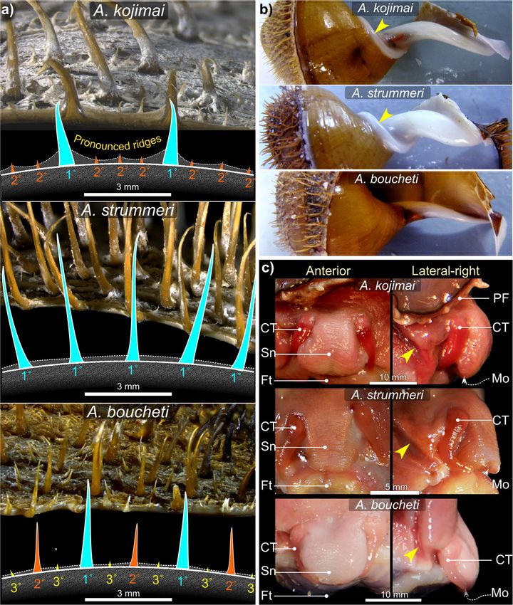
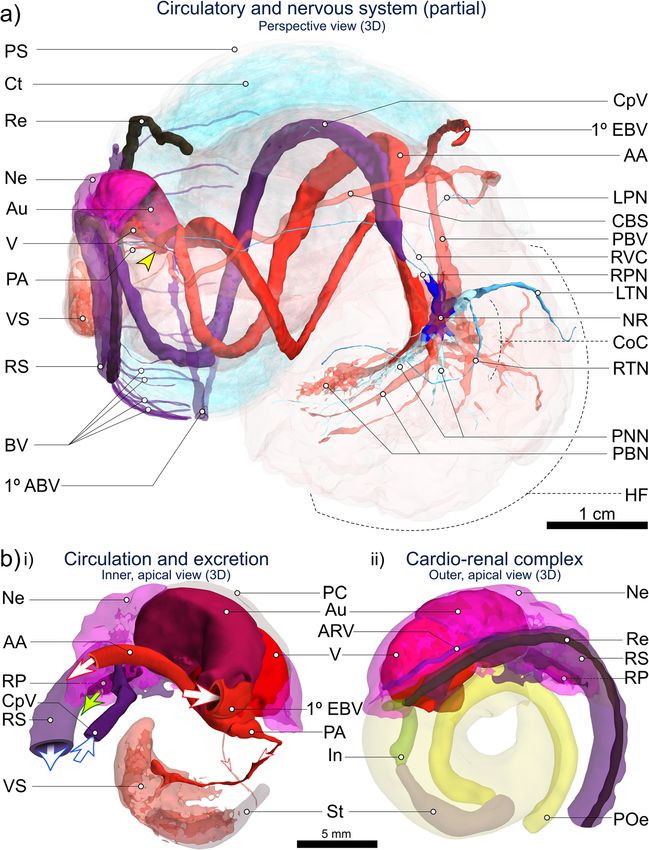
Laming et al. Frontiers in Zoology   (2020) 17:12                                                               Page 13 of 27

with the gastric shield and digestive-gland ducts located      Circulatory, excretory and nervous system
more posteriorly (Fig. 5c). Much of the external stomach       The heart’s single ventricle and auricle are relatively
surface is obscured from view, being almost entirely envel-    small, together occupying around 0.5% of the soft-tissue
oped in two digestive glands composed of furcate digestive     volume in A. kojimai and in A. strummeri (derived from
diverticula (Fig. 5b-vi). Histological analyses revealed the   CT segment volumes of the heart and the total soft tis-
presence of particulate material both between the digest-      sue). Oxygenated blood from the lacunae of branchial
ive diverticula and in the layer of connective tissue found    filaments appears to drain into the primary efferent
between diverticula and the stomach lining, similar in         branchial blood vessel (Fig. 9a), which widens consider-
appearance to the granular connective tissue in the cepha-     ably as it travels the length of the ctenidium posteriorly
lopedal haemocoel (Fig. 7d). The boundaries of each            on its left side – parallel to the gill axis – until finally,
digestive gland are poorly delineated but the digestive        it makes an abrupt turn dorsally to enter the auricle
ducts – one for each gland – were readily identifiable in      (Fig. 8b, note that blood-vessel openings in this figure
dissections (Fig. 5c-i) and during CT analyses (Figs. 4, 6,    are hand-drawn and indicative only). Blood flows from
Additional files 1 and 2 + models). The opening of the         the reservoir in the auricle into the muscularised ven-
adaxially orientated, posterior digestive duct is immedi-      tricle chamber (Additional file 5c), where the contrac-
ately anterior to that of the lower posterior oesophagus,      tion of a dense matrix of muscle fibres – evident in
both located in the posterior-most region of the stomach       histological section (Additional file 5d) – drives the
(Fig. 5c). The opening of the much-larger anterior digest-     blood via the large anterior aorta towards the head-
ive duct is located posterior to the style sac. It communi-    foot, and via the much smaller posterior aorta towards
cates with the stomach adaxially but then extends              the visceral sinus (Fig. 8, Additional file 5b, but also
anteriorly, parallel to the descending lower posterior         Additional files 1 and 2 + models). In the buccal cavity,
oesophagus along its adapical side (Figs. 4, 5c, Additional    the anterior aorta branches into a blood network that
files 1 and 2 + models). Internally, the anterior digestive    radiates from the oesophageal nerve ring towards: the
duct opens opposite to the sorting area (Fig. 5c-iii) and is   arterial pedal sinus (ventrally); the buccal mass (anteri-
demarcated by the beginning of the minor and major             orly); the cephalic tentacles (laterally) and – via a vessel
typhosoles (Fig. 5c-iv). The minor typhosole only extends      that doubles back dorso-posteriorly – towards the left
half the length of the style sac but the major typhosole       side of the pallial margin to supply the co-marginal
persists as a simple fold along the interior of the S-shaped   blood sinus (Fig. 8a). The posterior aorta supplies blood
intestinal tract with which the style sac communicates. In     to the arterial visceral sinus (Fig. 8b), which extends as
the current study, the end of this fold is considered the      several branching channels among the digestive diver-
juncture between the intestine and the rectum, where the       ticula visible on the right side of the visceral hump
rectal sinus begins (Fig. 8c). The rectum and rectal sinus     (Additional file 5b). Blood returns to the nephridium
return anteriorly for most of the length of the animal         from the head-foot via the prominent cephalopedal vein
enclosed in the mantle, which thickens to meet the pallial     (Fig. 8b) and presumably, from the venous visceral
floor on the right (Fig. 4, Additional files 1 and 2 +         sinus via a visceral vein, however the latter was not ap-
models). Regions of moderate rectal distension are found       parent during dissections could not be readily identified
intermittently along its length where contents are present     in CT scans.
inside, particularly towards the anus.                            The nephridium forms a cradle around the right half
   The foregut (anterior to the stomach) mainly con-           of the pericardial cavity, slightly invading the pallial skirt
tained mucus identifiable in preserved A. kojimai and          anteriorly, abutting the most posterior branchial fila-
A. strummeri specimens as fluffy, slightly compacted           ments (Fig. 8, Additional files 1 and 2 plus models,
material, occasionally punctuated with particulate mat-        Additional file 5c). It is composed of a dense arrange-
ter of various forms. Few breaks in these contents were        ment of primary and secondary folds, the former evident
found suggesting the intake of mucus had occurred              in cross-section (Fig. 9f). The cephalopedal vein commu-
prior to fixation, perhaps as a result of handling stress.     nicates with the nephridium’s anterior wall and appears
Similar but patchy contents were also found in the             to merge with the afferent renal vein, which runs the
stomach, however, the style-sac region and the hindgut         length of the nephridium’s right wall (Fig. 8b and
beyond were characterised by contents punctuated               Additional-file-1 model), parallel to the nephridial gland,
with crystalline debris, particularly evident in CT            with a blind terminus in the posterior-most region of
volume data (voxels with greyscale levels comparable           the nephridium. The renal pore, from which waste
to calcareous shell, Additional file 4). In fact, the          compounds from the nephridium are normally excreted
presence of this crystalline material made it relatively       (Fig. 8b, green arrow), is located near to the origin of the
easy to follow the rectum’s progress anteriorly in CT          afferent renal vein, visible as a compressed slit in the
volume data.                                                   right, posterior-most region of the pallial cavity. The
Laming et al. Frontiers in Zoology       (2020) 17:12                                                                                        Page 14 of 27

 Fig. 8 CT-based 3D visualisation of the circulatory, nervous and excretory systems in Alviniconcha. CT-scanned A. kojimai specimen: a 3D overview
 of cardio-renal complex, sinuses, vessels and nerves identified and visualised from CT volumes (N.B. pedal sinus not segmented, dorsal-most
 pallial vessels not shown, see Additional file 1); b Inner (i) and outer (ii) apical views of cardio-renal complex: i) Schematic derived from CT
 visualisation depicting the arrival of blood to the auricle (Au) via the primary efferent branchial vessel (1° EBV), after which blood exits the
 ventricle (V) via the larger anterior aorta (AA ~ > various cephalopedal sinuses) from which two smaller posterior vessels branch off, including the
 posterior aorta (PA ~ > visceral arterial sinus VS). Blood from rectal sinus (RS) later drains into the primary afferent branchial vessel – 1° ABV in a) –
 via lateral blood vessels (BV). Blood returning from head-foot to nephridium (Ne) arrives via the cephalopedal vein (CpV) and then passes to the
 afferent renal vein (ARV) visible in (ii) running along the right side of the nephridium, parallel with the start of the rectal sinus. Blood-vessel
 openings have been drawn by hand and are indicative only. Oxygenated blood represented by red vessels and arrows, deoxygenated blood by
 purple vessels and arrows. Oxygen content in rectal sinus likely decreases along its length. Green arrow in (i): excretion from the nephridium into
 the pallial cavity is via the renal pore (RP). Figure is of 3D renderings with exaggerated perspective, thus scales are approximate. Additional
 abbreviations: CBS Co-marginal blood sinus; CoC Circumoesophageal complex; Ct Ctenidium; HF Head-foot; In Intestine; LPN Left pallial nerve;
 LTN Left tentacular nerve; NR Oesophageal nerve ring; PBN Pedal blood network; PBV Pallial blood vessel; PNN Pedal neural network; POe Lower
 posterior oesophagus; PS Pallial skirt; Re Rectum; RPN Right pallial nerve; RTN Right tentacular nerve; RVC Right visceral connective; St Stomach
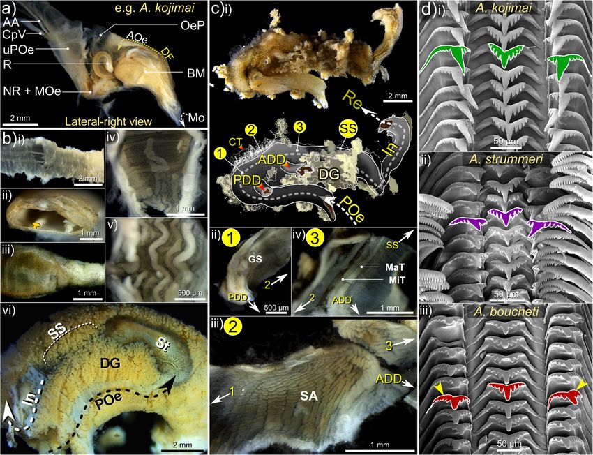
Laming et al. Frontiers in Zoology      (2020) 17:12                                                                                     Page 15 of 27

 Fig. 9 Summary of branchial anatomy and proximal tissues. Schematic in a depicts arrangement of gill filaments in relation to overall transverse
 cross-sectional anatomy at pallial cavity’s widest point (~ 0.25 whorls behind buccal cavity). Blood circulation is based on CT data, whole-filament
 histological staining (sections and unembedded) and details recorded during dissection. Though not depicted, areas in cephalopedal haemocoel
 not occupided by blood vessels or the alimnetary canal, were filled with spongy connective tissue full of particulate deposits. Depicted in b a
 branchial filament in sagittal section approximately located along the red line marked on the filament in a. The colour coding used in (a) and (b)
 is repeated in (c) and (d) for comparative purposes. Ctendium pictured in c is viewed ventrally: with most of filaments extending dorsally from
 the ventral support axes. One filament however has been displaced sideways and has been colour coded according to its abnormal orientation.
 Close up, these filaments appear as depicted in d. The micorgraph in e is of the dorsal face of the mantle on the far-right side of the animal,
 along which the the rectal sinus (RS) runs. Arrows indicate the blood vessels that deliver blood to the primary afferent branchial vessel (1° ABV) of
 the ctenidium from the rectal sinus. Image f is of the nephridium (Ne, with a transverse cut), which abuts directly with the posterior-most
 branchial filaments (Ct). Primary folds (PF) and the afferent renal vein (ARV) can be seen, along with traces of the digestive gland (DG). Additional
 abbreviations: 2° ABV secondary afferent branchial vessel; 1° / 2° EBV primary / secondary efferent branchial vessel; AA Anterior aorta; AOe
 Anterior oesophagus; CM Columellar muscle; Co Columella; CpV Cephalopedal Vein; CM columellar muscle; HF Head-foot; PC Pallial cavity; PF
 Pallial fringe; PS Pallial skirt; RS Rectal sinus

rectal sinus bathes the rectum in haemolymph for al-                         haemolymph is then thought to drain into individual
most its entire length, sending out dorso-lateral pallial                    branchial filaments to be re-oxygenated (Fig. 9a).
blood vessels leftwards towards the primary afferent                           The nervous system and neuronal characters were not
branchial vessel located medially in the pallial skirt (Figs.                examined by histology but observations during dissec-
8a, 9a, e and Additional-file-1 model), where                                tions and visualisations based on CT analyses suggest it
Laming et al. Frontiers in Zoology   (2020) 17:12                                                                     Page 16 of 27

is epiathroid, as described previously in Alviniconcha [3].        adhered region (Figs. 4, 9a, c, Additional file 1 + model,
The presumptive arrangements of the pleural, cerebral              Additional file 5f). Each filament is comprised of a rigid
and pedal ganglia (informed in part by previous studies)           supporting rod along its ventral edge, a narrow ciliated
are indicated in the model in Additional file 1. However,          zone and a much wider membranous region, which is
further analyses using histology are necessary to estab-           delicate, plicate and highly vascularised (Fig. 9b, c, d). It is
lish whether these are true ganglia, i.e. paired neuron-           in this latter region that bacteriocytes are found, housing
cell cortices with centrically directed neurites (neuro-           the bacterial symbionts (Fig. 9b). The primary afferent
phil) and peripheral neuronal somata, connected to one             branchial vessel delivers blood to secondary afferent blood
another by neurite bundles (e.g. connectives, commis-              vessels, which run along the dorsal edge of each filament,
sures). Dense nerve bundles were observed to extend                while secondary efferent blood vessels lie along the
ventrally from the oesophageal nerve ring into the foot’s          boundary between the ciliated and bacteriocyte zones
metapodium, mesopodium and anterior-most propo-                    (Fig. 9a, d). Thus, the secondary branchial vessels
dium, based on dissections and CT visualisations (Fig.             frame the bacteriocyte region and are visibly linked
8a). Three closely packed nerves extend anteriorly from            by a vertical network of delicate, dorsoventral blood
each side of the oesophageal nerve ring and innervate              vessels (Fig. 9a, d). Histological examination of these
four points on the buccal mass, two posterio-dorsally              blood vessels reveals the coagulation and transport of
and two anterio-ventrally, as well as the two cephalic             lysed biological material of unknown origin from the
tentacles (Fig. 8a). The dorsal, posterior-most region of          bacteriocyte region to secondary efferent blood vessels
the oesophageal nerve ring appears disproportionately              (Fig. 7a), providing putative evidence for bacterial
long on its left side. Several nerves also extend out more         assimilation.
posteriorly (minimum of two from right side, four from
left), of which the right and left pallial nerves (tracked         Species-specific characteristics
towards mantle, Fig. 8a) and the right visceral connective         Filaments in A. strummeri, and particularly A. kojimai, are
could be confidently identified (the last, progressing             wide dorsoventrally (Additional file 5d); the dorsal half of
along the cephalopedal vein, Fig. 8a).                             each are creamy white in appearance and engorged (par-
                                                                   tially visible in Additional file 5b). In A. boucheti, filaments
Ctenidium and associated tissues                                   are notably darker (lacking engorged tissue, Fig. 2b), more
General observations                                               mucous and narrower dorsoventrally (Additional file 5d).
The ctenidium – or gill – is hypertrophied and represents
about 60% of the total, uncoiled body length (based on             Reproductive system
dissections) and a little over 10% of the animal’s total vol-      Gonad size varied – in terms of its extent anterio-
ume (based on CT data). The ctenidium extends the full             posteriorly – from being restricted to the posterior
length of the pallial cavity (e.g. Fig. 2b, Additional file 3b),   region of the cephalopedal haemocoel, to extending as
fused with the pallial margin anteriorly and attached to           far as the anterior end of the columellar muscle. Obser-
the far left of the pallial roof along its axis. Its widens for    vations during dissections gave the impression that this
0.5 whorls and then tapers posteriorly, terminating just           variability was not a function of size, however the num-
prior to the pallial skirt, at which point the primary effer-      ber of specimens dissected was relatively low and gonad
ent branchial vessel enters the pericardial cavity and the         size was not quantitatively assessed. The smallest indi-
auricle of the heart (Fig. 8b, Additional files 1 and 2 +          vidual dissected however, an A. kojimai specimen with
models). The osphradium, an olfactory sensory organ, ex-           SH 37.8 mm, had no visible gonad. Limited data avail-
tends from the pallial margin’s inhalant left side for about       able from the current study indicate that these Alvini-
half the pallial cavity, approximately parallel to, and left of    concha spp. are gonochoric. In those specimens that
the ctenidium’s axis (Fig. 9a, and Additional files 2 +            were reliably sexed, SH for each sex were as follows: A.
model and Additional file 5e). It takes the form of a cen-         kojimai ♀ 41.5–78.8 mm (n = 8), ♂ 52.6–77.4 mm (n =
tral flange bordered by two much-less pronounced ridges.           5); A. strummeri ♀ 53.7 mm, ♂ 46.1–51.6 mm (n = 4)
Tight ranks of branchial filaments extend laterally from           and; A. boucheti ♀ 65.9–77.6 mm [n = 3], ♂ 59.8 mm. In
the ctenidium’s axis. Most filaments terminate right of the        frozen individuals, gonads confirmed by tissue smear are
pallial cavity’s midline; at their longest – above the             creamy white and opaque in females, while gonads in
anterior-most dorsal hump of the cephalopedal haemocoel            males are brown-to-orange and some internal detail is
– they occupy around 80% of the cavity’s width (Fig. 4,            visible through the epithelium. Mean defrosted oocyte
Additional files 1 and 2 + models). Filaments are adhered          diameters were small at 50.69 μm ± 4.5, measured in A.
to the pallial epithelium for their proximal half, free-           kojimai only (three individuals for which eggs were
floating distally, where the afferent branchial vessel             intact, minimum of five replicates per individual).
demarcates the right, medio-dorsal boundary of this                However, the meiotic status of these oocytes was not
You can also read