CLINICAL CORRELATIONS OF THE BILIOPANCREATIC CARREFOUR IN DOGS AND CATS
←
→
Page content transcription
If your browser does not render page correctly, please read the page content below
Cercetãri Experimentale & Medico-Chirurgicale
)
Anul XIII l Nr.1/2006 l Pag. 75-67 Cercetari
e x p e r i m e n ta le &
m edico-chirurgicale
CLINICAL CORRELATIONS OF THE
BILIOPANCREATIC CARREFOUR IN DOGS AND CATS
Gheorghe M. Constantinescu1, Summary: Complete understanding of the biliopancreatic carrefour (BPC) in dogs and cats
Fred Anthony Mann2 is crucial for successful surgical management of hepatobiliary disease and diseases
Ileana A. Constantinescu1 requiring upper gastrointestinal resection.
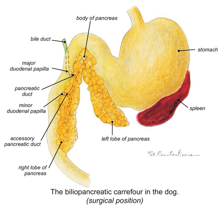
In the dog the choledochus differs in shape and relationship to the pancreatic duct compared
to the same structure of the cat. The openings of the ducts in the major duodenal papilla also
differ between the dog and the cat. In the cat a hepatopancreatic ampulla (HPA) similar to
that in humans protects the openings of both ducts. The ducts are provided with sphincters;
an additional sphincter of the HPA is present in the cat. The clinical relevance of the close
proximity of the choledochus and pancreatic duct in both dogs and cats is manifest as biliary
obstruction secondary to swelling or scarring as a result of pancreatitis. The hepatic artery,
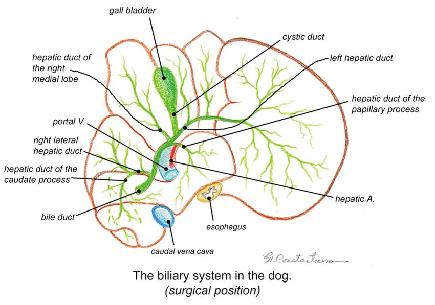
portal vein and bile duct are together enclosed in a peritoneal fold of the hepatoduodenal
ligament forming the ventral rim of the omental foramen. Surgeons exploit this knowledge of
anatomy by performing the Pringle maneuver to control hepatic hemorrhage. In dorsal
recumbency the BPC is approached surgically from the right side using the mesoduodenum
as an anatomical retractor to reflect the abdominal viscera to the left. Common surgical
interventions requiring complete knowledge of the BPC include cholecystoduodenostomy,
partial gastrectomy with upper duodenal resection (Bilroth II procedure), cholecystectomy,
cholecystotomy, and partial pancreatectomy.
A perfect knowledge of the anatomy of the BPC is also needed for accurate diagnostic
investigations using endoscopy, ultrasonography, computed tomography, and magnetic
Received for publication: resonance imaging.
Revised:
Keywords: type 1 diabetes mellitus, pancreatic autoantibodies, prediction
1 - Biomedical Sciences, College of Veterinary Medicine, University of Missouri-Columbia
2 - Veterinary Medicine and Surgery, College of Veterinary Medicine, University of Missouri-Columbia
Introduction
n An increasing incidence of hepatobiliary n Unfamiliarity with the anatomy of the biliary
abnormalities in dogs and cats and need for precise system and surrounding structures will hamper
diagnosis procedures is recently mentioned in the confidence in surgical manipulations necessary to
literature (Fahie, Martin,1995; Leveille, Biller, treat some of these abnormalities.
Shiroma, 1996; Newell et al, 1996; Barnhart,
Rasmussen, 1996; Ludwig, 1997; Rivers et al,
1997; Bromel et al, 1998; Voros et al, 2001; Eich,
Ludwig et al, 2002; Mayhew et al, 2002; Owens et
al, 2003; Bacon, White, 2003; Savary-Bataille et al,
2003; Holt et al, 2004; Worley, Hottinger,
Lawrence, 2004; Mehler et al, 2004 etc. etc.).
Correspondence to: Gheorghe M. Constantinescu , College of Veterinary Medicine, University of Missouri,
1600 E. Rollins, Columbia, Missouri, 65211, USA
75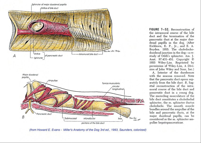
76
ANATOMICAL DIFFERENCES BETWEEN THE DOG AND THE CAT
 n   In the dog the choledochus differs in shape and           ducts. An additional sphincter of the HPA is present
     relationship to the pancreatic duct compared to the       in the cat.
     same structure of the cat. The openings of the        n   The sphincter of Oddi in the dog is illustrated in
     ducts in the major duodenal papilla provided with         detail.
     sphincters also differ between the dog and the cat.   n   The distribution of the hepatic branches of vagus N.
     In the cat a hepatopancreatic ampulla (HPA) similar       in the dog follows (Stanley L. Chiu, 1943).
     to that in humans protects the openings of both

                                                                                                                77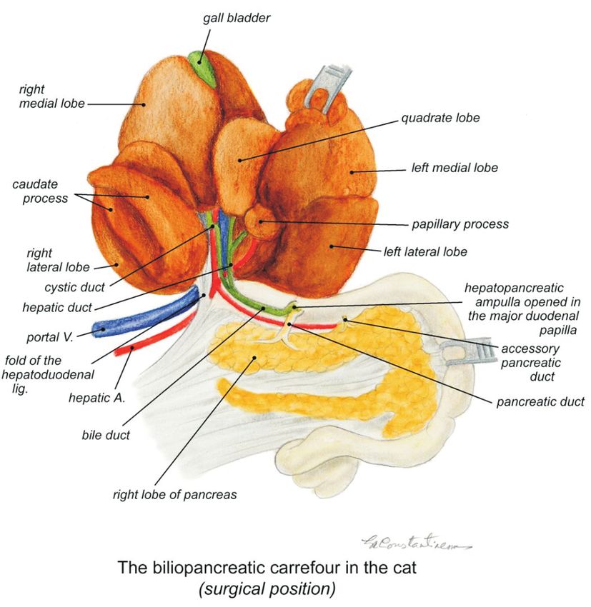
muscular septum into two compartments, one for
the bile duct and one for the pancreatic duct. The
submucosal part of ductus choledochus is mainly
formed by the major duodenal papilla. This is
provided with a sphincter (sphincter of Oddi). The
sphincter of Oddi is composed of various layers of
smooth muscle embedded in connective tissue
with complex relations to other muscular
structures of the choledocho-duodenal junction.
Crucial need for strong knowledge of biliary
anatomy in the area of:
n Diagnostic imaging
o Abdominal ultrasonography
o Biliary scintigraphy
n Treatment of various conditions
o Cholangitis, necrotizing cholecystitis,
cholelithiasis, biliary mucoceles, biliary
obstruction
ANATOMICAL DIFFERENCES BETWEEN THE n Successful surgical and endoscopic procedures
DOG AND THE CAT o Cholecystoenterostomy, cholecystectomy,
n According to Kyosola and Rechardt (1974), in the laparoscopic biliary procedures
cat the HPA gives rise to concentric retrograde o Upper gastrointestinal resections
saccules around the ampulla and the terminal part
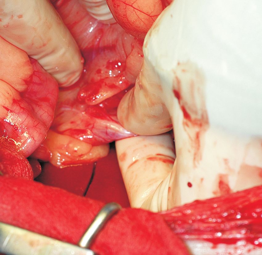
Making the correct intra-operative decision
of the bile duct. The Fenestra choledocha, the
Bile leaking into the abdominal cavity:
slit-like opening in the circular muscle layer of the
Cholecystoenterostomy vs. Cholecystectomy ?
duodenum pierces the rigth side posterior wall of
Gall bladder rupture:
the duodenum.
n Bile duct obstruction?
n Boyden in 1957 divided the intramural part of
o Cholecystoenterostomy is indicated.
choledochus into a proximal infundibular part and a
n Necrotizing cholecystitis?
distal submucosal part. The infundibular part is
o Cholecystectomy is indicated.
enclosed in a muscular funnel divided by a
The wrong choice would likely be fatal
78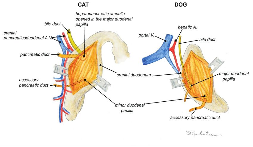
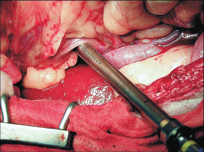
n   Surgeons exploit the knowledge of anatomy by
Clinical relevance                                               performing the Pringle maneuver to control hepatic
  n   The clinical relevance of the close proximity of the       hemorrhage.
      choledochus and pancreatic duct in both dogs and       n   Common surgical interventions requiring complete
      cats is manifest as biliary obstruction secondary to       knowledge of the BPC include cholecysto-
      swelling or scarring as a result of pancreatitis.          duodenostomy, partial gastrectomy with upper
                                                                 duodenal resection (Bilroth II procedure),
Clinical correlates                                              cholecystectomy, cholecystotomy, and partial
                                                                 pancreatectomy.
  n   In dorsal recumbency the BPC is approached
      surgically from the right side using the
      mesoduodenum as an anatomical retractor to
      reflect the abdominal viscera to the left.

                                                                              Mesoduodenum

                         Cranial

                                                             Mesocolon

                                                                                                                79
Hepatic artery

                                                                Vena cava

                             Cranial

     Conclusions
        A complete understanding and perfect knowledge of
     the anatomy of the BPC in dogs and cats is crucial for
     precise diagnosis and successful surgical interventions:
        n for accurate diagnostic investigations using

          endoscopy, echoendoscopy, ultrasonography,
          computed tomography, scintigraphy, and
          magnetic resonance imaging
        n for performing successful surgical interventions in

          conditions such as hepatobiliary disease and
          various pancreatic ducts conditions

80
References:
1. Bacon NJ, White RA. Extrahepatic biliary tract surgery in the cat: a case series and review. J Small Anim Pract. 2003
    May;44(5):231-5.
2. Barnhart MD, Rasmussen LM. Pleural effusion as a complication of extrahepatic biliary tract rupture in a dog. J Am Anim Hosp
    Assoc. 1996 Sep-Oct;32(5):409-12.
3. Boyden EA. The sphincter of Oddi in man and certain representative animals. Surgery 1937 1:25-37
4. Bromel C, Leveille R, Scrivani PV, Smeak DD, Podell M, Wagner SO. Gallbladder perforation associated with cholelithiasis and
    cholecystitis in a dog. J Small Anim Pract. 1998 Nov;39(11):541-4.
5. Casas AP. Contribution a l’etude du sphincter d’Oddi chez Canis familiaris. Acta Anat 1958 34 1-2 :130-153
6. Chiu SL. The superficial hepatic branches of the vagi and their distribution to the extrahepatic biliary tract in certain mammals. The
    Anat Rec 1943 86:149-155
7. Dupre G. Chirurgie des voies biliaires du chien. 1ere partie ; anatomie, physiologie et pathologie chirurgicale. Le Point Veterinaire
    1988 20 112 :7(95)-14(102)
8. Dupre G. Chirurgie des voies biliaires du chien. 2eme partie : techniques chirurgicales. Le Point Veterinaire 1988 20
    113 :17(193)-24(200)
9. Eich CS, Ludwig LL. The surgical treatment of cholelithiasis in cats: a study of nine cases. J Am Anim Hosp Assoc. 2002
    May-Jun;38(3):290-6.
10. Eichhorn EP Jr., Boyden EA. The choledochoduodenal junction in the dog – a restudy of Oddi’s sphincter. Am J Anat 1955
    97:431-451
11. Evans H. 1993. Miller’s Anatomy of the dog 3rd ed. W.B.Saunders
12. Fahie MA, Martin RA. Extrahepatic biliary tract obstruction: a retrospective study of 45 cases (1983-1993). J Am Anim Hosp
    Assoc. 1995 Nov-Dec;31(6):478-82
13. Halpert B. The choledocho-duodenal junction. A morphological study in the dog. The Anat Rec 53 1:83-102
14. Hitt ME, Jones BD, Constantinescu GM, 1987, The feline liver: What a practitioner needs to know. Vet. Med. 82:129-138
15. Holt DE, Mehler S, Mayhew PD, Hendrick MJ., Canine gallbladder infarction: 12 cases (1993-2003) Vet Pathol. 2004
    Jul;41(4):416-8.
16. Kyosola K., Rechardt L. The anatomy and innervation of the sphincter of Oddi in the dog and cat. Am J Anat 1974 140:497-522
17. Leveille R, Biller DS, Shiroma JT. Sonographic evaluation of the common bile duct in cats. J Vet Intern Med. 1996
    Sep-Oct;10(5):296-9.
18. Ludwig LL, McLoughlin MA, Graves TK, Crisp MS. Surgical treatment of bile peritonitis in 24 dogs and 2 cats: a retrospective study
    (1987-1994). Vet Surg. 1997 Mar-Apr;26(2):90-8
19. Mann FC, Brimhall SD, Foster JP. The extrahepatic biliary tract in common domestic and laboratory animals. The Anat Rec 1920
    18:47-66
20. Markowitz J, Rappaport A. The function of the hepatic artery in the dog. The Am J Digest Diseases 1949 16 10:344-348
21. Mayhew PD, Holt DE, McLear RC, Washabau RJ. Pathogenesis and outcome of extrahepatic biliary obstruction in cats. J Small
    Anim Pract. 2002 Jun;43(6):247-53.
22. Mehler SJ, Mayhew PD, Drobatz KJ, Holt DE. Variables associated with outcome in dogs undergoing extrahepatic biliary surgery:
    60 cases (1988-2002). Vet Surg. 2004 Nov-Dec;33(6):644-9.Morita Y, Takiguchi M, Yasuda J, Kitamura T, Syakalima M, Eom
    K-D, Hashimoto A. Endoscopic ultrasonography of the pancreas in the dog. Vet Radiol & Ultrasound 1998 39:552-556
23. Newell SM, Selcer BA, Roberts RE, Cornelius LM, Mahaffey EA. Hepatobiliary scintigraphy in the evaluation of feline liver disease.
    J Vet Intern Med. 1996 Sep-Oct;10(5):308-15.
24. Nielsen SW, Bishop EJ. The duct system of the canine pancreas. Am J Vet Res 1954 XV 55:266-271
25. Owens SD, Gossett R, McElhaney MR, Christopher MM, Shelly SM., Three cases of canine bile peritonitis
26. with mucinous material in abdominal fluid as the prominent cytologic finding. Vet Clin Pathol. 2003;32(3) :114-20.
27. Revell DG. The pancreatic ducts in the dog. The Am J Anat 1901-1902 1:443-457
28. Rivers BJ, Walter PA, Johnston GR, Merkel LK, Hardy RM. Acalculous cholecystitis in four canine cases: ultrasonographic
    findings and use of ultrasonographic-guided, percutaneous cholecystocentesis in diagnosis. J Am Anim Hosp Assoc. 1997
    May-Jun;33(3):207-14.
29. Savary-Bataille KC, Bunch SE, Spaulding KA, Jackson MW, Law JM, Stebbins ME. Percutaneous ultrasound-guided
    cholecystocentesis in healthy cats. J Vet Intern Med. 2003 May-Jun;17(3):298-303.
30. Vlad M., 1999, Carrefour-ul Biliopancreatic. Ed. Modelism, Bucuresti
31. Voros K, Nemeth T, Vrabely T, Manczur F, Toth J, Magdus M, Perge E., Ultrasonography and surgery of canine biliary diseases.
    Acta Vet Hung. 2001;49(2):141-54.
32. Worley DR, Hottinger HA, Lawrence HJ. Surgical management of gallbladder mucoceles in dogs: 22 cases (1999-2003). J Am
    Vet Med Assoc. 2004 Nov 1;225(9):1418-22.

                                                                                                                                      81
You can also read