Clinoptilolite supported feeding reduces excessive iron in thalassemia rat model created with iron loading
←
→
Page content transcription
If your browser does not render page correctly, please read the page content below
Available online at www.medicinescience.org

                                                                                                                                              Medicine Science
                                                                     ORIGINAL ARTICLE                                                         International
                                                                                                                                              Medical Journal
                                                           Medicine Science 2021;10(3):1031-8

   Clinoptilolite supported feeding reduces excessive iron in thalassemia rat model created
                                       with iron loading

                           Durdane Serap Kuruca1,                Kadriye Akgun Dar2,               Ayesegul Kapucu2,             Dilsad Ozerkan3
                                           1
                                            Istanbul University Faculty of Medicine, Department of Physiology, Istanbul, Turkey
                                              2
                                               Istanbul University Faculty of Science, Department of Biology, Istanbul, Turkey
                              3
                                Istinye University, Health Sciences Faculty, Department of Physiotherapy and rehabilitation, Istanbul, Turkey

                                                          Received 09 December 2020; Accepted 12 March 2021
                                                  Available online 09.05.2021 with doi: 10.5455/medscience.2020.12.248

                                                    Copyright@Author(s) - Available online at www.medicinescience.org
                           Content of this journal is licensed under a Creative Commons Attribution-NonCommercial 4.0 International License.

Abstract

There is no regulatory mechanism for the removal of iron that accumulates in the body. Thalassemia patients are most affected by iron overload. The method often used
in the treatment of these patients is iron chelation therapy, which involves removing excess iron from the bloodstream, but it is insufficient. Nutritional supplements and
herbal remedies are complementary tools that may help improve the health of an individual with iron load. Here, we tried to develop a method that affecting intestinal
absorption in an animal model with clinoptilolite feeding to reduce the iron load in the circulation. 32 rats were divided into 4 groups as control, Cli, Iron, Cli+Iron. Iron
and Cli+Iron groups received iron at a dose of 250 mg/kg/day for 10 days, and Cli and Cli+Iron groups were fed a diet with 50% clinoptilolite for one month. Histological
preparation and Fe2+, Cu2+, Zn2+ measurements were done in all tissues. Consequently, clinoptilolite significantly lowered the iron level in the stomach. On the contrary,
iron absorption was increased in the small intestine, but iron transportation to the blood was decreased by clinoptilolite. The iron levels of clinoptilolite groups (Cli and
Cli+Iron) were reduced in the tissues of heart, lung, liver, kidney, and spleen because of the different level of iron necessities of each tissue compared to the group with iron
overload. The clinoptilolite could preserve organs against iron toxicity by enhancing the absorption of iron in the small intestine but lowering the iron level in blood. Even
though there was no detailed information regarding the mechanism of reduction iron overload by the clinoptilolite, serum and chyme could help to make some helpful
inferences to elucidate the mechanism of iron chelation.

Keywords: Clinoptilolite, Iron overload, heavy metal accumulation, atomic absorption spectrophotometry

Introduction                                                                              per year [1]; in those who are transfused, this load is doubled
                                                                                          and primarily liver, heart and endocrine glands are affected.
The thalassemia types are a group of anemias that result from
inherited defects in the production of hemoglobin. The anemia                             Iron chelation therapy aims to prevent harmful iron accumulation
is treated by frequent erythrocyte transfusions. This therapy                             and remove excess iron in patients with thalassemia, but this
results in the accumulation of iron overload that is exacerbated                          treatment is not always successful. Many clinical investigations
by the breakdown products of hemoglobin and the increased                                 have addressed pharmacokinetic efficacy of some effective
iron uptake but ineffective erythrocyte production. As a                                  nutraceuticals for β-thalassemia. Supplementation diet including
result, organ damage and even death may occur. In patients                                functional food such as vitamins, vegetables, fruits are shown to
with thalassemia, regular blood transfusions are performed to                             have beneficial effects in a lot of studies. Curcumin significantly
maintain normal hemoglobin levels in the blood. Even in non-                              reduced iron overload in iron-loaded rats’ liver and kidney [2].
transfusion thalassemia intermedia patients, iron load is 2-5 g                           It has been determined that green tea extract blocks dietary iron
                                                                                          absorption and chelating [3-6]. Administration of nutraceuticals
                                                                                          to patients with thalassemia in addition to the regular application
                                                                                          of iron chelation therapy can significantly reduce organ damage
                                                                                          and dysfunction. However, using these foods that have antioxidant
                                                                                          effects is not sufficient alone. In addition, the body does not have
*Corresponding Author: Dilsad Ozerkan, Istinye University, Health Sciences                a physiological mechanism that can reduce the excess iron load
Faculty. Bölüm belirtiniz, Istanbul, Turkey, E-mail: dilsadokan@gmail.com
                                                                                          by itself. Nowadays, it is known that chelation therapy applied in

                                                                                                                                                                         1031
doi: 10.5455/medscience.2020.12.248				                                   		                       Med Science 2021;10(3):1031-8

patients with thalassemia is insufficient to reduce iron overload.     years to treat life-threatening injuries. Therefore, it is inevitable
                                                                       to develop zeolite-based nanoparticles to achieve hemostasis [22].
Zeolites are aluminum silicates with crystalline hydration. They
were formed millions of years ago by the chemical reaction of          Various experiments have been conducted investigating the
ash and lava that emerged from the mixture of volcanic eruptions       biological and chemical effects of natural zeolites on humans and
with lake or sea waters [7]. This means the zeolites have crystal      animals. In many of these studies, it was aimed to benefit from the
lattice and inorganic structure which makes them minerals. As          adsorption and ion exchange properties of clinoptilolite. However,
porous materials, zeolites are known as selective ion exchangers,      the effects of natural zeolites in reducing iron load in vivo are
adsorbents, and molecular sieves. That is why the zeolites are         still controversial. Therefore, in our study, we planned to use the
utilized in various areas, from industrial applications to medical     thalassemia rat model created by iron loading and to investigate
uses [8]. For example, some zeolites, including clinoptilolite,        the effectiveness of Clinoptilolite obtained from Manisa-Gördes/
F-type and W-type, have the property of administering hemodialysis     Turkey on this model.
in ammonia ion exchange systems where a major challenge in
developing a hemodialysis system is the removing of ammonia            Material and Methods
from a recirculating dialysate stream [9]. Also, much effort has
been made to improve the adsorption efficiency of zeolites             Animals
against toxins in various environments [10-12]. It has also been
                                                                       This study with rats was performed after taking permission
suggested that the ability of zeolite (FAU13 × and FERCP914C)
                                                                       numbered 2011/94 from Istanbul University Animal Experiments
to decrease ROS accumulation in extracorporeal cycles in patients
                                                                       Local Ethics Committee. 32 adult male Wistar albino rats
with chronic renal failure undergoing dialysis is as beneficial as
                                                                       weighing 200-250 g were used. The subjects were divided into 4
antioxidant supplementation (i.e. vitamin E) and leads to reduce
                                                                       experimental groups which were the control group (n=7, Group
mortality [13]. Several studies have investigated whether zeolites
                                                                       1), clinoptilolite-supported feeding group (n=7, Group 2), iron-
can be used as food supplements in poultry houses and farms to
                                                                       overloaded group, (n=9, Group 3), clinoptilolite-supported feeding
improve their performance and efficiency by removing heavy
                                                                       group with iron overload (n=8, Group 4). The rats in Group 3 and
metals from animals' bodies. A group of scientists used zeolites
                                                                       4 received iron (Ferro III hydroxide polymaltose) by gavage at a
as dietary supplements for dairy cows and observed that long-
                                                                       dose of 250 mg/kg/day for 10 days, and Group 2 and 4 were fed a
term feeding of zeolites increased milk yield and that zeolites
                                                                       diet 50% clinoptilolite for one month as mentioned before [23, 24].
had no significant negative effect on cows liver function and
hematological parameters [14]. On the other hand, a South Korean       Chemicals
scientist postulated and corrected the hypothesis of using zeolites
as an effective nutraceutical to lower contaminants in poultry         Clinoptilolite used in this experiment is (Ca, K2, Na2,
and pig manure [15]. However, even after these experiments and         Mg)4Al8Si40O96.24H2O, and has a 7.0-8.0 pH value. The
studies on animals, people still thought zeolites to be carcinogenic   clinoptilolites’ mean particle size is
doi: 10.5455/medscience.2020.12.248 Med Science 2021;10(3):1031-8
of trace elements. Blood collection and serum separation were 5.0). Experimental groups were compared with respect to using
performed in a dust-free environment. Fe2+, Cu2+, Zn2+ levels Bonferroni’s multiple comparison test by applying the one-way
were determined by using flame atomic absorption spectrometry variance analysis. All results were expressed as means±SD and
(Shimadzu AA-680). p≤0.05 was regarded as significant.
Tissue sample collection and histological slide preparation Results
The rats were decapitated at the end of the experiment. Their Histological examination of tissues obtained from all groups
stomachs, small intestines, large intestines, livers, spleens, kidneys, Small Intestine
heart, and lungs were removed, fixed with 10% neutral formalin,
and then embedded in paraffin. Four-micrometer paraffin tissue The most significant alteration observed from the examination of
sections were mounted on the slides which were then deparaffinized. small intestine section was morphological changes of villi in the
The sections were stained with hematoxylin and eosin (H&E) and Iron group. These changes were an increase in number of villi and
Prussian Blue [26]. The slides were microscopically examined shortening of them. Also, there were invaginations in epithelial
and captured by using Kameram 390CU Imaging system (Micro tissue which was on the luminal side of small intestine. There was
System Ltd. Turkey). very little reaction in the sections which were stained with Prussian
blue. The villi’s structure from clinoptilolite group was slightly
Statistical Analysis changed; in contrast, there were some significant changes such as
increase in number and lengthening of the villi from the Cli+Iron
First, whether the data show a normal distribution or not was
group; therefore, it has been observed that the villi were in a tight
evaluated primarily through the histogram graph. The normality
order as small intestine’s luminal surface was enlarged, and there
test was carried out because of the graphics that seemed contrary
were lymph nodes as well. Furthermore, the Prussian blue reaction
to the normal distribution. Results were analyzed non-parametric
in the Cli+Iron group was more severe than the reaction in the Iron
student’s t test with the Graphad Prism statistical program (version
group (Figure 1a).
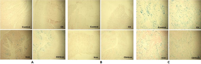
Figure 1. The tissue sections from the control and experimental groups were randomly chosen from the subjects of experiment and were stained by Prussian Blue (n=9
per groups). A. The small intestine B. The large intestine C. The spleen.
Large Intestine of epithelial cells in the LI2 region was greater than that of LI1.
In this study, the large intestine was examined in three sections as
The amount of connective tissue in the LI2 region increased more
LI1; LI2; LI3. In the control group, normal intestinal histology was
with the LI1 region than the group given zeolite. The brush border
observed in all three sections. The epithelium had striated margins
could not be seen in the LI2 region, the increase in leukocyte was
and the crypts were smooth.
observed the most in this group. While examining the sections,
The sections were stained darker in the group given clinoptilolite, leukocyte infiltration was detected in 3-4 areas in the LI3 region,
the number of blood vessels and leukocytes increased compared one of which was quite large. The eosinophil count was increased.
to the control. Connective tissue was increased in all parts of the
intestine, mostly in the LI3 region. Leukocyte infiltration was
present in all departments, mostly in the LI3 region (Figure 1b). In the group where iron and clinoptilolite were applied together,
fragmentation in the apical of the intestinal epithelium was
significantly reduced in all three regions compared to the iron-
In the iron treated group, In the LI1 region, the epithelium was
treated group. Crypta were seen to be smooth. Brush border
fragmented in patches and the integrity of the striated margin
integrity could be traced. The connective tissue was significantly
was disrupted, but it was visible. The crypts were corrupted.
reduced in the LI1 and LI2 regions compared to the iron given
An increase in the number of blood vessels was observed. An
group but was slightly more in the LI3 group. The eosinophil count
increase in connective tissue was determined, but this increase
was lower than the iron-treated group (Table 1).
was less than in the zeolite group. The fragmentation in the apical
1033doi: 10.5455/medscience.2020.12.248 Med Science 2021;10(3):1031-8
Table 1. Histological changes in the large intestine in all groups are summarized.
Disintegration
Connective Number of Number of Number of
in the apical Brush border Cyripta
tissue blood vessels leukocytes eosonophils
epithelium
LI1 - steady normal normal normal normal normal
Control LI2 - steady normal normal normal normal normal
LI3 - steady normal normal normal normal normal
LI1 - steady normal increased + increased + increased + increased +
Cli LI2 - steady normal ++ + + +
LI3 - steady normal +++ ++ ++ +
Partially steady
LI1 + Degraded + + +++ ++ +
+
Iron LI2 ++ Not seen Degraded +++ ++++ +++ +++ +
LI3 ++++ Not seen Degraded +++ ++++ +++ +++ +++
Similar to Similar to
LI1 + + + + +
normal normal
Similar to Similar to
Cli+Iron LI2 Rare + + + +
normal normal
Similar to Similar to
LI3 Rare + + + +
normal normal
Liver
The Kupffer cells got visually more obvious and there was an
increase in the number of them. Also, accumulation in the Cli+Iron
group was less than the Iron group. On the other hand, the number
and appearance of the Kupffer cells in the control and Cli groups
were similar. In addition, there was no reaction in the liver sections
stained by Prussian blue from any of the experimental groups and
control group.
Spleen
According to the histological examinations, it has been observed Figure 2. Amount of Fe2+ in the control and experimental groups. The bars represent
that the red pulp areas of spleen tissue were stained by Prussian the amount of Fe2+ measured by atomic absorption spectrometry for the control and
blue due to high intensity of iron in the control and the experimental experimental groups of each tissue section type (n=9 per groups). Data were shown
groups. as mean ± S.E.M. *Pdoi: 10.5455/medscience.2020.12.248 Med Science 2021;10(3):1031-8
We observed that the iron amount is higher in Cli+Iron group than
Iron group for stomach content. Following the analysis of serum
levels, this situation has been reversed. Zn2+ and Cu2+ displayed
different results separately. According to the Iron group, higher
levels of Zn2+ were determined in the Cli+Iron group in both
stomach content and serum. Contrary to this, Cu2+ content was
established in reduced values (Figure 5).
Discussion
Figure 4. Level of Cu2+ in the control and experimental groups. The bars represent
the amount of Cu2+ measured by atomic absorption spectrometry in each tissue There is no regulatory mechanism for the removal of iron that
section type of the control and experimental groups (n=9 per groups). The level of accumulates in our body. The organism is programmed to protect
Cu2+ was measured as µg/g of the wet tissue. Data were shown as mean ± S.E.M.
*Pdoi: 10.5455/medscience.2020.12.248 Med Science 2021;10(3):1031-8
serum and the chyme of rats with iron supplementation was proved diminished with clinoptilolite implement in Kupffer cells [36].
to cause iron deposition. Thus, it was shown diet with clinoptilolite The iron level of spleen of Cli+Iron group was significantly lower
was very effective to reduce the iron overload. In this study we than the iron level of Iron group. Also, the iron level of the spleen
gave Fe3+ to rats orally and calculated the accumulation of Iron by of Cli+Iron group was lower than the iron level of control group.
two ways. During AAS measurements, it is possible to detect total Furthermore, the spleen, the iron deposit of body, produces the
iron (in Fe2+ form) as the tissues are ruptured with nitric acid. In erythrocytes, and this could be correlated to the highest staining
addition, since the proteins are not broken down with the Prussian of the spleen tissues by Prussian blue in comparison with the level
Blue, it stains Fe3+ bound to the proteins and the stored iron in the of staining in all the other organ tissues. In the other organs such
tissues is shown in this way. When we examine the tissues in detail; as kidney, heart, and lung Cli+Iron application decreased iron
although staining with Prussian blue in the iron group was negative levels when compared to Iron group. Even though there was no
in all groups, iron in the stomach tissue was found to be quite high detailed information regarding the mechanism of reduction of
in AAS measurements. Although the iron absorption occurs in the iron overload by the clinoptilolite, the metal ion measurements
duodenum, this can change due to severe Iron overload and iron by atomic absorption spectrophotometry and the histological
deposits might be seen in gastric mucosa [35]. In our experiment examination on the tissue sections, serum and chyme could help to
the rats fed with the clinoptilolite-diet during the iron-overloading make some helpful inferences to elucidate the mechanism of iron
process, did not undergo any alteration in the level of any metal in chelation in our experiment.
the stomach tissues. In the Cli+Iron group, iron decreased in the
stomach tissue and the increase in iron in the chyme of this group In this study, besides iron accumulation, we wanted to understand
indicates that clinoptilolite succeed the retention of iron. the changes of levels of zinc and copper which are toxic in
excessive levels and related to the mechanism of iron metabolism
Although there is intense staining around the lumen of small in the clinoptilolite implemented iron groups of rats. It was
intestine in iron and Cli+Iron groups; especially, the iron level interesting that Zn increased especially in tissues with decreased
of Cli+Iron group was significantly higher than control and iron Fe. It confirms that DMT1 is common in all tissues and increases in
groups by AAS . Also, the iron level of Cli group was higher heterochromatosis [37]. The clinoptilolite treatment on the rats with
than control group. These results in small intestine tissue showed iron-overload decreased the copper level of the serum. Whereas
that iron absorption increased in Cli and Cli+Iron groups and the iron and copper levels in the Cli group were not changed in
accumulated in the tissue but not observed in the blood. While Fe2+ comparison to the control group for the stomach tissues, the zinc
is high in serum of Iron group, Fe2+ decreases in serum of Cli+Iron level in the Cli group was significantly larger than the zinc level
group. High serum Fe2+ level resulting from iron overload in the of the control group. In the Cli group, the clinoptilolite adsorbed
Iron group decreases in intestinal absorption via the feedback route, the iron ions, which had their normal level in the body, during its
and iron level in the small intestine tissue diminishes. Enterocytes travel in the digestive tract, and that is why the zinc ions could bind
work in conjunction with other cells that consume iron stores, such to the DMT1 receptors on the small intestine due to the paucity of
as erythroid precursors, or are involved in storing excess iron, such iron ions, so the zinc level was increased. However, in the case
as hepatocytes, to regulate iron homeostasis [27]. According to of iron overload such as the Iron group, once the clinoptilolite-
the histological examination of the tissues, the increased level of supported diet was given to the rats with iron-overload, the iron
invagination, the shortening in the length of villi and an increase level was not decreased enough that the DMT1 receptors was
in the number of villi showed that the absorption surface on the competitively bound by the iron ions rather than the zinc ions.
intestine was enlarged. The iron level in large intestine tissue of all Therefore, in this case, the zinc level was decreased, but this was
other groups was smaller than control group. In all the experimental not statistically significant. Then, the zinc levels were increased
groups, we see that iron does not accumulate in the large intestine because of its competitive binding to the DMT1 receptors because
because it is not absorbed. In the small and large intestines, an of the scarcity of iron ions.
increase in the lymphoid tissue and the number of eosinophils
suggested that there was an increase in the immunological and The toxic metals like Cu and Zn are normally found in natural
allergic reactions. This could be the immunomodulatory impact of water because of industrial waste and some of them cannot be
Clinoptilolite which leads to regulation of human homeostasis by removed [38]. According to the ion exchange and adsorption
intestine’s local detoxification feature [18]. features of Clinoptilolite; it is used to remove heavy metals from
wastewaters. This mechanism is a very complex process, because
An imbalance in iron level occurs when iron intake is consistently of the variability of particle surface site affinity with metal ions
higher than normal iron loss. In high iron intake, excess iron is [39]. So, removal capacity of heavy metal can differ due to this
first stored in the reticuloendothelial system. When the capacity situation. For example, the maximum exchange capacity of Cu was
of these stores is exceeded, iron accumulates in various organs observed more than Zn in the usage of different zeolites [40,41].
and joints such as heart, kidney lung, pancreas, thyroid gland. Our results represent that Clinoptilolite reduced the Zn levels in
In our study, although the iron group was found to be high in all tissues of iron-overloaded group. The opposite was determined
tissues such as spleen, liver, heart, kidney and lung, the increase for serum and stomach content. Zn adsorption may be affected by
in stomach and liver was more significant. When tissues outside pH variability of tissues that leads degradation of crystal structure
the gastrointestinal system are examined, we see that iron does and competition of Zn ions by hydrogen ions. Also, lower pH is
not accumulate in the Cli and Cli + Iron groups. This is related more effective for that [42]. On the other hand, Cu levels were
to the low level of iron in the serum of these groups. It is related diminished in large intestine, spleen lung, serum, and stomach
to the retention of excess iron by Kupffer cells. The same results content.
were established in an experiment proved that iron aggregates
1036doi: 10.5455/medscience.2020.12.248 Med Science 2021;10(3):1031-8
Conclusion 16. Oczypok EA, Sanchez MS, Van Orden DR, et al. Case Report Erionite-
associated malignant pleural mesothelioma in Mexico. Int J Clin Exp
Pathol. 2016:9;5722-32.
It can be concluded that Clinoptilolite are successful for decreasing
iron overloading according to histochemical results and AAS 17. Carbone M, Baris YI, Bertino P, et al. Erionite exposure in North Dakota
and Turkish villages with mesothelioma. Proc Natl Acad Sci U S A.
measurements. So, it can be used as natural supplemental material 2011;108:13618-23.
in medicine, but more experimental research should be done to
18. Kraljević Pavelić S, Simović Medica J, Gumbarević D, et al. Critical review
understand this mechanism. on zeolite clinoptilolite safety and medical applications in vivo. Front
Pharmacol. 2018;9:1350.
Acknowledgement
19. Mastinu A, Kumar A, Maccarinelli G, et al. Zeolite clinoptilolite: therapeutic
virtues of an ancient mineral. Molecules. 2019;24:1517.
We would like to thank to Rota Mining Corporation for clinoptilolite
20. Rodriguez-Fuentes G, Barrios MA, Iraizoz A, et al. Enterex: anti-diarrheic
supplied in this study, and to Mustafa Gönenç Aydoğan for his drug based on purified natural clinoptilolite. Zeolites. 1997;19:441–8.
contributions in experiments. Finally, thank you for the meticulous
21. Potgieter W, Samuels CS, Snyman JR. Potentiated clinoptilolite: artificially
and careful editing of Ekin Altepe. enhanced aluminosilicate reduces symptoms associated with endoscopically
negative gastroesophageal reflux disease and nonsteroidal anti-inflammatory
Conflict of interests drug induced gastritis. Clin Exp Gastroenterol. 2014;7:215–20.
The authors declare that they have no competing interests.
22. Li Y, Liao X, Zhang X, et al. In situ generated thrombin in the protein
Financial Disclosure corona of zeolites: relevance of the functional proteins to its biological
impact. Nano Res. 2014;7:1457–65.
All authors declare no financial support.
23. Seymen O, Seven A, Candan G, et al. The effect of iron supplementation
Ethical approval on GSH levels, GSH-Px, and SOD activities of erythrocytes in L-thyroxine
This study with rats was performed after taking permission numbered 2011/94 from administration. Acta Medica Okayama. 1997;51:129-33.
Istanbul University Animal Experiments Local Ethics Committee. 24. Seymen HO, Ozcelik D, Gulyasar T, et al. Effect of iron overloading on the
tissue levels of iron. Cerrahpaşa J Med. 1990;30:207-13.
References
25. Delves, HT. Atomic absorption spectroscopy in clinical analysis. Annals
1. JHershko C, Konijn AM, Link G. Iron chelators for thalassaemia. Br J Clin Biochemist. 1987;24:529-51.
Haematol. 1998; 101:399-406.
26. Humason GL, Animal tissue techniques. 3rd ed. W. H. Freeman and
2. Badria FA, Ibrahim AS, Badria AF, et al. Curcumin attenuates iron Company, New York. 1962: p. 282.
accumulation and oxidative stress in the liver and spleen of chronic iron-
overloaded rats. PLoS ONE. 2015; 10:e0134156. 27. Dev S, Babitt, JL. Overview of iron metabolism in health and disease.
Hemodialysis Int. 2017;21:6-20.
3. Srichairatanakool S, Ounjaijean S, Thephinlap C, et al. Iron-chelating and
free-radical scavenging activities of microwave-processed green tea in iron 28. Sprynskyy M, Buszewski B, Terzyk AP, Namieśnik J. Study of the selection
overload. Hemoglobin. 2006; 30:311-27. mechanism of heavy metal (Pb2+, Cu2+, Ni2+, and Cd2+) adsorption on
clinoptilolite. J Colloid Interface Sci. 2006:304:21-8.
4. Thephinlap C, Ounjaijean S, Khansuwan U, et al. Epigallocatechin-3-gallate
and epicatechin-3-gallate from green tea decrease plasma non-transferrin 29. Zorpas AA, Vassilis I, Loizidou M, Grigoropoulou H. Particle size effects
bound iron and erythrocyte oxidative stress. Med Chem. 2007;3:289-96. on uptake of heavy metals from sewage sludge compost using natural
zeolite clinoptilolite. J Colloid Interface Sci. 2002;250:1-4.
5. Ounjaijean S, Thephinlap C, Khansuwan U, et al. Effect of green tea on iron
status and oxidative stress in iron-loaded rats. Med Chem. 2008;4:365-70. 30. Kurama H, Zimmer A, Reschetilowski W. Chemical modification effect
on the sorption capacities of natural clinoptilolite. Chem Eng Technol.
6. Saewong T, Ounjaijean S, Mundee Y, et al. Effects of green tea on iron 2002;25:301-5.
accumulation and oxidative stress in livers of iron-challenged thalassemic
mice. Med Chem. 2010;6:57-64. 31. Hecht K. Clinoptilolite-zeolite and montmorillonite minerals rich in SiO2:
What are they? What can they achieve? Why are they so important for
7. Hong M, Yu L, Wang Y, et al. Heavy metal adsorption with zeolites: The human health? Science Without, Science Without Borders. Transactions of
role of hierarchical pore architecture. Chem Eng J. 2019;359:363-72. the ICSD/IAS H&E. 2017/2019;4:71-99.
8. Grancarić AM, Tarbuk A, Kovaček I. Nanoparticles of activated natural 32. Gesquiere I., Matthys C, Van der Schueren B, Chapter 53: Iron and Bariatric
zeolite on textiles for protection and therapy. CI&CEQ 2009;15:203-10. Surgery, in: Preedy VR, Rajendram R, Martin CR. (Editors), Metabolism
and Pathophysiology of Bariatric Surgery: Nutrition, Procedures, Outcomes
9. Patzer JF, Yao SJ, Wolfson S. Zeolitic ammonium ion exchange for portable and Adverse Effects. Academic Press, Massachusetts. 2017, pp. 499-508.
hemodialysis dialysate regeneration. ASAIO J. 1995;41:221–6.
33. Miret S, Simpson RJ, McKie AT. Physiology and molecular biology of
10. Bergé-Lefranc D, Vagner C, Calaf R, et al. In vitro elimination of protein dietary iron absorption. Annu Rev Nutr. 2003;23:283-301.
bound uremic toxin p-cresol by MFI-type zeolites. Microporous Mesoporous
Mater. 2012;153:288–93. 34. Papanikolaou G, Tzilianos M, Christakis JI, et.al. Hepcidin in iron overload
disorders. Blood. 2005; 105:4103-5.
11. Narasimhan L, Boulet P, Kuchta B, et al. Adsorption of paracresol in
silicalite-1 and pure silica faujasite. A comparison study using molecular 35. Schmidt U, Sundermann U, Preu E, et al. Untersuchungen zur
simulation. Appl Surf Sci. 2010;256:5470–4. diagnostischen Bedeutung der quantitativen Eisenbestimmung in der
Magenschleimhaut bei Eisenüberladung [Diagnostic significance of
12. Wernert V, Schäf O, Faure V, et al. Adsorption of the uremic toxin p-cresol quantitative iron determination in gastric mucosa in iron overload]. Dtsch Z
onto hemodialysis membranes and microporous adsorbent zeolite silicalite. Verdau Stoffwechselkr. 1988;48:10-3.
J Biotechnol. 2006;123:164–73.
36. Fan X, McLaughlin C, Ravasini J, et al. Zeolite protects mice from iron-
13. Pellegrino P, Mallet B, Delliaux S, et al. Zeolites are effective ROS- induced damage in a mouse model trial. FEBS Open Bio. 2018;8:1773-81.
scavengers in vitro. Biochem Biophys Res Commun. 2011;410:478–83.
37. Nadadur SS, Srirama K, Mudipalli A. Iron transport & homeostasis
14. Katsoulos PD, Panousis N, Roubies N, et al. Effects of long-term feeding mechanisms: their role in health & disease. Indian J Med Res. 2008;128:533-
of a diet supplemented with clinoptilolite to dairy cows on the incidence of 44.
ketosis, milk yield and liver function. Vet Rec. 2006;159:415-8.
38. Thakur LS, Parmar M. Adsorption of heavy metal (Cu2+, Ni2+ and Zn2+)
15. Nahm KH. Efficient feed nutrient utilization to reduce pollutants in poultry from synthetic wastewater by tea waste adsorbent. Int J Chem Phys.
and swine manure. Crit Rev Environ Sci Technol. 2002;32:1-16. 2013;2:6-19.
1037doi: 10.5455/medscience.2020.12.248 Med Science 2021;10(3):1031-8
39. Trgo M, Perić J. Interaction of the zeolitic tuff with Zn-containing simulated 41. Perić J, Trgo M, Vukojević Medvidović N. Removal of zinc, copper, and
pollutant solutions. J Colloid Interface Sci. 2003;260:166-75. lead by natural zeolite-a comparison of adsorption isotherms. Water Res.
2004;38:1893-9.
40. Qiu W, Zheng Y. Removal of lead, copper, nickel, cobalt, and zinc from
water by a cancrinite-type zeolite synthesized from fly ash. Chem Eng J.
2009;145:483-8.
1038You can also read