Establishment of incontinence associated dermatitis rat models and assessment of the therapeutic effects of zinc oxide, painless skin protective ...
←
→
Page content transcription
If your browser does not render page correctly, please read the page content below
EXPERIMENTAL AND THERAPEUTIC MEDICINE 22: 1058, 2021

   Establishment of incontinence‑associated dermatitis rat models
       and assessment of the therapeutic effects of zinc oxide,
          painless skin protective film and silicone dressing
                     GUIYUAN CHEN1, LIQUAN HUANG1, YINGXUN CHEN2, SHUFENG ZHENG3,
                                    LOUYING ZHU4 and MINGXING DING5

   1
     Nursing Faculty, School of Medicine, Jinhua Polytechnic, Jinhua, Zhejiang 321007; 2Department of General Medicine,
                Jinhua Municipal Central Hospital; 3Department of Gastroenterology, Jinhua People's Hospital;
    4
      Jinhua Center of Laboratory Animals, Jinhua Municipal Food and Drug Inspection Institute, Jinhua, Zhejiang 321000;
    5
      Medical Molecular Biology Laboratory, School of Medicine, Jinhua Polytechnic, Jinhua, Zhejiang 321007, P.R. China

                                         Received February 1, 2021; Accepted July 1, 2021

                                                    DOI: 10.3892/etm.2021.10492

Abstract. The aim of the present study was to construct               tissue structure of IAD rats both in trypsin model group and
incontinence‑associated dermatitis (IAD) rat models and               joint model group was severely damaged, the wounds were
observe the therapeutic effects of zinc oxide, painless skin          not covered by epidermis, and numerous inflammatory cell
protective film and silicone dressing on IAD. A total of 54           infiltrations were observed. After treatment, dermatitis was
rats were randomly divided into nine groups: i) Control               improved. Skin tissue from the trypsin and joint IAD models
group; ii) trypsin model group; iii) model + zinc oxide group;        had higher MHC‑II, NF‑κ B p65, p‑NF‑κ B p65, STAT1 and
iv) model + painless skin protective film group; v) model +           p‑STAT1 expression than controls, which was decreased
silicon dressing group; vi) synthetic urine combined with             by protective film and silicon dressing. Zinc oxide reduced
trypsin model group (joint model group); vii) joint model +           NF‑κ B p65, p‑NF‑κ B p65, STAT1 and p‑STAT1 expres‑
zinc oxide group; viii) joint model + painless skin protective        sion. However, no significant differences were observed in
film group; and ix) joint model + silicone dressing group. A          NF‑κ B/p‑NF‑κ B ratio and STAT1/p‑STAT1 ratio among
total of 4 days after applying the zinc oxide, protective film        groups. Furthermore, serum IFN‑γ, IL‑1β, IL‑2 and TNF‑α
or silicon dressing intervention, IAD scores and pH values in         levels were significantly elevated in trypsin and joint IAD
skin tissues were examined. Skin tissues and blood samples            rats. The upregulation of these cytokines was significantly
were collected. Hematoxylin and eosin staining, immuno‑               inhibited after all three treatments. Among the three treat‑
histochemical staining of major histocompatibility complex            ment methods, silicone dressing had the best therapeutic
class II (MHC‑II) and western blot analysis of MHC‑II,                effect. Thus, these findings revealed that zinc oxide, painless
NF‑ κ B/p65, phosphorylated (p)‑NF‑ κ B/p65, STAT1 and                skin protective film and silicone dressing could ameliorate
p‑STAT1 were carried out in skin tissue. Serum IFN‑γ, IL‑1β,          the severity of IAD rat models, and that silicone dressing
IL‑2 and TNF‑α levels were determined using ELISA. The                possessed the best therapeutic effect.
results demonstrated that IAD scores and pH values were
both higher in the model groups than the control, which               Introduction
were significantly ameliorated by silicone dressing. The skin
                                                                      Incontinence‑associated dermatitis (IAD) is inflammation
                                                                      of the skin caused by long‑term exposure of the perineum or
                                                                      other skin to urine or feces (1). It is clinically manifested as
                                                                      erythema, with or without erosion or secondary infection (2). It
Correspondence to: Dr Mingxing Ding, Medical Molecular                widely occurs in bedridden patients, the elderly and critically
Biology Laboratory, School of Medicine, Jinhua Polytechnic,           ill patients (3,4). Risk factors for IAD include incontinence
1188 Wuzhou Street, Wucheng, Jinhua, Zhejiang 321007, P.R. China      chemical irritants (such as proteases and lipases that digest
E‑mail: mtd5tc@163.com
                                                                      intestinal enzymes), changes in skin surface pH and associ‑
Abbreviations: IAD, incontinence‑associated dermatitis; H&E,          ated micro‑organisms (such as fungal infections caused by
hematoxylin and eosin                                                 Candida albicans), repeated skin cleansing activities, and
                                                                      occluded perineal environment (such as the use of airtight
Key words: incontinence‑associated dermatitis, zinc oxide, painless   nursing pads) and mechanical factors such as friction (5).
skin protective film, silicone dressing, major histocompatibility     IAD is recognized as a risk factor for pressure ulcers, causing
complex class II, NF‑κ B, STAT1                                       serious inconvenience and pain to patients (6). Therefore, the
                                                                      development of preventive and therapeutic methods for IAD is
                                                                      urgently needed.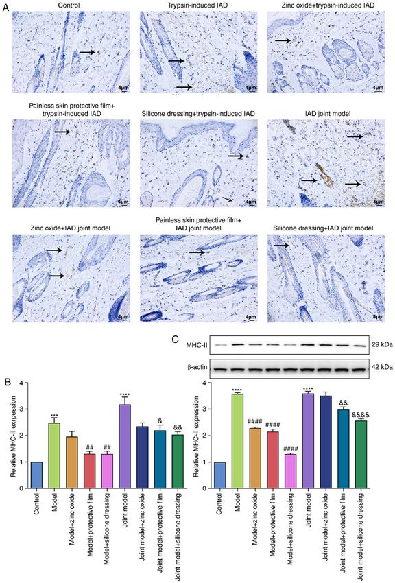
2                                CHEN et al: ESTABLISHMENT AND TREATMENT OF IAD MODELS

    Currently, the prevention and treatment of IAD include:        back of the rat. IAD scores and skin pH values were exam‑
i) Correcting the causes of diarrhea and incontinence;             ined every day. After 4 days, the bandage was removed and
ii) reducing urine and stool irritation; iii) correct cleaning,    the severity of IAD was observed, including the size of the
moisturizing and skin care; iv) maintaining the skin pH; and       dermatitis occurrence area and the IAD score of severity. The
v) regular observation and evaluation (7‑9). Since the presence    rats were anesthetized using an intraperitoneal injection of
of high moisture and corrosive enzymes in the intestinal juice     sodium pentobarbital. All procedures were strictly in line with
can cause destructive damage to the skin, leading to peeling       The Guide for the Care and Use of Laboratory Animals of the
and erosion of the cortex, barrier products such as petrolatum,    National Institutes of Health. The control group was treated
polydimethylsiloxane and zinc oxide ointment are necessary to      with cotton balls soaked in saline.
protect patients with IAD (10). Although several studies on the
prevention and treatment of IAD have been conducted, there         Assessment of the IAD model. According to the IAD severity
is significant heterogeneity and low comparability between         assessment tool scale described by Borchert et al (14), the rats
results (11‑13). It is difficult for clinical medical staff to     were observed and evaluated once a day before and after the
determine the relative performance of these barrier materials.     intervention for 4 days. Furthermore, the degree of recovery
Indeed, due to deficiencies in knowledge and clinical evidence,    from dermatitis was also evaluated after the intervention
product selection is still a challenge faced by clinical medical   according to the severity of the rash and missing skin. IAD was
staff when preventing and managing IAD. The aim of the             evaluated based on the degree of skin redness in the back, skin
present study was to identify a simple and easy‑to‑replicate rat   loss and rash. The scoring criteria were as follows: i) None,
model of IAD, and to study the effect of zinc oxide, painless      0 point; ii) erythema, 1 point; iii) edema, 2 points; iv) papule,
skin protective film and silicone dressing on the healing of       3 points; and v) erosion and superficial ulcer, 4 points.
IAD and to provide experimental evidence for the treatment
of IAD.                                                            Animal groups. After successful modeling, 54 rats were
                                                                   randomly divided into 9 groups according to the random
Materials and methods                                              number table (n=6 in each group): i) Control group; ii) trypsin
                                                                   model group; iii) model + zinc oxide group; iv) model + pain‑
Animals. 63 male Sprague‑Dawley rats (weight, 150‑220 g)           less skin protective film group; v) model + silicon dressing
aged 7‑8 weeks were provided by The Jinhua Center of               group; vi) synthetic urine combined with trypsin model group
Laboratory Animals (Zhejiang, China). Animal experiments           (joint model group); vii) joint model + zinc oxide (Guangzhou
were carried out in The Jinhua Food and Drug Inspection            Baiyunshan Pharmaceutical Co., Ltd.) group; viii) joint
and Testing Research Institute (Zhejiang, China). Before the       model + painless skin protective film (3M company) group;
experiment, all rats were adaptively fed for one week. All         and ix) joint model + silicone dressing (Jiangsu Youchuang
procedures were performed following the recommendations            Biomedical Technology Co., Ltd.) group.
of the Guide for the Care and Use of Laboratory Animals of             For rats in the zinc oxide group, zinc oxide ointment was
the National Institutes of Health. This study was approved by      evenly applied >1 cm away from the urine‑ and feces‑contam‑
The Ethics Committee of The School of Medicine, Jinhua             inated skin, twice a day. For rats in the painless skin
Polytechnic (approval no. 2019018).                                protective film group, the skin protective film was sprayed
                                                                   twice a day. The distance was 10‑15 cm from the urine‑ and
Construction of an IAD model. In an initial experiment, 9 rats     stool‑contaminated skin and l cm beyond the urine and stool
were randomly divided into three groups according to a random      contaminated skin. The painless skin protective film was
number table: i) Control group; ii) trypsin‑induced IAD (model)    applied on the affected skin. For rats in the silicone dressing
group; and iii) synthetic urine combined with trypsin‑induced      group, the silicone dressing was applied twice a day to the
IAD (joint model) group. The artificial urine was generated        affected skin.
as follows: i) 25 g urea (purity >99%); ii) 9 g sodium chloride        IAD severity assessment and skin pH test were performed
(>99%); iii) 3 g ammonium chloride (>99.9%); iv) 3 g sodium        for each group every 4 days after applying the zinc oxide,
sulfite (>98%); v) 2.5 g anhydrous disodium hydrogen ortho‑        protective film or silicon dressing intervention. After the rats
phosphate (>99%); and vi) 2 g creatinine (>99%). These were        had been anesthetized by intraperitoneal injection of pentobar‑
dissolved in 1,000 ml Ultrapure water prepared using a Direct      bital sodium (60 mg/kg), blood samples were collected from
Q5 purification system (EMD Millipore). Subsequently, 25%          the eyeball. All rats were then sacrificed by intraperitoneal
ammonium hydroxide solution was added into 25 ml of the            injection of an overdose of pentobarbital sodium (200 mg/kg).
above mixture, thus resulting in synthetic urine solution with     Skin tissue samples were then collected.
1% ammonium hydroxide. Sodium hydroxide was added into
4 g/100 ml trypsin liquid or synthetic urine combined with         Hematoxylin and eosin (H&E) staining. Fresh skin tissue
trypsin, thus adjusting pH to 7.5‑8.5.                             samples were fixed in 4% paraformaldehyde (Wuhan Google
    The selected area on the back of the rat was covered with      Biotechnology Co., Ltd.) for 24 h at 4˚C. After dehydration and
a cotton ball containing the trypsin solution or the synthetic     paraffin embedding, tissue sections were cut to 4‑µm thickness.
urine with trypsin and adhesive tape (3M company) and fixed        After the paraffin sections were deparaffinized, H & E staining
with an elastic bandage. The cotton ball was maintained            was performed. The sections were stained with hematoxylin
for 4 days. Once a day in the morning and afternoon, 5 ml          (cat. no. B600020; ProteinTech Group) at 37˚C for 5 min and
corresponding solution was added to the cotton ball of the rat     0.5% eosin solution (Sigma‑Aldrich; Merck KGaA) at 37˚C
to keep the cotton ball moist and continuously covered the         for 2 min. After dehydration, the sections were mounted with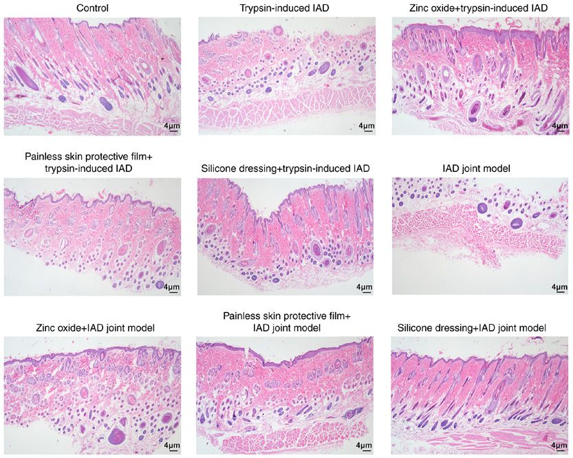
EXPERIMENTAL AND THERAPEUTIC MEDICINE 22: 1058, 2021                                            3

neutral gum and placed under a light microscope (BX53;             ANOVA followed by Tukey's post hoc test was used. P
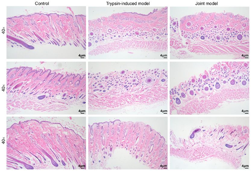
4                                       CHEN et al: ESTABLISHMENT AND TREATMENT OF IAD MODELS

Figure 1. Construction of IAD rat models. Hematoxylin and eosin staining was used to assess the pathological changes in skin tissue samples from the control,
trypsin‑induced IAD model and synthetic urine combined with trypsin‑induced IAD model groups. Magnification, x40. IAD, incontinence‑associated dermatitis.

damage and edema. Among them, rats in the silicone dressing                     NF‑κ B and STAT1 were not affected in the two IAD models.
treatment group displayed the best recovery.                                    After treatment with painless skin protective film (P
EXPERIMENTAL AND THERAPEUTIC MEDICINE 22: 1058, 2021 5
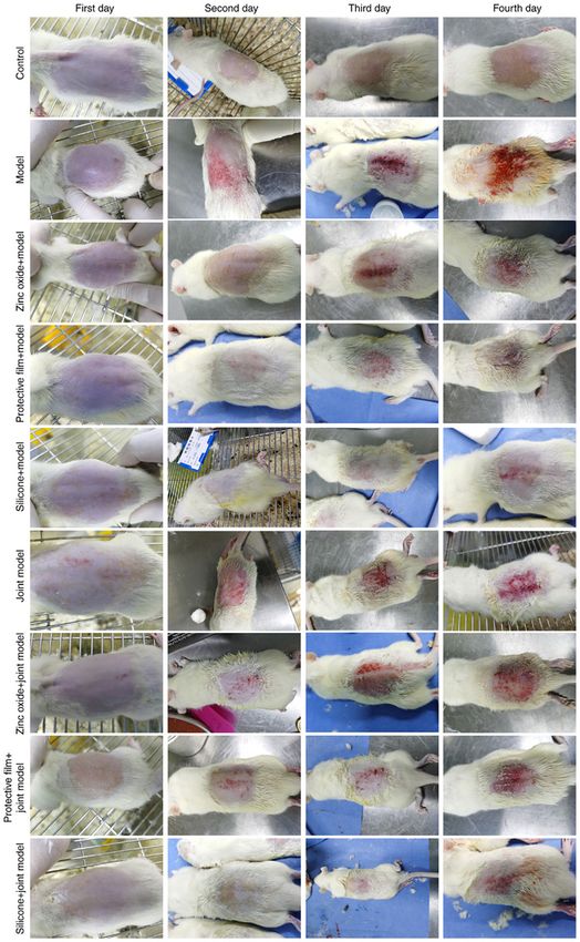
Figure 2. Photographs of the nine experimental groups on day 1, 2, 3 and 4 after treatment. There was significant dermatitis in trypsin‑induced and synthetic
urine combined with trypsin‑induced IAD rats compared to rats in the control group. Following treatment with zinc oxide, painless skin protective film or
silicone dressing for 1, 2, 3 and 4 days, the dermatitis of IAD rats was significantly ameliorated. IAD, incontinence‑associated dermatitis.
Discussion patients' quality of life and increased the economic burden of
patients (15). Therefore, the development of preventive and
IAD has a high incidence in the community, nursing homes and therapeutic methods for IAD is urgently needed. In the present
long‑term care institutions, as well as in the clinical departments study, rat models of IAD were established using two methods
and intensive care units of hospitals. It has severely reduced the (trypsin model and synthetic urine combined with trypsin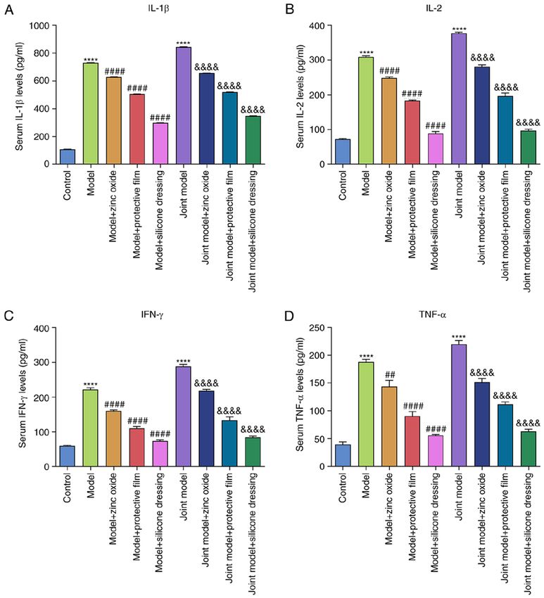
6                                      CHEN et al: ESTABLISHMENT AND TREATMENT OF IAD MODELS

Figure 3. Effects of zinc oxide, painless skin protective film and silicone dressing on the IAD score and skin pH of IAD rats induced by trypsin or
synthetic urine combined with trypsin. (A) Assessment of IAD scores in each group. (B) Determination of pH values in each group. **P
EXPERIMENTAL AND THERAPEUTIC MEDICINE 22: 1058, 2021                                                                7

Figure 5. Evaluation of MHC‑II expression in the skin tissue of rats in the control, trypsin‑induced IAD model and synthetic urine combined with trypsin‑induced
IAD model groups. (A and B) Immunohistochemical detection of MHC‑II expression in skin tissue samples from each group. Magnification, x200. MHC‑II
expression is indicated by the arrows. (C) Western blot analysis of MHC‑II expression in the skin tissue from each group. ***P
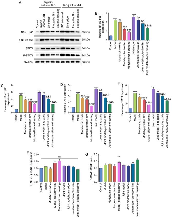
8                                       CHEN et al: ESTABLISHMENT AND TREATMENT OF IAD MODELS

Figure 6. Western blot analysis of the expression of NF‑κ B/p65, p‑NF‑κ B/p65, STAT1 and p‑STAT1 in the skin tissue of rats from the control,
trypsin‑induced IAD model and synthetic urine combined with trypsin‑induced IAD model groups. (A) Representative images of western blots of each
group. (B) NF‑κ B p65, (C) p‑NF‑κ B p65, (D) STAT1 and (E) p‑STAT1 expression levels were quantified according to their gray values. (F) p‑NF‑κ B/p65 to
NF‑κ B/p65 ratio. (G) p‑STAT1/STAT1 ratio. ****P
EXPERIMENTAL AND THERAPEUTIC MEDICINE 22: 1058, 2021                                                         9

Figure 7. ELISA of the serum levels of IFN‑γ, IL‑1β, IL‑2 and TNF‑ α in the control, trypsin‑induced IAD model and synthetic urine combined with
trypsin‑induced IAD model groups. (A) IL‑1β, (B) IL‑2, (C) IFN‑γ and (D) TNF‑α levels were examined in each group using ELISA. ****P
10 CHEN et al: ESTABLISHMENT AND TREATMENT OF IAD MODELS
an important role in the pathogenesis of dermatitis (27‑29). References
Inhibiting the expression of NF‑κ B p65, p‑NF‑κ B p65, STAT1
1. Beele H, Smet S, Van Damme N and Beeckman D: Incontinence‑
and p‑STAT1 can effectively reduce the severity of derma‑ associated dermatitis: Pathogenesis, contributing factors,
titis (30‑32). Compared with the control group, the expression prevention and management options. Drugs Aging 35: 1‑10, 2018.
of MHC‑Ⅱ, NF‑κ B p65, p‑NF‑κ B p65, STAT1 and p‑STAT1 2. Koudounas S, Bader DL and Voegeli D: Knowledge gaps in the
etiology and pathophysiology of incontinence‑associated derma‑
in the skin tissues of the model groups increased significantly. titis: A scoping review. J Wound Ostomy Continence Nurs 47:
After treatment, the expression of the above proteins was 388‑395, 2020.
significantly reduced. The reduction in the silicone dressing 3. Raepsaet C, Fourie A, Van Hecke A, Verhaeghe S and
Beeckman D: Management of incontinence‑associated derma‑
group was the most apparent. These results indicated that titis: A systematic review of monetary data. Int Wound J 18:
zinc oxide, painless skin protective film and silicone dress‑ 79‑94, 2021.
ings can effectively inhibit inflammation‑related pathways in 4. Zhang Y, Leng M, Guo J, Duan J and Wang Z: The effectiveness
of faecal collection devices in preventing incontinence‑associated
IAD, thereby improving the symptoms of dermatitis. Further dermatitis in critically ill patients with faecal incontinence: A system‑
studies are needed to examine the molecular mechanisms of atic review and meta‑analysis. Aust Crit Care 34: 103‑112, 2021.
zinc oxide, painless skin protective film or silicone dressings 5. Lichter feld‑Kot t ner A, El Genedy M, La h ma n n N,
Blume‑Peytavi U, Büscher A and Kottner J: Maintaining skin
in the treatment of IAD. integrity in the aged: A systematic review. Int J Nurs Stud 103:
In conclusion, zinc oxide, painless skin protective film 103509, 2020.
and silicone dressings significantly ameliorated dermatitis in 6. Barakat‑Johnson M, Basjarahil S, Campbell J, Cunich M,
Disher G, Geering S, Ko N, Lai M, Leahy C, Leong T, et al:
rats with IAD. Among these treatment modalities, silicone Implementing best available evidence into practice for
dressings exhibited the best therapeutic effects. Thus, these incontinence‑associated dermatitis in Australia: A multisite
intervention methods warrant further validation. multimethod study protocol. J Tissue Viability 30: 67‑77, 2021.
7. Gates BP, Vess J, Long MA and Johnson E: Decreasing incon‑
tinence‑associated dermatitis in the surgical intensive care unit:
Acknowledgements A quality improvement project. J Wound Ostomy Continence
Nurs 46: 327‑331, 2019.
8. Kottner J, Hahnel E, El Genedy M, Neumann K and Balzer K:
Not applicable. Enhancing SKIN health and safety in aged CARE (SKINCARE
Trial): A study protocol for an exploratory cluster‑randomized
Funding pragmatic trial. Trials 20: 302, 2019.
9. Phipps L, Gray M and Call E: Time of onset to changes in skin
condition during exposure to synthetic urine: A prospective
This work was funded by The Basic Public Welfare Research study. J Wound Ostomy Continence Nurs 46: 315‑320, 2019.
Program of Zhejiang Province (grant no. LGD20C040002) 10. Acton C, Ivins N, Bainbridge P and Browning P: Management of
incontinence‑associated dermatitis patients using a skin protec‑
and The Science and Technology Project of Jinhua City in tant in acute care: A case series. J Wound Care 29: 18‑26, 2020.
China (grant no. 2019‑4‑074). 11. Van Damme N, Van Hecke A, Himpens A, Verhaeghe S and
Beeckman D: Design and psychometric testing of the attitude
towards the prevention of incontinence‑associated dermatitis
Availability of data and materials instrument (APrIAD). Int Wound J 16: 492‑502, 2019.
12. Gray M: Context for practice: Prevention of pressure injury and
The datasets used and/or analyzed during the current study are incontinence‑associated dermatitis. J Wound Ostomy Continence
Nurs 44: 406‑408, 2017.
available from the corresponding author on reasonable request. 13. Kayser SA, Phipps L, VanGilder CA and Lachenbruch C:
Examining prevalence and risk factors of incontinence‑associ‑
Authors' contributions ated dermatitis using the international pressure ulcer prevalence
survey. J Wound Ostomy Continence Nurs 46: 285‑290, 2019.
14. Borchert K, Bliss DZ, Savik K and Radosevich DM: The
MD conceived and designed the study. GC and LH incontinence‑associated dermatitis and its severity instrument:
conducted most of the experiments and data analysis and Development and validation. J Wound Ostomy Continence
Nurs 37: 527‑535, 2010.
wrote the manuscript. YC, SZ and LZ conducted a number 15. Arnold‑Long M and Johnson E: Epidemiology of inconti‑
of experiments and data analysis, and contributed to the nence‑associated dermatitis and intertriginous dermatitis
writing and revision of the manuscript. MD and GC confirm (Intertrigo) in an acute care facility. J Wound Ostomy Continence
Nurs 46: 201‑206, 2019.
the authenticity of all the raw data. All authors read and 16. Minematsu T, Yamamoto Y, Nagase T, Naito A, Takehara K,
approved the final manuscript. Iizaka S, Komagata K, Huang L, Nakagami G, Akase T, et al:
Aging enhances maceration‑induced ultrastructural altera‑
tion of the epidermis and impairment of skin barrier function.
Ethics approval and consent to participate J Dermatol Sci 62: 160‑168, 2011.
17. Been RA, Bernatchez SF, Conrad‑Vlasak DM, Asmus RA,
Ekholm BP and Parks PJ: In vivo methods to evaluate a new skin
The study was approved by The Ethics Committee of protectant for loss of skin integrity. Wound Repair Regen 24:
The School of Medicine, Jinhua Polytechnic (approval 851‑859, 2016.
no. 2019018). 18. Mugita Y, Minematsu T, Huang L, Nakagami G, Kishi C,
Ichikawa Y, Nagase T, Oe M, Noguchi H, Mori T, et al:
Histopathology of incontinence‑associated skin lesions: Inner
Patient consent for publication tissue damage due to invasion of proteolytic enzymes and
bacteria in macerated rat skin. PLoS One 10: e0138117, 2015.
19. Wen Z, Zhu W, Liu Q, Zhang H, Mei B and Shen M: Development
Not applicable. of an animal model for inducing various degrees of severity of
incontinence‑associated dermatitis. J Wound Ostomy Continence
Competing interests Nurs 44: 578‑582, 2017.
20. Biçer Ş, Sayar İ, Gürsul C, Işık A, Aydın M, Peker K and
Demiryilmaz İ: Use of ozone to treat ileostomy dermatitis in an
The authors declare that they have no competing interests. experimental rat model. Med Sci Monit 22: 757‑765, 2016.EXPERIMENTAL AND THERAPEUTIC MEDICINE 22: 1058, 2021 11
21. Hoedl M and Eglseer D: Which characteristics of fecal incon‑ 28. Aquino M and Rosner G: Systemic contact dermatitis. Clin Rev
tinence predispose incontinence‑associated dermatitis? A Allergy Immunol 56: 9‑18, 2019.
classification and regression tree analysis. Adv Skin Wound 29. Kolesnik M, Franke I, Lux A, Quist SR and Gollnick HP: Eczema
Care 34: 103‑108, 2021. in psoriatico: An important differential diagnosis between
22. Koudounas S, Mugita Y, Minematsu T, Nakagami G, Weller C chronic allergic contact dermatitis and psoriasis in palmoplantar
and Sanada H: Does the presence of bacterial urinary infection localization. Acta Derm Venereol 98: 50‑58, 2018.
contribute to the development of incontinence‑associated derma‑ 30. Gil TY, Kang YM, Eom YJ, Hong CH and An HJ: Anti‑atopic
titis? A scoping review. J Tissue Viability 30: 256‑261, 2021. dermatitis effect of seaweed fulvescens extract via inhibiting the
23. Hödl M, Blanař V, Amir Y and Lohrmann C: Association STAT1 pathway. Mediators Inflamm 2019: 3760934, 2019.
between incontinence, incontinence‑associated dermatitis and 31. Yang BY, Cheng YG, Liu Y, Liu Y, Tan JY, Guan W, Guo S and
pressure injuries: A multisite study among hospitalised patients Kuang HX: Datura Metel L. Ameliorates imiquimod‑induced
65 years or older. Australas J Dermatol 61: e144‑e146, 2020. psoriasis‑like dermatitis and inhibits inflammatory cyto‑
24. Coyer F, Campbell J and Doubrovsky A: Efficacy of inconti‑ kines production through TLR7/8‑MyD88‑NF‑ κ B‑NLRP3
nence‑associated dermatitis intervention for patients in intensive Inflammasome pathway. Molecules 24: 2157, 2019.
care: An open‑label pilot randomized controlled trial. Adv Skin 32. Irrera N, Vaccaro M, Bitto A, Pallio G, Pizzino G, Lentini M,
Wound Care 33: 375‑382, 2020. Arcoraci V, Minutoli L, Scuruchi M, Cutroneo G, et al: BAY
25. Tay C, Yuh AS, Sheau Lan EL, Ong CE, Aloweni F and Lopez V: 11‑7082 inhibits the NF‑κ B and NLRP3 inflammasome pathways
Development and validation of the incontinence associated and protects against IMQ‑induced psoriasis. Clin Sci (Lond) 131:
dermatitis knowledge, attitude and practice questionnaire. 487‑498, 2017.
J Tissue Viability 29: 244‑251, 2020.
26. Werth SL and Justice R: Prevalence of moisture‑associated This work is licensed under a Creative Commons
skin damage in an acute care setting: Outcomes from a quality Attribution-NonCommercial-NoDerivatives 4.0
improvement project. J Wound Ostomy Continence Nurs 46: International (CC BY-NC-ND 4.0) License.
51‑54, 2019.
27. Park KH: The effect of a silicone border foam dressing for preven‑
tion of pressure ulcers and incontinence‑associated dermatitis
in intensive care unit patients. J Wound Ostomy Continence
Nurs 41: 424‑429, 2014.You can also read