Common Fractures and their management. Part A : NOF# - Dr. Mujtaba Shaukat.
←
→
Page content transcription
If your browser does not render page correctly, please read the page content below
Common Fractures and their
     management.
     Part A : NOF#
     Dr. Mujtaba Shaukat.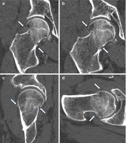
Neck of Femur fractures
• A fractured neck of femur (NOF) is a very
  common orthopaedic presentation.
• Over 65,000 hip fractures each year are
  recorded in the UK and they are
  becoming increasingly frequent due to an
  aging population.
• The mortality of a femoral neck fracture is up
  to 30% in one year
Definition
• A neck of Femur fracture is a break just below
  the Femoral head either in the capsular region
  or in the extracapsular part.
Cause
• Neck of femur fractures are typically caused
  either by low energy injuries (the most
  common type), such as a fall in frail older
  patient, or high energy injuries, such as a road
  traffic collision or fall from height and are
  often associated with other significant
  injuries.
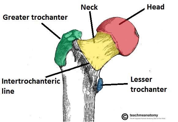
• The neck of femur can be considered to have two
  distinct areas, which are described relative to the joint
  capsule:
• Intra-capsular – from the subcapital region of the
  femoral head to basocervical region of the femoral
  neck, immediately proximal to the trochanters
• Extra-capsular – outside the capsule, subdivided into:
   – Inter-trochanteric, which are between the greater
     trochanter and the lesser trochanter
   – Sub-tronchanteric, which are from the lesser trochanter to
     5cm distal to this point
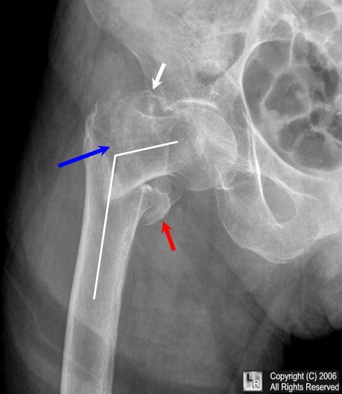
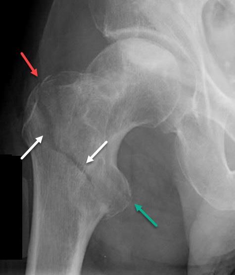
Intracapsular fractured neck of femur
Extracapsular fractured neck of femur
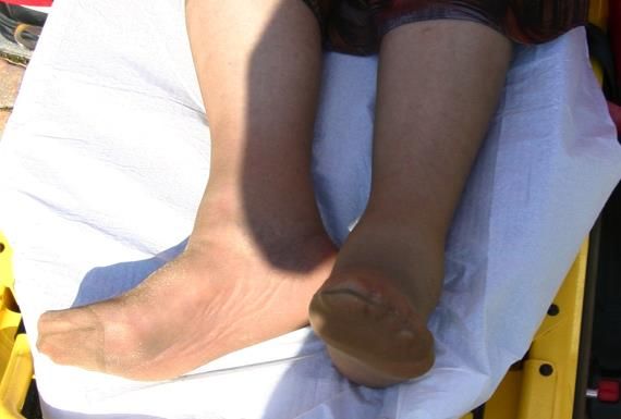
Clinical Features

• The leading symptom is trauma, often low-
  energy, which is followed by pain and
  an inability to weight bear. Pain is felt
  predominantly in the groin, thigh or,
  commonly in the elderly, referred to the knee.
• On examination, the leg is
  characteristically shortened and externally
  rotated, due to the pull of the short external
  rotators.
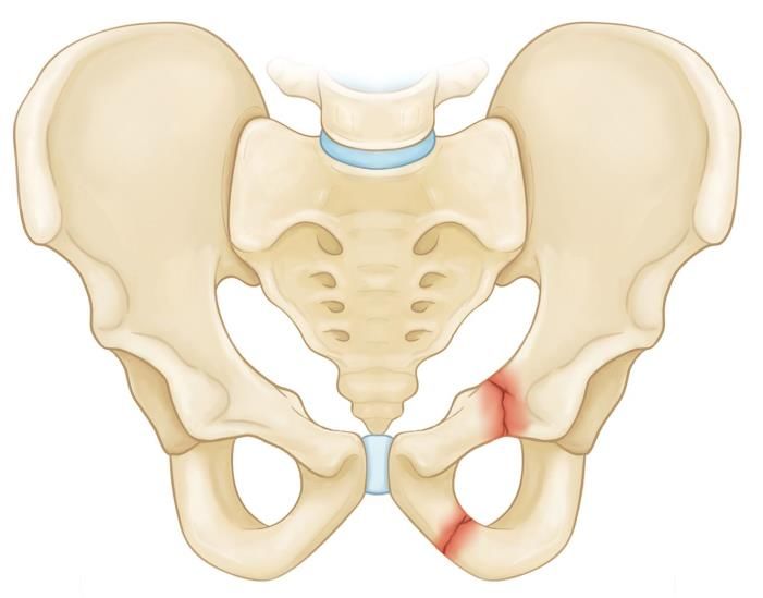
Differential Diagnoses

• Alternative fractures, such as of the pelvis
  (especially pubic ramus fractures),
  acetabulum, femoral head and femoral
  diaphysis, all need to be considered.
• Pathological fractures should be considered if
  there is not a significant history of trauma e.g.
  cancer.
Fractured pubic rami

                       Acetabular
                       fracture

                             Pathological
                             fracture
Investigations • Initial plain-film radiographic imaging should include antero-posterior (AP) and lateral views of the affected hip, as well as an AP pelvis (useful to assess the contralateral normal hip for pre- operative planning and templating). Obtain full length femoral radiographs too, if there is suspicion of a pathological fracture. • If clinical features are sugestive of a #NOF but the x-ray is inconclusive, consider bilateral hip CT.
CT of neck of femur fracture
Bloods :
• Basic routine blood tests, including FBC (to
  look at Hb pre-op), U&Es, and coagulation
  screen, are required alongside two seperate
  Group and Saves.
• If a long lie time could have occurred,
  a creatinine kinase (CK) level would be
  recommended to assess for any significant
  rhabdomyolysis.
Other investigations :
• Chest radiograph (CXR), Urine Mc&s and ECG
  are all necessary in order to carry out a
  complete assessment of the older patient
  group, especially for pre-operative assessment
  and peri-operative management.
• As mentioned, further work-up as to the
  cause of the fall is also essential including
  social history and baseline mobility.
Management

• Initial management of a neck of femur
  fracture should consist of an A to
  E approach to stabilise the patient and treat
  any immediately life- or limb- threatening
  problems as this cohort of patients will likely
  sustain concurrent injuries (even in low-
  impact cases). The most common example of
  this is head injuries e.g. sub- dural
  hematoma's
• Ensure adequate analgesia is provided, as hip
  fractures are very painful. This can be either
  as opioid analgesia and / or regional
  analgesia (such as a fascia-iliaca block)
• Urinary catheter should be inserted as
  patients will be unable to mobilise to the
  toilet and this will prevent further skin and
  bone damage and provide an accurate fluid
  balance.
Definitive management :
•  Definitive management is surgical.
•  Surgical option depends on type of fracture, as
   the blood supply to the head of the femur is via
   the neck.
• Non-operative conservative management is
  rarely recommended, as the benefits of surgical
  intervention nearly always outweigh the potential
  conservative management due to mortality being
  almost always 100% in non operative patients.
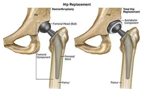
Intracapsular neck of femur fracture
• Displaced intracapsular fracture = Hip hemiarthroplasty or total hip
  replacement.
• Hip Hemiarthroplasty = Replacement of the femoral head and neck via a
  femoral component fixed in the proximal femur.
• Total hip replacement = acetabular component is included with the
  hemiarthroplasty.
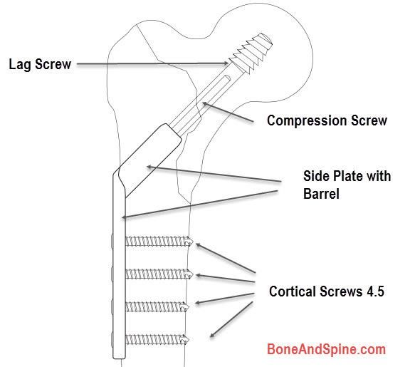
Extracapsular neck of femur fracture

• Inter-trochanteric and Basocervical = Dynamic hip screw.
• Dynamic hip screw consists of a lag screw into the neck, a
  sideplate, and bicortical screws. The lag screw is able to slide
  through the sideplate, allowing for compression and primary
  healing of the bone.
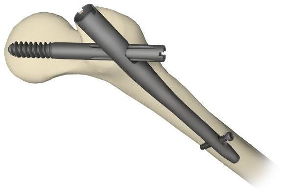
Extracapsular neck of femur fracture
• Sub-trochanteric = Intramedullary Femoral Nail.
The titanium rod is placed through the medullary cavity of the femur
for stabilisation.
Post-op planning.
• Early mobilisation/physiotherapy.
• Analgesia.
• Hemodynamic stability including being vigilant
  about chest infections, bleeding and wound
  problems.
• Occupational therapy.
Thank you!
Any questions ?
You can also read