Comparing methods to secure a tracheal tube placed via a surgical cricothyroidotomy: a randomised controlled study in cadavers
←
→
Page content transcription
If your browser does not render page correctly, please read the page content below
Groombridge et al. Scandinavian Journal of Trauma, Resuscitation and Emergency Medicine
(2021) 29:104
https://doi.org/10.1186/s13049-021-00925-y
ORIGINAL RESEARCH Open Access
Comparing methods to secure a tracheal
tube placed via a surgical
cricothyroidotomy: a randomised controlled
study in cadavers
Christopher J Groombridge1,2,4*, Amit Maini1,2, Joseph Mathew1,2,4, Yesul Kim1,4, Mark Fitzgerald1,4,
De Villiers Smit1,2,3 and Gerard O’Reilly1,2,3
Abstract
Objective: In the ‘can’t intubate can’t oxygenate’ scenario, techniques to achieve front of neck access to the airway
have been described in the literature but there is a lack of guidance on the optimal method for securing the
tracheal tube (TT) placed during this procedure. The aim of this study was to compare three different methods of
securing a TT to prevent extubation following a surgical cricothyroidotomy.
Methods: A randomised controlled trial was undertaken. The population studied were emergency physicians (EPs)
attending a cadaveric airway course. The intervention was securing a TT placed via a surgical cricothyroidotomy by
suture. The comparison was securing the TT using fabric tape with two different tying techniques. The primary
outcome was the force required to extubate the trachea. The trial was registered with ANZCTR.org.au (ACTR
N12621000320853).
Results: 17 emergency physicians completed intubations using all three of the securing methods on 12 cadavers
for a total of 51 experiments. The mean extubation force was 6.54 KG (95 % CI 5.54–7.55) in the suture group
compared with 2.28 KG (95 % CI 1.91–2.64) in the ‘Wilko tie’ group and 2.12 KG (95 % CI 1.63–2.60) in the ‘Lark’s foot
tie’ group; The mean difference between the suture and fabric tie techniques was significant (p < 0.001).
Conclusions: Following a surgical cricothyroidotomy in cadavers, EPs were able to effectively secure a TT using a
suture technique, and this method was superior to tying the TT using fabric tape.
Keywords: Intubation, Surgical airway
Background tracheal tube (TT) placed through the cricothyroid
The final step in the ‘can’t intubate can’t oxygenate’ membrane has not been clearly established.
(CICO) scenario is front of neck access (FONA)[1] and In the emergency department (ED), TTs placed during
whilst there are recommendations on the technique to orotracheal intubation may be secured using fabric tape
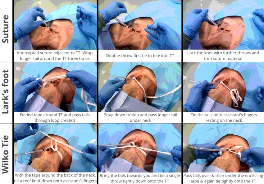
be employed the best method of securing a standard tied using a ‘Lark’s foot’ knot (Fig. 1)[2] or alternative
knots such as the ‘the Wilko tie’ (Fig. 1). Both these
methods, however, require the tape to be passed around
* Correspondence: christopher.groombridge@monash.edu the neck and clinicians may be reluctant to utilize these
1
National Trauma Research Institute, Melbourne, Australia methods to secure a surgical airway, for fear of occlud-
2
Emergency & Trauma Centre, The Alfred Hospital, Melbourne, Australia ing venous return from the head. To avoid this risk
Full list of author information is available at the end of the article
© The Author(s). 2021 Open Access This article is licensed under a Creative Commons Attribution 4.0 International License,
which permits use, sharing, adaptation, distribution and reproduction in any medium or format, as long as you give
appropriate credit to the original author(s) and the source, provide a link to the Creative Commons licence, and indicate if
changes were made. The images or other third party material in this article are included in the article's Creative Commons
licence, unless indicated otherwise in a credit line to the material. If material is not included in the article's Creative Commons
licence and your intended use is not permitted by statutory regulation or exceeds the permitted use, you will need to obtain
permission directly from the copyright holder. To view a copy of this licence, visit http://creativecommons.org/licenses/by/4.0/.
The Creative Commons Public Domain Dedication waiver (http://creativecommons.org/publicdomain/zero/1.0/) applies to the
data made available in this article, unless otherwise stated in a credit line to the data.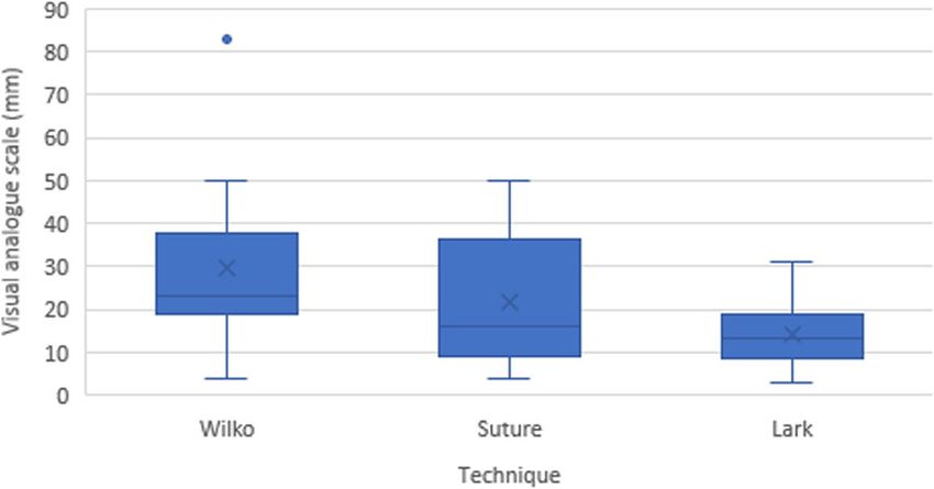
Groombridge et al. Scandinavian Journal of Trauma, Resuscitation and Emergency Medicine   (2021) 29:104                Page 2 of 5

 Fig. 1 Three methods for securing the tracheal tube.

clinicians may opt for a suture ‘drain stitch’ (Fig. 1) to             from The Airway Course, a one-day cadaveric airway
maintain TT position.                                                  management course for critical care clinicians.

  Our aim is to provide clarity on an optimal method
for securing a surgical airway which can be taught along-              Intervention
side FONA techniques. In this study we have chosen to                  Prior to the experiment, participants received instruction
compare the force required to extubate a fresh-frozen                  on the securing methods as part of The Airway Course.
cadaver where the surgical cricothyroidotomy TT has                    Each EP performed all three techniques on three differ-
been secured by either a Lark’s foot knot, using fabric                ent cadavers amounting to 51 experiments, the order of
tape, a ‘Wilko tie’, again using fabric tape, and a ‘drain             which was randomised in advance by computer random-
stitch’, using a silk suture.                                          isation to minimise learning effects (https://www.
                                                                       randomizer.org).
                                                                         The TT was a Portex Choice size 6.5mm cuffed tra-
Methods                                                                cheal tube. The securing tape was a standard white fab-
Study design and participants                                          ric 12mm wide tape cut to 1 m lengths. The suture was
This randomised controlled trial was approved by the                   a 1 Sofsilk™ 75 cm braided silk suture with a cutting nee-
University of Melbourne research ethics committee (ap-                 dle from Covidien™.
proval number 1,648,354). The trial was registered be-                   For all the experiments the starting condition was with
fore clinician enrolment at Australian New Zealand                     the supine cadaver intubated with a TT placed through
Clinical Trials Registry (ACTRN12621000320853).                        a surgical airway performed as part of the course educa-
Twelve fresh-frozen cadavers were provided by the Uni-                 tion (midline longitudinal incision, followed by horizon-
versity of Melbourne’s department of human anatomy.                    tal cricothyroid puncture as part of a scalpel, finger,
Seventeen emergency physicians (EP) were recruited                     bougie technique).
Groombridge et al. Scandinavian Journal of Trauma, Resuscitation and Emergency Medicine         (2021) 29:104                         Page 3 of 5

Table 1 Characteristics of participants
Characteristic                                               n = 17 (%)
Gender
Female                                                       6 (35 %)
Male                                                         11 (65 %)
Year’s experience as EM specialist
< 5 years                                                    4 (23.5 %)
5–10 years                                                   10 (58.8 %)
11–15 years                                                  1 (5.9 %)
> 15 years                                                   2 (11.8 %)     Fig. 3 Visual Analogue Scale of technique difficulty. The boxplots
EM = Emergency Medicine                                                     show medians (solid line), means (X) and interquartile ranges (IQR).
                                                                            The whiskers give the range except for “outliers” that are more than
  After the TT was deemed secure by the participant,                        ± 1.5 times the IQR.
the cadaver was draped such that the investigator was
unable to determine the securing method used.                              80 % and a small difference in extubation force (200 g)
                                                                           between procedures. A sample size of 15 participants
Outcomes                                                                   was then recruited to ensure complete data for the
The force required to extubate the cadaver, the primary                    analysis.
outcome, was estimated using a digital force gauge                            Symmetrical numerical data has been summarised
(Dr.meter digital scale ES-PS01) attached to the TT. The                   using the mean (SD); skewed numerical data has been
same investigator, blinded to the securing method, per-                    summarised using the median (IQR); and categorical
formed all measurements by applying slow gradual up-                       data has been summarised using frequencies (%). The
wards traction on the meter, until the TT cuff was                         statistical significance of these measures of association
completely through the surgical wound, and recording                       were tested using paired statistical testing procedures,
the maximum value. Assuming Force = Mass x Acceler-                        i.e., Repeated Measures Analysis of Variance (ANOVA).
ation, and given the slow traction technique, where ac-                    P values < 0.05 were considered statistically significant.
celeration was considered negligible (and consistent
across experiments), the weight measurement of the
gauge was taken to be a surrogate for the extubation                       Results
force.                                                                     The planned sample size was achieved with 17 clinicians
  After performing each technique the participant was                      completing the three techniques on 12 cadavers for a
asked to mark a visual analogue scale (VAS) to indicate                    total of 51 experiments. Characteristics of the partici-
how easy or hard the technique had been to undertake                       pants are presented in Table 1. The twelve cadavers had
following minimal instruction.                                             a mean height 150 cm, mean weight 66KG, and 7 were
                                                                           female.
Statistical analysis                                                         Extubation force (Fig. 2) and VAS of technique diffi-
A minimum of 10 clinicians had been determined by a                        culty (Fig. 3) are summarised in Table 2.
power analysis based on an alpha of 0.05, a power of

                                                                             The mean extubation force was highest for the suture
                                                                           technique, followed by the Wilko Tie and then Lark’s
                                                                           foot technique, which had the lowest force required. The
                                                                           difference between these mean values was significant
                                                                           using a repeated measures ANOVA (p < 0.001), as seen
                                                                           in Table 2. Post-hoc pairwise comparison using a Bon-
                                                                           ferroni adjustment demonstrated a significant mean dif-
                                                                           ference in force between the suture technique and the
                                                                           Wilko (mean difference 4.27KG; 95 % CI 3.02–5.52; p <
                                                                           0.001) and Lark’s foot technique (mean difference
 Fig. 2 Extubation force (Kg). The boxplots show medians (solid line),     4.43KG; 95 % CI 3.44–5.42; p < 0.001). There was no sig-
 means (X) and interquartile ranges (IQR). The whiskers give the           nificant difference identified between the Wilko Tie and
 range except for “outliers” that are more than ± 1.5 times the IQR.
                                                                           Lark’s foot techniques.
Groombridge et al. Scandinavian Journal of Trauma, Resuscitation and Emergency Medicine (2021) 29:104 Page 4 of 5
Table 2 Technique performance outcomes
Lark’s foot Wilko Tie Suture p-value
Extubation force (KG) 2.12 (1.63–2.60) 2.28 (1.91–2.64) 6.54 (5.54–7.55) < 0.001
Visual Analogue Scale of difficulty (mm) 14.35 (9.91–18.80) 29.53 (20.10–38.96) 21.59 (13.69–29.49) < 0.05
The mean VAS was highest for the Wilko Tie tech- which is able to resist a mean extubation force equiva-
nique, followed by the suture technique and then the lent to at least 6 KG.
Lark’s foot technique, which was reported as the easiest There are limitations to this study. Firstly, we used ca-
of the techniques. The difference between these mean davers rather than anaesthetised ED patients and so the
values was significant using a repeated measures results cannot be automatically extrapolated to this pa-
ANOVA (p = 0.016), as seen in Table 2. Post-hoc pair- tient group. Fresh frozen cadavers represent the best
wise comparison using a Bonferroni adjustment demon- existing alternative model for this type of research, how-
strated a significant difference between the Wilko Tie ever, particularly when compared with manikin models.
and Lark’s foot technique (mean difference 15.18mm; Secondly, the force applied to the TT in this experiment
95 % CI 3.42–26.94; p = 0.012). There was no significant may not be representative of accidental extubations
difference identified between the other techniques. in vivo, which can involve sudden application of force in
any direction. Lastly, the study’s participants were a rela-
Discussion tively experienced group of EPs who had chosen to
This randomised controlled trial compared the force re- undertake a cadaveric airway course, and this may also
quired to extubate a TT placed via a surgical cricothyr- limit the generalisability of the findings.
oidotomy, using three securing techniques, and found
that a suture ‘drain stitch’ method was able to resist a Conclusions
greater extubation force than two fabric tape tying Following a surgical cricothyroidotomy in fresh frozen
methods. The suture technique was also not reported to cadavers, EPs were able to effectively secure a TT using
be more difficult to perform than the fabric tape tie a simple suture technique and this method was superior
techniques. to tying the TT using fabric tape.
One of the concerns with the fabric tape tie techniques
is that the tape must be passed around the neck with the Acknowledgements
We would like to thank Paramedic Christopher Wilkinson who invented the
associated risk of occluding venous return which may ‘Wilko Tie’.
affect intracranial pressure, particularly in head injured
patients[3]. The technique taught in this study involved Authors' contributions
All authors made substantial contributions. CG, AM, and GO did the
tying down onto an assistant’s three fingers, placed onto conception and design of the study. CG, AM, JM did the data acquisition.
the anterolateral neck, such that when their fingers are CG, GO, and MF did data analysis and interpretation of data. CG, JM, YK and
removed the tape is not constricting the neck. This add- DVS drafted the article. All authors revised it critically for intellectual content.
All authors have given final approval of the version submitted.
itional ‘slack’ in the system may have allowed the TT
cuff to be pulled from the cricothyroidotomy and does Funding
not reflect the likely success of these tying methods to This work received no funding.
secure an orally placed TT. Prior studies have assessed
Availability of data and materials
different options for securing a TT placed orally, with The anonymised datasets generated and/or analysed during the current
both different types of knot[4], as well as proprietary study are available from corresponding author.
tube holding devices[5], and adhesive tape[6, 7]. Most of
these studies, however, were undertaken in mannequins Declarations
and none assessed methods of securing a TT placed via Ethics approval and consent to participate
surgical cricothyroidotomy. Ethical approval for this study was obtained from University of Melbourne
research ethics committee (approval number 1648354, dated 1st October
Surgical airways are performed rarely in the ED[8] and 2020). Written consent was obtained from all the participants.
as such may be stressful to perform. This has the poten-
tial to impair performance[9] and clear guidance on the Competing interests
optimal method for securing the TT after successful CG and AM run a cadaveric airway skills workshop to teach emergency
airway management to critical care clinicians.
completion of a surgical airway should form part of edu-
cation around this procedure. Author details
1
We have demonstrated that EPs with a brief training National Trauma Research Institute, Melbourne, Australia. 2Emergency &
Trauma Centre, The Alfred Hospital, Melbourne, Australia. 3School of Public
intervention are able to successfully secure a TT placed Health and Preventive Medicine, Monash University, Melbourne, Australia.
via a surgical cricothyroidotomy using a silk suture, 4
Central Clinical School, Monash University, Melbourne, Australia.Groombridge et al. Scandinavian Journal of Trauma, Resuscitation and Emergency Medicine (2021) 29:104 Page 5 of 5
Received: 12 June 2021 Accepted: 16 July 2021
References
1. Higgs A, McGrath BA, Goddard C, Rangasami J, Suntharalingam G, Gale R,
Cook TM. Difficult Airway Society; Intensive Care Society; Faculty of Intensive
Care Medicine; Royal College of Anaesthetists. Guidelines for the
management of tracheal intubation in critically ill adults. Br J Anaesth. 2018;
120(2):323–52.
2. Sandberg M, Nakstad AR, Berlac PA, Hyldmo PK, Boylan M. Airway
assessment and management. ABC Prehospital Emer Med. 2013;258:20.
3. Davies G, Deakin C, Wilson A. The effect of a rigid collar on intracranial
pressure. Injury. 1996;27(9):647–9.
4. Walters HR, Young HE, Young PJ. A Modified Tie Technique for Securing
Endotracheal Tubes. Respir Care. 2018;63(4):424–9.
5. Murdoch E, Holdgate A. A comparison of tape-tying versus a tube-holding
device for securing endotracheal tubes in adults. Anaesth Intensive Care.
2007;35(5):730–5.
6. Landsperger JS, Byram JM, Lloyd BD, Rice TW, Pragmatic Critical Care
Research Group. The effect of adhesive tape versus endotracheal tube
fastener in critically ill adults: the endotracheal tube securement (ETTS)
randomized controlled trial. Crit Care. 2019;7(1):161. 23(.
7. Shimizu T, Mizutani T, Yamashita S, Hagiya K, Tanaka M. Endotracheal tube
extubation force: adhesive tape versus endotracheal tube holder. Respir
Care. 2011;56(11):1825–9.
8. Groombridge C, Maini A, Olaussen A, Kim Y, Fitzgerald M, Mitra B, Smit V.
Impact of a targeted bundle of audit with tailored education and an
intubation checklist to improve airway management in the emergency
department: an integrated time series analysis. Emerg Med J. 2020;37(9):
576–80.
9. Groombridge CJ, Kim Y, Maini A, Smit V, Fitzgerald MC. Stress and decision-
making in resuscitation: A systematic review. Resuscitation. 2019;144:115–22.
Publisher’s Note
Springer Nature remains neutral with regard to jurisdictional claims in
published maps and institutional affiliations.You can also read