Use of Foley Catheter in Control of Internal Carotid Hemorrhage during Endoscopic Endonasal Surgery
←
→
Page content transcription
If your browser does not render page correctly, please read the page content below
Folia Medica 63(5):809-14
                                                                                                                          DOI: 10.3897/folmed.63.e56461

                                                                   Case Report

Use of Foley Catheter in Control of Internal
Carotid Hemorrhage during Endoscopic
Endonasal Surgery
Matteo Alicandri-Ciufelli1, Francesco Maccarrone1, Cecilia Botti1,2, Giacomo Pavesi3, Livio Presutti1
1 Department of Otolaryngology - Head and Neck Surgery, University Hospital of Modena, Modena, Italy
2 PhD Program in Clinical and Experimental Medicine, University of Modena and Reggio Emilia, Italy
3 Department of Neurosciences, Neurosurgery Unit, University Hospital of Modena, Modena, Italy

Corresponding author: Cecilia Botti, Department of Otolaryngology - Head and Neck Surgery, University Hospital of Modena, Via del Pozzo, 41125,
Modena, Italy; Email: botceci@gmail.com; Tel.: +390594222402

Received: 13 July 2020 ♦ Accepted: 6 Oct 2020 ♦ Published: 31 Oct 2021

Citation: Alicandri-Ciufelli M, Maccarrone F, Botti C, Pavesi G, Presutti L. Use of Foley catheter in control of internal carotid hemor-
rhage during endoscopic endonasal surgery. Folia Med (Plovdiv) 2021;63(5):809-14. doi: 10.3897/folmed.63.e56461.

Abstract
Internal carotid artery (ICA) injuries during endoscopic endonasal surgery (EES) are rare life-threatening events. We describe a tech-
nique to manage ICA injuries based on the use of Foley catheters.
A 26-year-old female underwent endoscopic transnasal trans-sphenoidal removal of pituitary adenoma. Cerebrospinal fluid leak
occurred 4 days postoperatively. During repair procedure, accidental injury of ICA occurred. Emergency nasal packing through
positioning of four Foley urologic catheters was successfully performed to stop bleeding. The patient did not report neurologic deficits.
In author’s opinion, Foley catheters are suitable to obtain immediate bleeding control since they are rapidly available and easily usable.

Keywords
balloon nasal packing, endoscopic endonasal surgery, internal carotid injury, internal carotid hemorrhage, pituitary surgery

INTRODUCTION
Endoscopic endonasal surgery (EES) is a mini-invasive pro-                      fibrosis and alteration of anatomical landmarks, are risk
cedure, first introduced in the management of paranasal                         factors for ICA injuries. The massive bleeding is hard to
sinus disease. It became progressively popular in surgical                      manage in EES because it is a one-hand technique in most
management of anterior skull base disease and pituitary ade-                    cases, and the bleeding reduces visibility in the surgical
noma. Complications of EES have been widely described: ce-                      field by dirtying the tip.2
rebrospinal fluid leak, orbital hematoma, damage to nasola-                         Approaches for immediate management of ICA injuries
crimal duct, optic nerve damage, diplopia, and hemorrhage.1                     are nasal packing, endoscopic clipping, bipolar coagulation
   Injury of internal carotid artery (ICA) is the most severe                   and intraoperative angiography.3-5
and frightening complication which can result in stroke,                            The aim of this report was to describe a technique to first
cranial nerve palsy and death. Revision surgery, radiation,                     line emergency treatment of ICA injuries during EES by
previous bromocriptine treatment and acromegaly, causing                        using Foley urologic catheters.

Copyright by authors. This is an open access article distributed under the terms of the Creative Commons Attribution License (CC-BY 4.0),
which permits unrestricted use, distribution, and reproduction in any medium, provided the original author and source are credited.

                                                                                                                                                   809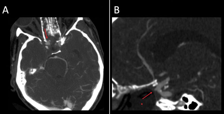
M. Alicandri-Ciufelli et al

CASE REPORT                                                           recent operation. A preliminary exploration and landmark
                                                                      identification from the right nasal fossa were performed, and
A 26-year-old female from North Africa presented at the               during demucosization of the previously uncovered lamina
Neurosurgical Department with a history of headache and               papyracea, a marked bleeding was verified. The bleeding was
visual loss. During an access to the Emergency Department,            provoked by elevation of granulation tissue by a suction can-
she underwent a brain CT scan that documented a large                 nula, in the region between lateral wall of sphenoid sinus and
mass involving sellar and parasellar region with marked ero-          lamina papyracea. Surgical field was immediately obscured
sion of sphenoid sinus walls (Fig. 1A).                               by blood that could not be suctioned by any kind of aspira-
    A brain MR confirmed the diagnosis, and documented                tion cannula. The suction tube without any aspiration can-
a massive encasement of carotid arteries, cerebral posterior          nula was then used, directly inside the right nasal fossa, but
and basilar arteries (Fig. 1B). At blood testing, a marked            also in this case, the surgical field was not cleared. At this
increase of growth hormone (GH) was found (GH 24.83 ng/               point, any kind of packaging (cottonoids, Merocel, coagula-
mL; IGF-1 543 ng/mL). At a close clinical evaluation, the             tion) was deemed impossible, due to the presence of the as-
patient showed physical features partially consistent with            piration tube and the insufficient view of the surgical field.
gigantism/acromegaly, with tall height and increase of foot           In this extremely urgent conditions, it was then decided to use
and finger dimensions. Laboratory and radiological findings           a balloon packaging by 14 Fr Foleys urologic catheters, using
were consistent with GH-secreting pituitary macroadenoma              the technique herein described
(3.0×2.5×2.2 cm), with parasellar extension (Hardy’s clas-
sification of pituitary tumors: grade E) and encasement of            Operative technique description: Foley
intracavernous ICA (Knosp classification system: grade 4).            catheter packaging for endonasal
The CT-scan showed Keros type 2 olfactory fossa and thick-            carotid bleeding
ening of the base of skull.
    After multidisciplinary evaluation, surgical removal was          After removing the aspiration tube from nasal cavity, the first
planned. A transnasal, trans-sphenoidal endoscopic removal            two Foleys are rapidly placed to close the posterior nasal fossa
was performed. The procedure was regular, without signifi-            at the level of the choanae (Fig. 2A). To perform this, is can
cant complications. At post-operative CT scan, a residual of          be very useful to overcome the nasal fossa, and feeling the
the pathology lateral to left carotid artery was found (Fig.          caudal bending of the catheter. Then the catheter is filled with
1C). At 4 days postoperatively, the patient showed an inter-          10-15 cc of water (Fig. 2B), and then pulled back to occlude
mittent cerebrospinal fluid (CSF) leak with watery rhinor-            the ipsilateral choana (Fig. 2C). The same procedure is per-
rhea, and persistent pneumocephalus. Hence, a CSF leak                formed for the contralateral part. At this point, nasal fossae
repair with fat obliteration of the sphenoid sinus and closure        are separated completely from other cavities (i.e. the nasop-
with nasoseptal flap was planned.                                     harynx). The catheter already positioned helps and guides
    During surgery, granulation tissue and inflammatory               the further ones to be placed in the sphenoethmoidal region,
material was found at the surgical site, consistent with the          since the most inferior part of the nasal fossa is occupied by

Figure 1. A. Preoperative brain CT scan, showing a mass involving sellar and parasellar regions. Erosion of sphenoid sinus walls is
present; B. Preoperative brain MR showing the mass encasing the carotid, cerebral posterior and basilar arteries; C. Postoperative brain
CT scan: residual lesion can be noticed lateral to left carotid artery.

810                                                                                           Folia Medica I 2021 I Vol. 63 I No. 5
Foley Catheter in Internal Carotid Hemorrhage
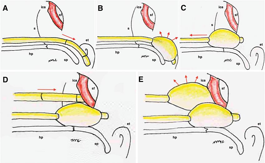
Figure 2. Foley catheter packaging procedure during endonasal carotid bleeding. The first Foley catheter is placed through each nasal
cavity until it reaches the rhinopharynx (A), and it is filled with 10-15 cc of water in the rhinopharynx (B). The catheter is then pulled
back to occlude the choana (C). A second Foley is put in the nasal fossa (D) and then it is filled with saline solution (E) to obtain the
hemostasis. The procedure is made bilaterally. S: nasal septum; ica: internal carotid artery; sf: sphenoidal sinus; et: Eustachian tube; hp:
hard palate; sp: soft palate.
the first ones. A third Foley can so be placed and filled with
saline in the nasal fossa of bleeding (Figs 2D, 2E), having the
true hemostatic purpose. The fourth Foley is placed on the
contralateral side to avoid displacement of the previous one.
The final result is shown in Fig. 3.
At the end of the procedure, the bleeding stopped immedi-
ately. The patients underwent at first angiography that failed
to identify the origin of bleeding, most probably because of
the packaging in place. An angio-CT scan was performed
two days later and a pseudoaneurysm, created by surgical
trauma, was identified (Fig. 4A). The position of the Foley
compared to the lesion was also verified (Fig. 4B). A stent-
ing was then performed with success, although an occlusion
of the carotid was verified 48 hours after the procedure. The
patient had a good contralateral supply through the ante-
rior communicating artery, so she did not show any brain
ischemia. On the third day after the event, a repair of the
fistula was performed as planned in advance, by obliterat-
ing the sphenoid sinus with adipose tissue and then covering
the repair with mucoperichondrial graft obtained from nasal
septum. The patient did not report any neurologic deficit af-
ter the procedures and was discharged 20 days after the event, Figure 3. Complete packaging with four Foley catheters. S: nasal
with indications to endocrinology and radiologic follow-up septum; rf: rhinopharynx; ica: internal carotid artery; sf: sphenoi-
for her residual. dal sinus; et: Eustachian tube.
Folia Medica I 2021 I Vol. 63 I No. 5 811M. Alicandri-Ciufelli et al
Figure 4. Angio-CT scan showing the cuffed Foley (*), a pseudoaneurysm (arrow) of the ICA, and the close relationship between the
pseudoaneurysm and the ending of the Foley. A. Axial section. B. Sagittal section.
DISCUSSION The most common segment injured is the cavernous
segment due to its anatomic intimate relationship with
EES is considered worldwide the gold-standard techni- the lateral wall of the sphenoid sinus. The lateral wall of
que for diseases of nasal cavity, paranasal sinus and ante- the sphenoid sinus can be thinned because of anatomical
rior skull base, since it offers significant advantages over conformities or because of the expansion of the pathology.
open approaches including lack of external incisions, Incautious surgical maneuvers in the sphenoid sinus and
direct access to the tumour without frontal lobe retracti- anterior skull base can lead to a damage of ICA.3
on, improved visualization using high-definition endosco- Chin et al.5 recently described all possible surgical
pes with dynamic endoscopy, less postoperative pain, and interventions in case of ICA rupture, reporting nasal
reduced length of hospital stay. However, the expansion of packing (72%) using various material (Surgicel, cottonoid,
the surgical field in EES exposes more of the ICA, incre- Merocel), endoscopic clip sacrifice (16%), bipolar cautery
asing the potential risks of its injury and, hence, massive (8%).
intraoperative bleeding. Moreover, profuse bleeding during Immediate cessation of the procedure to perform
EES can cause loss of intraoperative orientation and obscu- direct hemostasis should be followed by immediate intra-
ration of surgical field.5,6 operative or early post-operative angiography to assess ICA
Management of catastrophic hemorrhage requires the injury.5 Angiographic findings can show either carotid
skills of an experienced skull base surgeon familiar with cavernous fistula or a pseudo-aneurism, stenosis, dis-
the endoscopic hemostatic techniques and an experienced section, and thrombosis of the ICA. In the case report-
anesthesiology and radiology team familiar with the medi- ed, angiography failed to identify the site of injury, and
cal and surgical management of cerebral ischemia includ- angio-CT scan was necessary to identify a pseudo-an-
ing endovascular stenting and revascularization. Risk eurism which was then treated by stenting. If stenting is
factors for ICA injury have been identified by numerous unsuccessful, endovascular balloon occlusion or coil em-
groups and include the following: prior surgery, radiation, bolization should be the next treatment, with high risk of
prolonged bromocriptine, acromegaly (tortuous arteries), ischemic complications.5
anatomical variations (dehiscent carotid), and invasive In the case reported, successful control of bleeding was
tumours.5 achieved by placing four Foley catheters through nasal cav-
Prevention of ICA injury is the best surgical strategy. ity to occupy the entire nasopharynx and sphenoid cavity.
The knowledge of the anatomical intracranial course of the The technique is inspired by classic posterior nasal pack-
ICA and preoperative imaging evaluation are fundamental ing used by otolaryngologists to stop bleedings from sphe-
requirements to minimize the risk of iatrogenic ICA inju- nopalatine artery branches. In those cases, it is nowadays
ries. popular to use balloon packing (e.g. Bivona®, Epistax™, etc.),
812 Folia Medica I 2021 I Vol. 63 I No. 5Foley Catheter in Internal Carotid Hemorrhage
but only one catheter per time is placed in a nasal fossa. Ethical statement
Actually, the drawback of those epistaxis catheters is that
they have two cuffs (one anterior and larger and one pos- This research was conducted in accordance with the Wor-
terior and smaller). The anterior cuff makes the catheter ld Medical Association Declaration of Helsinki (2002) and
bulkier compared to a standard Foley urologic catheter, with the ethical standards of the institutional research com-
and this prevents the placement of two devices in a nasal mittee of the University Hospital of Modena.
fossa at the same time. On the contrary, Foley catheter has
a single cuff, and the body of the device is thin and pli-
able. So, it is possible to place up to four Foley catheters ICMJE Statement
in total, the first two occupying the nasopharynx and the
second two the sphenoid cavity and they can be cuffed All authors meet the ICMJE authorship criteria.
with water until complete hemostasis is reached. Further-
more, positioning of Foley catheters can be obtained even
in cases of low visibility caused by massive bleeding, while REFERENCES
other techniques, such as endoscopic clip position, require
clear visibility of the surgical field which is hard to reach, 1. Battaglia P, Lambertoni A, Castelnuovo P. Transnasal endoscopic sur-
especially in a one-hand technique. Last but not least, it is gery: surgical techniques and complications. Adv Otorhinolaryngol
widely and rapidly available in every operative room. 2020; 84:46–55.
2. Alicandri-Ciufelli M, Pingani L, Mariano D, et al. Rating surgi-
cal field quality in endoscopic ear surgery: proposal and validation
CONCLUSIONS of the “Modena Bleeding Score”. Eur Arch Otorhinolaryngol 2019;
276(2):383–8.
Inadvertent intraoperative injury to ICA during EES 3. Lum SG, Gendeh BS, Husain S, et al. Internal carotid artery injury
represents a life-threatening event. Approaches for the im- during endonasal sinus surgery: our experience and review of the lit-
mediate management of ICA injuries are nasal packing, erature. Acta Otorhinolaryngol Ital 2019; 39(2):130–6.
endoscopic clipping, bipolar coagulation and intraopera- 4. Zhang Y, Tian Z, Li C, et al. A modified endovascular treatment pro-
tive angiography. However, these procedures are not always tocol for iatrogenic internal carotid artery injuries following endo-
feasible. scopic endonasal surgery. J Neurosurg 2019; 132(2):343–50.
Foley catheter can be a safe, low-cost and widely acces- 5. Chin OY, Ghosh R, Fang CH, et al. Internal carotid artery injury in
sible alternative in the immediate bleeding control in case endoscopic endonasal surgery: A systematic review. Laryngoscope
of injury of ICA during EES. 2016; 126(3):582–90.
Folia Medica I 2021 I Vol. 63 I No. 5 813M. Alicandri-Ciufelli et al Использование катетера Фолея для контроля внутреннего каротидного кровотечения во время эндоскопической эндоназальной хирургии Матео Аликандри-Чиуфели1, Франческо Макароне1, Сесилия Боти1,2, Джакомо Павеси3, Ливио Пресути1 1 Кафедра отоларингологии- хирургии головы и шеи, Университетская больница Модены, Модена, Италия 2 Аспирантская программа клинической и экспериментальной медицины, Университет Модены и Реджио-Эмилия, Италия 3 Кафедра неврологии, Отделение нейрохирургии, Университетская больница Модены, Модена, Италия Адрес для корреспонденции: Сесилия Боти, Кафедра отоларингологии- хирургии головы и шеи, Университетская больница Модены, Вия дел Поццо, 41125 Модена, Италия; Email: botceci@gmail.com; Тел.: +390594222402 Дата получения: 13 июля 2020 ♦ Дата приемки: 6 октября 2020 ♦ Дата публикации: 31 октября 2021 Образец цитирования: Alicandri-Ciufelli M, Maccarrone F, Botti C, Pavesi G, Presutti L. Use of Foley catheter in control of inter- nal carotid hemorrhage during endoscopic endonasal surgery. Folia Med (Plovdiv) 2021;63(5):809-14. doi: 10.3897/folmed.63.e56461. Резюме Повреждения внутренней сонной артерии (ВСА) во время эндоскопической эндоназальной хирургии (ВЭХ) представляют собой редкие опасные для жизни случаи. Мы описываем методику контроля повреждений BCA с помощью катетеров Фолея. Женщине 26 лет выполнено эндоскопическое трансназальное трансфеноидальное удаление аденомы гипофиза. Произошло послеоперационное подтекание спинномозговой жидкости. Случайное повреждение ВСА произошло во время процедуры восстановления. Для остановки кровотечения было успешно выполнено экстренное закрытие носа путём введения четырёх урологических катетеров Фолея. Пациент не сообщал о неврологических нарушениях. По мнению авторов, катетеры Фолея подходят для немедленной остановки кровотечения, поскольку они доступны и просты в использовании. Ключевые слова баллонная тампонада, эндоскопическая эндоназальная хирургия, повреждение внутренней сонной артерии, внутреннее кровоизлияние в сонную артерию, хирургия гипофиза 814 Folia Medica I 2021 I Vol. 63 I No. 5
You can also read